C-Jun N-terminal kinases

Last updated
mitogen-activated protein kinase 8
Mapk8.PNG
Identifiers
Symbol MAPK8
Alt. symbolsJNK1, PRKM8
NCBI gene 5599
HGNC 6881
OMIM 601158
RefSeq NM_002750
UniProt P45983
Other data
Locus Chr. 10 q11.2
Search for
Structures Swiss-model
Domains InterPro
mitogen-activated protein kinase 9
Identifiers
SymbolMAPK9
Alt. symbolsJNK2, PRKM9
NCBI gene 5601
HGNC 6886
OMIM 602896
RefSeq NM_002752
UniProt P45984
Other data
Locus Chr. 5 q35
Search for
Structures Swiss-model
Domains InterPro
mitogen-activated protein kinase 10
Identifiers
Symbol MAPK10
Alt. symbolsJNK3, PRKM10
NCBI gene 5602
HGNC 6872
OMIM 602897
RefSeq NM_002753
UniProt P53779
Other data
Locus Chr. 4 q22-q23
Search for
Structures Swiss-model
Domains InterPro

c-Jun N-terminal kinases (JNKs), were originally identified as kinases that bind and phosphorylate c-Jun on Ser-63 and Ser-73 within its transcriptional activation domain. They belong to the mitogen-activated protein kinase family, and are responsive to stress stimuli, such as cytokines, ultraviolet irradiation, heat shock, and osmotic shock. They also play a role in T cell differentiation and the cellular apoptosis pathway. Activation occurs through a dual phosphorylation of threonine (Thr) and tyrosine (Tyr) residues within a Thr-Pro-Tyr motif located in kinase subdomain VIII. Activation is carried out by two MAP kinase kinases, MKK4 and MKK7, and JNK can be inactivated by Ser/Thr and Tyr protein phosphatases. [1] It has been suggested that this signaling pathway contributes to inflammatory responses in mammals and insects. [ citation needed ]

Contents

Isoforms

The c-Jun N-terminal kinases consist of ten isoforms derived from three genes: JNK1 (four isoforms), JNK2 (four isoforms) and JNK3 (two isoforms). [2] Each gene is expressed as either 46 kDa or 55 kDa protein kinases, depending upon how the 3' coding region of the corresponding mRNA is processed. There have been no functional differences documented between the 46 kDa and the 55 kDa isoform, however, a second form of alternative splicing occurs within transcripts of JNK1 and JNK2, yielding JNK1-α, JNK2-α and JNK1-β and JNK2-β. Differences in interactions with protein substrates arise because of the mutually exclusive utilization of two exons within the kinase domain. [1]

c-Jun N-terminal kinase isoforms have the following tissue distribution:

Function

Inflammatory signals, changes in levels of reactive oxygen species, ultraviolet radiation, protein synthesis inhibitors, and a variety of stress stimuli can activate JNK. One way this activation may occur is through disruption of the conformation of sensitive protein phosphatase enzymes; specific phosphatases normally inhibit the activity of JNK itself and the activity of proteins linked to JNK activation. [4]

JNKs can associate with scaffold proteins JNK interacting proteins (JIP) as well as their upstream kinases JNKK1 and JNKK2 following their activation.

JNK, by phosphorylation, modifies the activity of numerous proteins that reside at the mitochondria or act in the nucleus. Downstream molecules that are activated by JNK include c-Jun, ATF2, ELK1, SMAD4, p53 and HSF1. The downstream molecules that are inhibited by JNK activation include NFAT4, NFATC1 and STAT3. By activating and inhibiting other small molecules in this way, JNK activity regulates several important cellular functions including cell growth, differentiation, survival and apoptosis.

JNK1 is involved in apoptosis, neurodegeneration, cell differentiation and proliferation, inflammatory conditions and cytokine production mediated by AP-1 (activation protein 1) such as RANTES, IL-8 and GM-CSF. [5]

Recently, JNK1 has been found to regulate Jun protein turnover by phosphorylation and activation of the ubiquitin ligase Itch.

Neurotrophin binding to p75NTR activates a JNK signaling pathway causing apoptosis of developing neurons. JNK, through a series of intermediates, activates p53 and p53 activates Bax which initiates apoptosis. TrkA can prevent p75NTR-mediated JNK pathway apoptosis. [6] JNK can directly phosphorylate Bim-EL, a splicing isoform of Bcl-2 interacting mediator of cell death (Bim), which activates Bim-EL apoptotic activity. JNK activation is required for apoptosis but c-jun, a protein involved in the JNK pathway, is not always required. [7]

Roles in DNA repair

The packaging of eukaryotic DNA into chromatin presents a barrier to all DNA-based processes that require recruitment of enzymes to their sites of action. To allow repair of double-strand breaks in DNA, the chromatin must be remodeled. [8] Chromatin relaxation occurs rapidly at the site of a DNA damage. [9] In one of the earliest steps, JNK phosphorylates SIRT6 on serine 10 in response to double-strand breaks (DSBs) or other DNA damage, and this step is required for efficient repair of DSBs. [10] Phosphorylation of SIRT6 on S10 facilitates the mobilization of SIRT6 to DNA damage sites, where SIRT6 then recruits and mono-phosphorylates poly (ADP-ribose) polymerase 1 (PARP1) at DNA break sites. [10] Half maximum accumulation of PARP1 occurs within 1.6 seconds after the damage occurs. [11] The chromatin remodeler Alc1 quickly attaches to the product of PARP1 action, a poly-ADP ribose chain, [9] allowing half of the maximum chromatin relaxation, presumably due to action of Alc1, by 10 seconds. [9] This allows recruitment of the DNA repair enzyme MRE11, to initiate DNA repair, within 13 seconds. [11]

Removal of UV-induced DNA photoproducts, during transcription coupled nucleotide excision repair (TC-NER), depends on JNK phosphorylation of DGCR8 on serine 153. [12] While DGCR8 is usually known to function in microRNA biogenesis, the microRNA-generating activity of DGCR8 is not required for DGCR8-dependent removal of UV-induced photoproducts. [12] Nucleotide excision repair is also needed for repair of oxidative DNA damage due to hydrogen peroxide (H2O2), and DGCR8 depleted cells are sensitive to H2O2. [12]

In aging

In Drosophila , flies with mutations that augment JNK signaling accumulate less oxidative damage and live dramatically longer than wild-type flies. [13] [14]

In the tiny roundworm Caenorhabditis elegans , loss-of-function mutants of JNK-1 have a decreased life span, while amplified expression of wild-type JNK-1 extends life span by 40%. [15] Worms with overexpressed JNK-1 also have significantly increased resistance to oxidative stress and other stresses. [15]

See also

Related Research Articles

<span class="mw-page-title-main">DNA repair</span> Cellular mechanism

DNA repair is a collection of processes by which a cell identifies and corrects damage to the DNA molecules that encode its genome. In human cells, both normal metabolic activities and environmental factors such as radiation can cause DNA damage, resulting in tens of thousands of individual molecular lesions per cell per day. Many of these lesions cause structural damage to the DNA molecule and can alter or eliminate the cell's ability to transcribe the gene that the affected DNA encodes. Other lesions induce potentially harmful mutations in the cell's genome, which affect the survival of its daughter cells after it undergoes mitosis. As a consequence, the DNA repair process is constantly active as it responds to damage in the DNA structure. When normal repair processes fail, and when cellular apoptosis does not occur, irreparable DNA damage may occur, including double-strand breaks and DNA crosslinkages. This can eventually lead to malignant tumors, or cancer as per the two hit hypothesis.

A mitogen-activated protein kinase is a type of protein kinase that is specific to the amino acids serine and threonine. MAPKs are involved in directing cellular responses to a diverse array of stimuli, such as mitogens, osmotic stress, heat shock and proinflammatory cytokines. They regulate cell functions including proliferation, gene expression, differentiation, mitosis, cell survival, and apoptosis.

<span class="mw-page-title-main">Protein kinase B</span> Set of three serine/threonine-specific protein kinases

Protein kinase B (PKB), also known as Akt, is the collective name of a set of three serine/threonine-specific protein kinases that play key roles in multiple cellular processes such as glucose metabolism, apoptosis, cell proliferation, transcription, and cell migration.

Casein kinase 2 (CK2/CSNK2) is a serine/threonine-selective protein kinase that has been implicated in cell cycle control, DNA repair, regulation of the circadian rhythm, and other cellular processes. De-regulation of CK2 has been linked to tumorigenesis as a potential protection mechanism for mutated cells. Proper CK2 function is necessary for survival of cells as no knockout models have been successfully generated.

<span class="mw-page-title-main">Mitogen-activated protein kinase 9</span> Protein-coding gene in the species Homo sapiens

Mitogen-activated protein kinase 9 is an enzyme that in humans is encoded by the MAPK9 gene.

<span class="mw-page-title-main">Death-associated protein 6</span> Protein found in humans

Death-associated protein 6 also known as Daxx is a protein that in humans is encoded by the DAXX gene.

<span class="mw-page-title-main">MAPK8</span> Protein-coding gene in the species Homo sapiens

Mitogen-activated protein kinase 8 is a ubiquitous enzyme that in humans is encoded by the MAPK8 gene.

<span class="mw-page-title-main">ETS1</span> Protein-coding gene in the species Homo sapiens

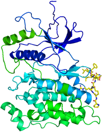
Protein C-ets-1 is a protein that in humans is encoded by the ETS1 gene. The protein encoded by this gene belongs to the ETS family of transcription factors.

<span class="mw-page-title-main">MAP2K4</span> Protein-coding gene in the species Homo sapiens

Dual-specificity mitogen-activated protein kinase kinase 4 is an enzyme that in humans is encoded by the MAP2K4 gene.

<span class="mw-page-title-main">MAP2K6</span> Protein-coding gene in the species Homo sapiens

Dual specificity mitogen-activated protein kinase kinase 6 also known as MAP kinase kinase 6 or MAPK/ERK kinase 6 is an enzyme that in humans is encoded by the MAP2K6 gene, on chromosome 17.

<span class="mw-page-title-main">MAP2K3</span> Protein-coding gene in the species Homo sapiens

Dual specificity mitogen-activated protein kinase kinase 3 is an enzyme that in humans is encoded by the MAP2K3 gene.

<span class="mw-page-title-main">DNA damage-inducible transcript 3</span> Human protein and coding gene

DNA damage-inducible transcript 3, also known as C/EBP homologous protein (CHOP), is a pro-apoptotic transcription factor that is encoded by the DDIT3 gene. It is a member of the CCAAT/enhancer-binding protein (C/EBP) family of DNA-binding transcription factors. The protein functions as a dominant-negative inhibitor by forming heterodimers with other C/EBP members, preventing their DNA binding activity. The protein is implicated in adipogenesis and erythropoiesis and has an important role in the cell's stress response.

<span class="mw-page-title-main">MAP2K7</span> Protein-coding gene in the species Homo sapiens

Dual specificity mitogen-activated protein kinase kinase 7, also known as MAP kinase kinase 7 or MKK7, is an enzyme that in humans is encoded by the MAP2K7 gene. This protein is a member of the mitogen-activated protein kinase kinase family. The MKK7 protein exists as six different isoforms with three possible N-termini and two possible C-termini.

<span class="mw-page-title-main">MDC1</span> Protein-coding gene in the species Homo sapiens

Mediator of DNA damage checkpoint protein 1 is a 2080 amino acid long protein that in humans is encoded by the MDC1 gene located on the short arm (p) of chromosome 6. MDC1 protein is a regulator of the Intra-S phase and the G2/M cell cycle checkpoints and recruits repair proteins to the site of DNA damage. It is involved in determining cell survival fate in association with tumor suppressor protein p53. This protein also goes by the name Nuclear Factor with BRCT Domain 1 (NFBD1).

<span class="mw-page-title-main">MAP3K12</span>

Mitogen-activated protein kinase 12 is an enzyme that in humans is encoded by the MAP3K12 gene.

<span class="mw-page-title-main">Protein phosphorylation</span> Process of introducing a phosphate group on to a protein

Protein phosphorylation is a reversible post-translational modification of proteins in which an amino acid residue is phosphorylated by a protein kinase by the addition of a covalently bound phosphate group. Phosphorylation alters the structural conformation of a protein, causing it to become either activated or deactivated, or otherwise modifying its function. Approximately 13000 human proteins have sites that are phosphorylated.

<span class="mw-page-title-main">Sirtuin 6</span> Protein-coding gene in the species Homo sapiens

Sirtuin 6 is a stress responsive protein deacetylase and mono-ADP ribosyltransferase enzyme encoded by the SIRT6 gene. In laboratory research, SIRT6 appears to function in multiple molecular pathways related to aging, including DNA repair, telomere maintenance, glycolysis and inflammation. SIRT6 is member of the mammalian sirtuin family of proteins, which are homologs to the yeast Sir2 protein.

<span class="mw-page-title-main">MAPK10</span> Protein-coding gene in the species Homo sapiens

Mitogen-activated protein kinase 10 also known as c-Jun N-terminal kinase 3 (JNK3) is an enzyme that in humans is encoded by the MAPK10 gene.

The Akt signaling pathway or PI3K-Akt signaling pathway is a signal transduction pathway that promotes survival and growth in response to extracellular signals. Key proteins involved are PI3K and Akt.

DNA damage is an alteration in the chemical structure of DNA, such as a break in a strand of DNA, a nucleobase missing from the backbone of DNA, or a chemically changed base such as 8-OHdG. DNA damage can occur naturally or via environmental factors, but is distinctly different from mutation, although both are types of error in DNA. DNA damage is an abnormal chemical structure in DNA, while a mutation is a change in the sequence of base pairs. DNA damages cause changes in the structure of the genetic material and prevents the replication mechanism from functioning and performing properly. The DNA damage response (DDR) is a complex signal transduction pathway which recognizes when DNA is damaged and initiates the cellular response to the damage.

References

  1. 1 2 Ip YT, Davis RJ (April 1998). "Signal transduction by the c-Jun N-terminal kinase (JNK)--from inflammation to development". Curr. Opin. Cell Biol. 10 (2): 205–19. doi:10.1016/S0955-0674(98)80143-9. PMID   9561845.
  2. Waetzig V, Herdegen T (2005). "Context-specific inhibition of JNKs: overcoming the dilemma of protection and damage". Br. J. Pharmacol. 26 (9): 455–61. doi:10.1016/j.tips.2005.07.006. PMID   16054242.
  3. 1 2 Bode AM, Dong Z (August 2007). "The Functional Contrariety of JNK". Mol. Carcinog. 46 (8): 591–8. doi:10.1002/mc.20348. PMC   2832829 . PMID   17538955. The protein products of jnk1 and jnk2 are believed to be expressed in every cell and tissue type, whereas the JNK3 protein is found primarily in brain and to a lesser extent in heart and testis
  4. Vlahopoulos S, Zoumpourlis VC (August 2004). "JNK: a key modulator of intracellular signaling". Biochemistry Mosc. 69 (8): 844–54. doi:10.1023/B:BIRY.0000040215.02460.45. PMID   15377263. S2CID   39149612.
  5. Oltmanns U, Issa R, Sukkar MB, John M, Chung KF (July 2003). "Role of c-jun N-terminal kinase in the induced release of GM-CSF, RANTES and IL-8 from human airway smooth muscle cells". Br. J. Pharmacol. 139 (6): 1228–34. doi:10.1038/sj.bjp.0705345. PMC   1573939 . PMID   12871843.
  6. Aloyz, R. S.; Bamji, S. X.; Pozniak, C. D.; Toma, J. G.; Atwal, J.; Kaplan, D. R.; Miller, F. D. (1998). "P53 is essential for developmental neuron death as regulated by the TrkA and p75 neurotrophin receptors". The Journal of Cell Biology. 143 (6): 1691–2303. doi:10.1083/jcb.143.6.1691. PMC   2132983 . PMID   9852160.
  7. Becker, E. B.; Howell, J.; Kodama, Y.; Barker, P. A.; Bonni, A. (2004). "Characterization of the c-Jun N-terminal kinase-BimEL signaling pathway in neuronal apoptosis". The Journal of Neuroscience. 24 (40): 8762–8770. doi:10.1523/JNEUROSCI.2953-04.2004. PMC   6729963 . PMID   15470142.
  8. Liu B, Yip RK, Zhou Z (2012). "Chromatin remodeling, DNA damage repair and aging". Curr. Genomics. 13 (7): 533–47. doi:10.2174/138920212803251373. PMC   3468886 . PMID   23633913.
  9. 1 2 3 Sellou H, Lebeaupin T, Chapuis C, Smith R, Hegele A, Singh HR, Kozlowski M, Bultmann S, Ladurner AG, Timinszky G, Huet S (2016). "The poly(ADP-ribose)-dependent chromatin remodeler Alc1 induces local chromatin relaxation upon DNA damage". Mol. Biol. Cell. 27 (24): 3791–3799. doi:10.1091/mbc.E16-05-0269. PMC   5170603 . PMID   27733626.
  10. 1 2 Van Meter M, Simon M, Tombline G, May A, Morello TD, Hubbard BP, Bredbenner K, Park R, Sinclair DA, Bohr VA, Gorbunova V, Seluanov A (2016). "JNK Phosphorylates SIRT6 to Stimulate DNA Double-Strand Break Repair in Response to Oxidative Stress by Recruiting PARP1 to DNA Breaks". Cell Rep. 16 (10): 2641–50. doi:10.1016/j.celrep.2016.08.006. PMC   5089070 . PMID   27568560.
  11. 1 2 Haince JF, McDonald D, Rodrigue A, Déry U, Masson JY, Hendzel MJ, Poirier GG (2008). "PARP1-dependent kinetics of recruitment of MRE11 and NBS1 proteins to multiple DNA damage sites". J. Biol. Chem. 283 (2): 1197–208. doi: 10.1074/jbc.M706734200 . PMID   18025084.
  12. 1 2 3 Calses PC, Dhillon KK, Tucker N, Chi Y, Huang JW, Kawasumi M, Nghiem P, Wang Y, Clurman BE, Jacquemont C, Gafken PR, Sugasawa K, Saijo M, Taniguchi T (2017). "DGCR8 Mediates Repair of UV-Induced DNA Damage Independently of RNA Processing". Cell Rep. 19 (1): 162–174. doi:10.1016/j.celrep.2017.03.021. PMC   5423785 . PMID   28380355.
  13. Wang MC, Bohmann D, Jasper H (2003). "JNK signaling confers tolerance to oxidative stress and extends lifespan in Drosophila". Dev. Cell. 5 (5): 811–6. doi: 10.1016/s1534-5807(03)00323-x . PMID   14602080.
  14. Wang MC, Bohmann D, Jasper H (2005). "JNK extends life span and limits growth by antagonizing cellular and organism-wide responses to insulin signaling". Cell. 121 (1): 115–25. doi: 10.1016/j.cell.2005.02.030 . PMID   15820683. S2CID   18365708.
  15. 1 2 Oh SW, Mukhopadhyay A, Svrzikapa N, Jiang F, Davis RJ, Tissenbaum HA (2005). "JNK regulates lifespan in Caenorhabditis elegans by modulating nuclear translocation of forkhead transcription factor/DAF-16". Proc. Natl. Acad. Sci. U.S.A. 102 (12): 4494–9. Bibcode:2005PNAS..102.4494O. doi: 10.1073/pnas.0500749102 . PMC   555525 . PMID   15767565.