Sequence motif

Last updated
A DNA sequence motif represented as a sequence logo for the LexA-binding motif. LexA gram positive bacteria sequence logo.png
A DNA sequence motif represented as a sequence logo for the LexA-binding motif.

In biology, a sequence motif is a nucleotide or amino-acid sequence pattern that is widespread and usually assumed to be related to biological function of the macromolecule. For example, an N-glycosylation site motif can be defined as Asn, followed by anything but Pro, followed by either Ser or Thr, followed by anything but Pro residue.

Contents

Overview

When a sequence motif appears in the exon of a gene, it may encode the "structural motif" of a protein; that is a stereotypical element of the overall structure of the protein. Nevertheless, motifs need not be associated with a distinctive secondary structure. "Noncoding" sequences are not translated into proteins, and nucleic acids with such motifs need not deviate from the typical shape (e.g. the "B-form" DNA double helix).

Outside of gene exons, there exist regulatory sequence motifs and motifs within the "junk", such as satellite DNA. Some of these are believed to affect the shape of nucleic acids [1] (see for example RNA self-splicing), but this is only sometimes the case. For example, many DNA binding proteins that have affinity for specific DNA binding sites bind DNA in only its double-helical form. They are able to recognize motifs through contact with the double helix's major or minor groove.

Short coding motifs, which appear to lack secondary structure, include those that label proteins for delivery to particular parts of a cell, or mark them for phosphorylation.

Within a sequence or database of sequences, researchers search and find motifs using computer-based techniques of sequence analysis, such as BLAST. Such techniques belong to the discipline of bioinformatics. See also consensus sequence.

Motif Representation

Consider the N-glycosylation site motif mentioned above:

Asn, followed by anything but Pro, followed by either Ser or Thr, followed by anything but Pro

This pattern may be written as N{P}[ST]{P} where N = Asn, P = Pro, S = Ser, T = Thr; {X} means any amino acid except X; and [XY] means either X or Y.

The notation [XY] does not give any indication of the probability of X or Y occurring in the pattern. Observed probabilities can be graphically represented using sequence logos. Sometimes patterns are defined in terms of a probabilistic model such as a hidden Markov model.

Motifs and consensus sequences

The notation [XYZ] means X or Y or Z, but does not indicate the likelihood of any particular match. For this reason, two or more patterns are often associated with a single motif: the defining pattern, and various typical patterns.

For example, the defining sequence for the IQ motif may be taken to be:

[FILV]Qxxx[RK]Gxxx[RK]xx[FILVWY]

where x signifies any amino acid, and the square brackets indicate an alternative (see below for further details about notation).

Usually, however, the first letter is I, and both [RK] choices resolve to R. Since the last choice is so wide, the pattern IQxxxRGxxxR is sometimes equated with the IQ motif itself, but a more accurate description would be a consensus sequence for the IQ motif.

Pattern description notations

Several notations for describing motifs are in use but most of them are variants of standard notations for regular expressions and use these conventions:

The fundamental idea behind all these notations is the matching principle, which assigns a meaning to a sequence of elements of the pattern notation:

a sequence of elements of the pattern notation matches a sequence of amino acids if and only if the latter sequence can be partitioned into subsequences in such a way that each pattern element matches the corresponding subsequence in turn.

Thus the pattern [AB] [CDE] F matches the six amino acid sequences corresponding to ACF, ADF, AEF, BCF, BDF, and BEF.

Different pattern description notations have other ways of forming pattern elements. One of these notations is the PROSITE notation, described in the following subsection.

PROSITE pattern notation

The PROSITE notation uses the IUPAC one-letter codes and conforms to the above description with the exception that a concatenation symbol, '-', is used between pattern elements, but it is often dropped between letters of the pattern alphabet.

PROSITE allows the following pattern elements in addition to those described previously:

  • The lower case letter 'x' can be used as a pattern element to denote any amino acid.
  • A string of characters drawn from the alphabet and enclosed in braces (curly brackets) denotes any amino acid except for those in the string. For example, {ST} denotes any amino acid other than S or T.
  • If a pattern is restricted to the N-terminal of a sequence, the pattern is prefixed with '<'.
  • If a pattern is restricted to the C-terminal of a sequence, the pattern is suffixed with '>'.
  • The character '>' can also occur inside a terminating square bracket pattern, so that S[T>] matches both "ST" and "S>".
  • If e is a pattern element, and m and n are two decimal integers with m <= n, then:
    • e(m) is equivalent to the repetition of e exactly m times;
    • e(m,n) is equivalent to the repetition of e exactly k times for any integer k satisfying: m <= k <= n.

Some examples:

  • x(3) is equivalent to x-x-x.
  • x(2,4) matches any sequence that matches x-x or x-x-x or x-x-x-x.

The signature of the C2H2-type zinc finger domain is:

  • C-x(2,4)-C-x(3)-[LIVMFYWC]-x(8)-H-x(3,5)-H

Matrices

A matrix of numbers containing scores for each residue or nucleotide at each position of a fixed-length motif. There are two types of weight matrices.

  • A position frequency matrix (PFM) records the position-dependent frequency of each residue or nucleotide. PFMs can be experimentally determined from SELEX experiments or computationally discovered by tools such as MEME using hidden Markov models.
  • A position weight matrix (PWM) contains log odds weights for computing a match score. A cutoff is needed to specify whether an input sequence matches the motif or not. PWMs are calculated from PFMs. PWMs are also known as PSSMs.

An example of a PFM from the TRANSFAC database for the transcription factor AP-1:

PosACGTIUPAC
016281R
023590S
0300017T
0400170G
0517000A
0601601C
073239T
084724N
099611M
104373N
116317W

The first column specifies the position, the second column contains the number of occurrences of A at that position, the third column contains the number of occurrences of C at that position, the fourth column contains the number of occurrences of G at that position, the fifth column contains the number of occurrences of T at that position, and the last column contains the IUPAC notation for that position. Note that the sums of occurrences for A, C, G, and T for each row should be equal because the PFM is derived from aggregating several consensus sequences.

Motif Discovery

Overview

The sequence motif discovery process has been well-developed since the 1990s. In particular, most of the existing motif discovery research focuses on DNA motifs. With the advances in high-throughput sequencing, such motif discovery problems are challenged by both the sequence pattern degeneracy issues and the data-intensive computational scalability issues.

Process of discovery

A flowchart depicting the process of motif discovery Sequence Motif Discovery.jpg
A flowchart depicting the process of motif discovery

Motif discovery happens in three major phases. A pre-processing stage where sequences are meticulously prepared in assembly and cleaning steps. Assembly involves selecting sequences that contain the desired motif in large quantities, and extraction of unwanted sequences using clustering. Cleaning then ensures the removal of any confounding elements. Next there is the discovery stage. In this phase sequences are represented using consensus strings or Position-specific Weight Matrices (PWM). After motif representation, an objective function is chosen and a suitable search algorithm is applied to uncover the motifs. Finally the post-processing stage involves evaluating the discovered motifs. [2]

De novo motif discovery

There are software programs which, given multiple input sequences, attempt to identify one or more candidate motifs. One example is the Multiple EM for Motif Elicitation (MEME) algorithm, which generates statistical information for each candidate. [3] There are more than 100 publications detailing motif discovery algorithms; Weirauch et al. evaluated many related algorithms in a 2013 benchmark. [4] The planted motif search is another motif discovery method that is based on combinatorial approach.

Phylogenetic motif discovery

Motifs have also been discovered by taking a phylogenetic approach and studying similar genes in different species. For example, by aligning the amino acid sequences specified by the GCM (glial cells missing) gene in man, mouse and D. melanogaster, Akiyama and others discovered a pattern which they called the GCM motif in 1996. [5] It spans about 150 amino acid residues, and begins as follows:

WDIND*.*P..*...D.F.*W***.**.IYS**...A.*H*S*WAMRNTNNHN

Here each . signifies a single amino acid or a gap, and each * indicates one member of a closely related family of amino acids. The authors were able to show that the motif has DNA binding activity.

A similar approach is commonly used by modern protein domain databases such as Pfam: human curators would select a pool of sequences known to be related and use computer programs to align them and produce the motif profile (Pfam uses HMMs, which can be used to identify other related proteins. [6] A phylogenic approach can also be used to enhance the de novo MEME algorithm, with PhyloGibbs being an example. [7]

De novo motif pair discovery

In 2017, MotifHyades has been developed as a motif discovery tool that can be directly applied to paired sequences. [8]

De novo motif recognition from protein

In 2018, a Markov random field approach has been proposed to infer DNA motifs from DNA-binding domains of proteins. [9]

Motif Discovery Algorithms

Motif discovery algorithms use diverse strategies to uncover patterns in DNA sequences. Integrating enumerative, probabilistic, and nature-inspired approaches, demonstrate their adaptability, with the use of multiple methods proving effective in enhancing identification accuracy.

Enumerative Approach: [2]

Initiating the motif discovery journey, the enumerative approach witnesses algorithms meticulously generating and evaluating potential motifs. Pioneering this domain are Simple Word Enumeration techniques, such as YMF and DREME, which systematically go through the sequence in search of short motifs. Complementing these, Clustering-Based Methods such as CisFinder employ nucleotide substitution matrices for motif clustering, effectively mitigating redundancy. Concurrently, Tree-Based Methods like Weeder and FMotif exploit tree structures, and Graph Theoretic-Based Methods (e.g., WINNOWER) employ graph representations, demonstrating the richness of enumeration strategies.

Probabilistic Approach: [2]

Diverging into the probabilistic realm, this approach capitalizes on probability models to discern motifs within sequences. MEME, a deterministic exemplar, employs Expectation-Maximization for optimizing Position Weight Matrices (PWMs) and unraveling conserved regions in unaligned DNA sequences. Contrasting this, stochastic methodologies like Gibbs Sampling initiate motif discovery with random motif position assignments, iteratively refining the predictions. This probabilistic framework adeptly captures the inherent uncertainty associated with motif discovery.

Advanced Approach: [2]

Evolving further, advanced motif discovery embraces sophisticated techniques, with Bayesian modeling [10] taking center stage. LOGOS and BaMM, exemplifying this cohort, intricately weave Bayesian approaches and Markov models into their fabric for motif identification. The incorporation of Bayesian clustering methods enhances the probabilistic foundation, providing a holistic framework for pattern recognition in DNA sequences.

Nature-Inspired and Heuristic Algorithms: [2]

A distinct category unfolds, wherein algorithms draw inspiration from the biological realm. Genetic Algorithms (GA), epitomized by FMGA and MDGA, [11] navigate motif search through genetic operators and specialized strategies. Harnessing swarm intelligence principles, Particle Swarm Optimization (PSO), Artificial Bee Colony (ABC) algorithms, and Cuckoo Search (CS) algorithms, featured in GAEM, GARP, and MACS, venture into pheromone-based exploration. These algorithms, mirroring nature's adaptability and cooperative dynamics, serve as avant-garde strategies for motif identification. The synthesis of heuristic techniques in hybrid approaches underscores the adaptability of these algorithms in the intricate domain of motif discovery.

This chart shows many different types of algorithms used in the discovery of sequence motifs and their categories Sequence motif algorithm figure.jpg
This chart shows many different types of algorithms used in the discovery of sequence motifs and their categories

Motif Cases

Three-dimensional chain codes

The E. coli lactose operon repressor LacI ( PDB: 1lcc chain A) and E. coli catabolite gene activator ( PDB: 3gap chain A) both have a helix-turn-helix motif, but their amino acid sequences do not show much similarity, as shown in the table below. In 1997, Matsuda, et al. devised a code they called the "three-dimensional chain code" for representing the protein structure as a string of letters. This encoding scheme reveals the similarity between the proteins much more clearly than the amino acid sequence (example from article): [12] The code encodes the torsion angles between alpha-carbons of the protein backbone. "W" always corresponds to an alpha helix.

3D chain codeAmino acid sequence
1lccATWWWWWWWKCLKWWWWWWGLYDVAEYAGVSYQTVSRVV
3gapAKWWWWWWGKCFKWWWWWWWRQEIGQIVGCSRETVGRIL

See also

Related Research Articles

<span class="mw-page-title-main">Alpha helix</span> Type of secondary structure of proteins

An alpha helix is a sequence of amino acids in a protein that are twisted into a coil.

<span class="mw-page-title-main">Bioinformatics</span> Computational analysis of large, complex sets of biological data

Bioinformatics is an interdisciplinary field of science that develops methods and software tools for understanding biological data, especially when the data sets are large and complex. Bioinformatics uses biology, chemistry, physics, computer science, computer programming, information engineering, mathematics and statistics to analyze and interpret biological data. The subsequent process of analyzing and interpreting data is referred to as computational biology.

<span class="mw-page-title-main">Sequence alignment</span> Process in bioinformatics that identifies equivalent sites within molecular sequences

In bioinformatics, a sequence alignment is a way of arranging the sequences of DNA, RNA, or protein to identify regions of similarity that may be a consequence of functional, structural, or evolutionary relationships between the sequences. Aligned sequences of nucleotide or amino acid residues are typically represented as rows within a matrix. Gaps are inserted between the residues so that identical or similar characters are aligned in successive columns. Sequence alignments are also used for non-biological sequences such as calculating the distance cost between strings in a natural language, or to display financial data.

Grammar theory to model symbol strings originated from work in computational linguistics aiming to understand the structure of natural languages. Probabilistic context free grammars (PCFGs) have been applied in probabilistic modeling of RNA structures almost 40 years after they were introduced in computational linguistics.

<span class="mw-page-title-main">Protein structure prediction</span> Type of biological prediction

Protein structure prediction is the inference of the three-dimensional structure of a protein from its amino acid sequence—that is, the prediction of its secondary and tertiary structure from primary structure. Structure prediction is different from the inverse problem of protein design. Protein structure prediction is one of the most important goals pursued by computational biology; it is important in medicine and biotechnology.

<span class="mw-page-title-main">Nucleic acid sequence</span> Succession of nucleotides in a nucleic acid

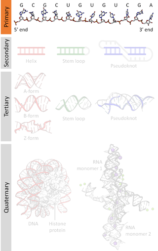
A nucleic acid sequence is a succession of bases within the nucleotides forming alleles within a DNA or RNA (GACU) molecule. This succession is denoted by a series of a set of five different letters that indicate the order of the nucleotides. By convention, sequences are usually presented from the 5' end to the 3' end. For DNA, with its double helix, there are two possible directions for the notated sequence; of these two, the sense strand is used. Because nucleic acids are normally linear (unbranched) polymers, specifying the sequence is equivalent to defining the covalent structure of the entire molecule. For this reason, the nucleic acid sequence is also termed the primary structure.

In bioinformatics, BLAST is an algorithm and program for comparing primary biological sequence information, such as the amino-acid sequences of proteins or the nucleotides of DNA and/or RNA sequences. A BLAST search enables a researcher to compare a subject protein or nucleotide sequence with a library or database of sequences, and identify database sequences that resemble the query sequence above a certain threshold. For example, following the discovery of a previously unknown gene in the mouse, a scientist will typically perform a BLAST search of the human genome to see if humans carry a similar gene; BLAST will identify sequences in the human genome that resemble the mouse gene based on similarity of sequence.

In a chain-like biological molecule, such as a protein or nucleic acid, a structural motif is a common three-dimensional structure that appears in a variety of different, evolutionarily unrelated molecules. A structural motif does not have to be associated with a sequence motif; it can be represented by different and completely unrelated sequences in different proteins or RNA.

<span class="mw-page-title-main">Structural bioinformatics</span> Bioinformatics subfield

Structural bioinformatics is the branch of bioinformatics that is related to the analysis and prediction of the three-dimensional structure of biological macromolecules such as proteins, RNA, and DNA. It deals with generalizations about macromolecular 3D structures such as comparisons of overall folds and local motifs, principles of molecular folding, evolution, binding interactions, and structure/function relationships, working both from experimentally solved structures and from computational models. The term structural has the same meaning as in structural biology, and structural bioinformatics can be seen as a part of computational structural biology. The main objective of structural bioinformatics is the creation of new methods of analysing and manipulating biological macromolecular data in order to solve problems in biology and generate new knowledge.

In molecular biology and bioinformatics, the consensus sequence is the calculated sequence of most frequent residues, either nucleotide or amino acid, found at each position in a sequence alignment. It represents the results of multiple sequence alignments in which related sequences are compared to each other and similar sequence motifs are calculated. Such information is important when considering sequence-dependent enzymes such as RNA polymerase.

Computational genomics refers to the use of computational and statistical analysis to decipher biology from genome sequences and related data, including both DNA and RNA sequence as well as other "post-genomic" data. These, in combination with computational and statistical approaches to understanding the function of the genes and statistical association analysis, this field is also often referred to as Computational and Statistical Genetics/genomics. As such, computational genomics may be regarded as a subset of bioinformatics and computational biology, but with a focus on using whole genomes to understand the principles of how the DNA of a species controls its biology at the molecular level and beyond. With the current abundance of massive biological datasets, computational studies have become one of the most important means to biological discovery.

In molecular biology, protein threading, also known as fold recognition, is a method of protein modeling which is used to model those proteins which have the same fold as proteins of known structures, but do not have homologous proteins with known structure. It differs from the homology modeling method of structure prediction as it is used for proteins which do not have their homologous protein structures deposited in the Protein Data Bank (PDB), whereas homology modeling is used for those proteins which do. Threading works by using statistical knowledge of the relationship between the structures deposited in the PDB and the sequence of the protein which one wishes to model.

A position weight matrix (PWM), also known as a position-specific weight matrix (PSWM) or position-specific scoring matrix (PSSM), is a commonly used representation of motifs (patterns) in biological sequences.

<span class="mw-page-title-main">Multiple sequence alignment</span> Alignment of more than two molecular sequences

Multiple sequence alignment (MSA) is the process or the result of sequence alignment of three or more biological sequences, generally protein, DNA, or RNA. These alignments are used to infer evolutionary relationships via phylogenetic analysis and can highlight homologous features between sequences. Alignments highlight mutation events such as point mutations, insertion mutations and deletion mutations, and alignments are used to assess sequence conservation and infer the presence and activity of protein domains, tertiary structures, secondary structures, and individual amino acids or nucleotides.

<span class="mw-page-title-main">Biomolecular structure</span> 3D conformation of a biological sequence, like DNA, RNA, proteins

Biomolecular structure is the intricate folded, three-dimensional shape that is formed by a molecule of protein, DNA, or RNA, and that is important to its function. The structure of these molecules may be considered at any of several length scales ranging from the level of individual atoms to the relationships among entire protein subunits. This useful distinction among scales is often expressed as a decomposition of molecular structure into four levels: primary, secondary, tertiary, and quaternary. The scaffold for this multiscale organization of the molecule arises at the secondary level, where the fundamental structural elements are the molecule's various hydrogen bonds. This leads to several recognizable domains of protein structure and nucleic acid structure, including such secondary-structure features as alpha helixes and beta sheets for proteins, and hairpin loops, bulges, and internal loops for nucleic acids. The terms primary, secondary, tertiary, and quaternary structure were introduced by Kaj Ulrik Linderstrøm-Lang in his 1951 Lane Medical Lectures at Stanford University.

<span class="mw-page-title-main">HMMER</span> Software package for sequence analysis

HMMER is a free and commonly used software package for sequence analysis written by Sean Eddy. Its general usage is to identify homologous protein or nucleotide sequences, and to perform sequence alignments. It detects homology by comparing a profile-HMM to either a single sequence or a database of sequences. Sequences that score significantly better to the profile-HMM compared to a null model are considered to be homologous to the sequences that were used to construct the profile-HMM. Profile-HMMs are constructed from a multiple sequence alignment in the HMMER package using the hmmbuild program. The profile-HMM implementation used in the HMMER software was based on the work of Krogh and colleagues. HMMER is a console utility ported to every major operating system, including different versions of Linux, Windows, and macOS.

<span class="mw-page-title-main">Nucleic acid secondary structure</span>

Nucleic acid secondary structure is the basepairing interactions within a single nucleic acid polymer or between two polymers. It can be represented as a list of bases which are paired in a nucleic acid molecule. The secondary structures of biological DNAs and RNAs tend to be different: biological DNA mostly exists as fully base paired double helices, while biological RNA is single stranded and often forms complex and intricate base-pairing interactions due to its increased ability to form hydrogen bonds stemming from the extra hydroxyl group in the ribose sugar.

<span class="mw-page-title-main">Gary Stormo</span> American geneticist (born 1950)

Gary Stormo is an American geneticist and currently Joseph Erlanger Professor in the Department of Genetics and the Center for Genome Sciences and Systems Biology at Washington University School of Medicine in St Louis. He is considered one of the pioneers of bioinformatics and genomics. His research combines experimental and computational approaches in order to identify and predict regulatory sequences in DNA and RNA, and their contributions to the regulatory networks that control gene expression.

PSI-blast based secondary structure PREDiction (PSIPRED) is a method used to investigate protein structure. It uses artificial neural network machine learning methods in its algorithm. It is a server-side program, featuring a website serving as a front-end interface, which can predict a protein's secondary structure from the primary sequence.

<span class="mw-page-title-main">Protein tandem repeats</span>

An array of protein tandem repeats is defined as several adjacent copies having the same or similar sequence motifs. These periodic sequences are generated by internal duplications in both coding and non-coding genomic sequences. Repetitive units of protein tandem repeats are considerably diverse, ranging from the repetition of a single amino acid to domains of 100 or more residues.

References

Primary sources

  1. Dlakić, Mensur; Harrington, Rodney E. (1996). "The Effects of Sequence Context on DNA Curvature". Proceedings of the National Academy of Sciences of the United States of America. 93 (9): 3847–3852. Bibcode:1996PNAS...93.3847D. doi: 10.1073/pnas.93.9.3847 . ISSN   0027-8424. JSTOR   39155. PMC   39447 . PMID   8632978.
  2. 1 2 3 4 5 Hashim, Fatma A.; Mabrouk, Mai S.; Al-Atabany, Walid (2019). "Review of Different Sequence Motif Finding Algorithms". Avicenna Journal of Medical Biotechnology. 11 (2): 130–148. ISSN   2008-2835. PMC   6490410 . PMID   31057715.
  3. Bailey TL, Williams N, Misleh C, Li WW (July 2006). "MEME: discovering and analyzing DNA and protein sequence motifs". Nucleic Acids Research. 34 (Web Server issue): W369-73. doi:10.1093/nar/gkl198. PMC   1538909 . PMID   16845028.
  4. Weirauch MT, Cote A, Norel R, Annala M, Zhao Y, Riley TR, et al. (February 2013). "Evaluation of methods for modeling transcription factor sequence specificity". Nature Biotechnology. 31 (2): 126–34. doi:10.1038/nbt.2486. PMC   3687085 . PMID   23354101.
  5. Akiyama Y, Hosoya T, Poole AM, Hotta Y (December 1996). "The gcm-motif: a novel DNA-binding motif conserved in Drosophila and mammals". Proceedings of the National Academy of Sciences of the United States of America. 93 (25): 14912–6. Bibcode:1996PNAS...9314912A. doi: 10.1073/pnas.93.25.14912 . PMC   26236 . PMID   8962155.
  6. "Modelling in Pfam". Pfam. Retrieved 14 December 2023.
  7. Siddharthan R, Siggia ED, van Nimwegen E (December 2005). "PhyloGibbs: a Gibbs sampling motif finder that incorporates phylogeny". PLOS Computational Biology. 1 (7): e67. Bibcode:2005PLSCB...1...67S. doi: 10.1371/journal.pcbi.0010067 . PMC   1309704 . PMID   16477324.
  8. Wong KC (October 2017). "MotifHyades: expectation maximization for de novo DNA motif pair discovery on paired sequences". Bioinformatics. 33 (19): 3028–3035. doi: 10.1093/bioinformatics/btx381 . PMID   28633280.
  9. Wong KC (September 2018). "DNA Motif Recognition Modeling from Protein Sequences". iScience. 7: 198–211. Bibcode:2018iSci....7..198W. doi:10.1016/j.isci.2018.09.003. PMC   6153143 . PMID   30267681.
  10. Miller, Andrew K.; Print, Cristin G.; Nielsen, Poul M. F.; Crampin, Edmund J. (2010-11-18). "A Bayesian search for transcriptional motifs". PLOS ONE. 5 (11): e13897. Bibcode:2010PLoSO...513897M. doi: 10.1371/journal.pone.0013897 . ISSN   1932-6203. PMC   2987817 . PMID   21124986.
  11. Che, Dongsheng; Song, Yinglei; Rasheed, Khaled (2005-06-25). "MDGA: Motif discovery using a genetic algorithm". Proceedings of the 7th annual conference on Genetic and evolutionary computation. GECCO '05. New York, NY, USA: Association for Computing Machinery. pp. 447–452. doi:10.1145/1068009.1068080. ISBN   978-1-59593-010-1. S2CID   7892935.
  12. Matsuda H, Taniguchi F, Hashimoto A (1997). "An approach to detection of protein structural motifs using an encoding scheme of backbone conformations" (PDF). Pacific Symposium on Biocomputing. Pacific Symposium on Biocomputing: 280–91. PMID   9390299.

Further reading

Primary sources