Protease inhibitor (biology)

Last updated

In biology and biochemistry, protease inhibitors, or antiproteases, [1] are molecules that inhibit the function of proteases (enzymes that aid the breakdown of proteins). Many naturally occurring protease inhibitors are proteins. [2]

Contents

In medicine, protease inhibitor is often used interchangeably with alpha 1-antitrypsin (A1AT, which is abbreviated PI for this reason). [3] A1AT is indeed the protease inhibitor most often involved in disease, namely in alpha-1 antitrypsin deficiency.

Classification

Protease inhibitors may be classified either by the type of protease they inhibit, or by their mechanism of action. In 2004 Rawlings and colleagues introduced a classification of protease inhibitors based on similarities detectable at the level of amino acid sequence. [4] This classification initially identified 48 families of inhibitors that could be grouped into 26 related superfamily (or clans) by their structure. According to the MEROPS database there are now 81 families of inhibitors. These families are named with an I followed by a number, for example, I14 contains hirudin-like inhibitors.

By protease

Classes of proteases are:

By mechanism

Classes of inhibitor mechanisms of action are:

Families

Inhibitor I4

This is a family of protease suicide inhibitors called the serpins. It contains inhibitors of multiple cysteine and serine protease families. Their mechanism of action relies on undergoing a large conformational change which inactivates their target's catalytic triad.

Inhibitor I9

Peptidase inhibitor I9
PDB 1spb EBI.jpg
subtilisin bpn' prosegment (77 residues) complexed with a mutant subtilisin bpn' (266 residues). crystal ph 4.6. crystallization temperature 20 c diffraction temperature-160 c
Identifiers
SymbolInhibitor_I9
Pfam PF05922
InterPro IPR010259
MEROPS I9
SCOP2 1gns / SCOPe / SUPFAM
Available protein structures:
Pfam   structures / ECOD  
PDB RCSB PDB; PDBe; PDBj
PDBsum structure summary

Proteinase propeptide inhibitors (sometimes referred to as activation peptides) are responsible for the modulation of folding and activity of the peptidase pro-enzyme or zymogen. The pro-segment docks into the enzyme, shielding the substrate binding site, thereby promoting inhibition of the enzyme. Several such propeptides share a similar topology, despite often low sequence identities. [5] [6] The propeptide region has an open-sandwich antiparallel-alpha/antiparallel-beta fold, with two alpha-helices and four beta-strands with a (beta/alpha/beta)x2 topology. The peptidase inhibitor I9 family contains the propeptide domain at the N-terminus of peptidases belonging to MEROPS family S8A, subtilisins. The propeptide is removed by proteolytic cleavage; removal activating the enzyme.

Inhibitor I10

Serine endopeptidase inhibitors
PDB 1ixu EBI.jpg
solution structure of marinostatin, a protease inhibitor, containing two ester linkages
Identifiers
SymbolInhibitor_I10
Pfam PF12559
InterPro IPR022217
Available protein structures:
Pfam   structures / ECOD  
PDB RCSB PDB; PDBe; PDBj
PDBsum structure summary

This family includes both microviridins and marinostatins. It seems likely that in both cases it is the C-terminus which becomes the active inhibitor after post-translational modifications of the full length, pre-peptide. It is the ester linkages within the key, 12-residue region that circularise the molecule giving it its inhibitory conformation.

Inhibitor I24

PinA peptidase inhibitor
Identifiers
SymbolInhibitor_I24
Pfam PF10465
InterPro IPR019506
MEROPS I24
Available protein structures:
Pfam   structures / ECOD  
PDB RCSB PDB; PDBe; PDBj
PDBsum structure summary

This family includes PinA, which inhibits the endopeptidase La. It binds to the La homotetramer but does not interfere with the ATP binding site or the active site of La.

Inhibitor I29

Cathepsin propeptide inhibitor domain (I29)
PDB 1cjl EBI.jpg
crystal structure of a cysteine protease proform
Identifiers
SymbolInhibitor_I29
Pfam PF08246
InterPro IPR013201
Available protein structures:
Pfam   structures / ECOD  
PDB RCSB PDB; PDBe; PDBj
PDBsum structure summary

The inhibitor I29 domain, which belongs to MEROPS peptidase inhibitor family I29, is found at the N-terminus of a variety of peptidase precursors that belong to MEROPS peptidase subfamily C1A; these include cathepsin L, papain, and procaricain. [7] It forms an alpha-helical domain that runs through the substrate-binding site, preventing access. Removal of this region by proteolytic cleavage results in activation of the enzyme. This domain is also found, in one or more copies, in a variety of cysteine peptidase inhibitors such as salarin. [8]

Inhibitor I34

Saccharopepsin inhibitor I34
PDB 1dp5 EBI.jpg
the structure of proteinase a complexed with an ia3 mutant inhibitor
Identifiers
SymbolInhibitor_I34
Pfam PF10466
InterPro IPR019507
MEROPS I34
Available protein structures:
Pfam   structures / ECOD  
PDB RCSB PDB; PDBe; PDBj
PDBsum structure summary

The saccharopepsin inhibitor I34 is highly specific for the aspartic peptidase saccharopepsin. In the absence of saccharopepsin it is largely unstructured, [9] but in its presence, the inhibitor undergoes a conformational change forming an almost perfect alpha-helix from Asn2 to Met32 in the active site cleft of the peptidase.

Inhibitor I36

Peptidase inhibitor family I36
PDB 1bhu EBI.jpg
the 3d structure of the streptomyces metalloproteinase inhibitor, smpi, isolated from streptomyces nigrescens tk-23, nmr, minimized average structure
Identifiers
SymbolInhibitor_I36
Pfam PF03995
Pfam clan CL0333
InterPro IPR007141
MEROPS I36
SCOP2 1bhu / SCOPe / SUPFAM
Available protein structures:
Pfam   structures / ECOD  
PDB RCSB PDB; PDBe; PDBj
PDBsum structure summary

The peptidase inhibitor family I36 domain is only found in a small number of proteins restricted to Streptomyces species. All have four conserved cysteines that probably form two disulphide bonds. One of these proteins from Streptomyces nigrescens, is the well characterised metalloproteinase inhibitor SMPI. [10] [11]

The structure of SMPI has been determined. It has 102 amino acid residues with two disulphide bridges and specifically inhibits metalloproteinases such as thermolysin, which belongs to MEROPS peptidase family M4. SMPI is composed of two beta-sheets, each consisting of four antiparallel beta-strands. The structure can be considered as two Greek key motifs with 2-fold internal symmetry, a Greek key beta-barrel. One unique structural feature found in SMPI is in its extension between the first and second strands of the second Greek key motif which is known to be involved in the inhibitory activity of SMPI. In the absence of sequence similarity, the SMPI structure shows clear similarity to both domains of the eye lens crystallins, both domains of the calcium sensor protein-S, as well as the single-domain yeast killer toxin. The yeast killer toxin structure was thought to be a precursor of the two-domain beta gamma-crystallin proteins, because of its structural similarity to each domain of the beta gamma-crystallins. SMPI thus provides another example of a single-domain protein structure that corresponds to the ancestral fold from which the two-domain proteins in the beta gamma-crystallin superfamily are believed to have evolved. [12]

Inhibitor I42

Chagasin family peptidase inhibitor I42
PDB 2fo8 EBI.jpg
solution structure of the trypanosoma cruzi cysteine protease inhibitor chagasin
Identifiers
SymbolInhibitor_I42
Pfam PF09394
InterPro IPR018990
MEROPS I42
Available protein structures:
Pfam   structures / ECOD  
PDB RCSB PDB; PDBe; PDBj
PDBsum structure summary

Inhibitor family I42 includes chagasin, a reversible inhibitor of papain-like cysteine proteases. [13] Chagasin has a beta-barrel structure, which is a unique variant of the immunoglobulin fold with homology to human CD8alpha. [14] [15]

Inhibitor I48

Peptidase inhibitor clitocypin
Identifiers
SymbolInhibitor_I48
Pfam PF10467
InterPro IPR019508
MEROPS I48
Available protein structures:
Pfam   structures / ECOD  
PDB RCSB PDB; PDBe; PDBj
PDBsum structure summary

Inhibitor family I48 includes clitocypin, which binds and inhibits cysteine proteinases. It has no similarity to any other known cysteine proteinase inhibitors but bears some similarity to a lectin-like family of proteins from mushrooms. [16]

Inhibitor I53

Thrombin inhibitor Madanin
Identifiers
SymbolInhibitor_I53
Pfam PF11714
InterPro IPR021716
MEROPS I53
Available protein structures:
Pfam   structures / ECOD  
PDB RCSB PDB; PDBe; PDBj
PDBsum structure summary

Members of this family are the peptidase inhibitor madanin proteins. These proteins were isolated from tick saliva. [17]

Inhibitor I67

Bromelain inhibitor VI
PDB 1bi6 EBI.jpg
nmr structure of bromelain inhibitor vi from pineapple stem
Identifiers
SymbolInhibitor_I67
Pfam PF11405
InterPro IPR022713
MEROPS I67
Available protein structures:
Pfam   structures / ECOD  
PDB RCSB PDB; PDBe; PDBj
PDBsum structure summary

Bromelain inhibitor VI, in the Inhibitor I67 family, is a double-chain inhibitor consisting of an 11-residue and a 41-residue chain.

Inhibitor I68

Carboxypeptidase inhibitor I68
PDB 1zli EBI.jpg
crystal structure of the tick carboxypeptidase inhibitor in complex with human carboxypeptidase b
Identifiers
SymbolInhibitor_I68
Pfam PF10468
InterPro IPR019509
MEROPS I68
Available protein structures:
Pfam   structures / ECOD  
PDB RCSB PDB; PDBe; PDBj
PDBsum structure summary

The Carboxypeptidase inhibitor I68 family represents a family of tick carboxypetidase inhibitors.

Inhibitor I78

Peptidase inhibitor I78 family
Identifiers
SymbolInhibitor_I78
Pfam PF11720
Pfam clan CL0367
InterPro IPR021719
MEROPS I78
Available protein structures:
Pfam   structures / ECOD  
PDB RCSB PDB; PDBe; PDBj
PDBsum structure summary

The peptidase inhibitor I78 family includes Aspergillus elastase inhibitor.

Compounds

See also

Related Research Articles

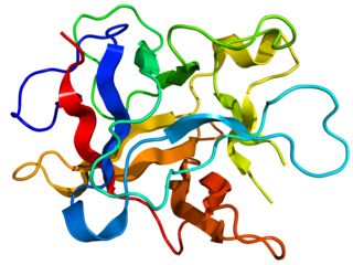
A metalloproteinase, or metalloprotease, is any protease enzyme whose catalytic mechanism involves a metal. An example is ADAM12 which plays a significant role in the fusion of muscle cells during embryo development, in a process known as myogenesis.

<span class="mw-page-title-main">Cystatin</span> Group of endogenous cysteine proteinase inhibitors

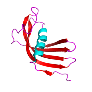
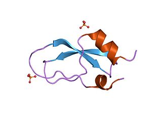
The cystatins are a family of cysteine protease inhibitors which share a sequence homology and a common tertiary structure of an alpha helix lying on top of an anti-parallel beta sheet. The family is subdivided as described below.

<span class="mw-page-title-main">ADAM (protein)</span>

ADAMs are a family of single-pass transmembrane and secreted metalloendopeptidases. All ADAMs are characterized by a particular domain organization featuring a pro-domain, a metalloprotease, a disintegrin, a cysteine-rich, an epidermal-growth factor like and a transmembrane domain, as well as a C-terminal cytoplasmic tail. Nonetheless, not all human ADAMs have a functional protease domain, which indicates that their biological function mainly depends on protein–protein interactions. Those ADAMs which are active proteases are classified as sheddases because they cut off or shed extracellular portions of transmembrane proteins. For example, ADAM10 can cut off part of the HER2 receptor, thereby activating it. ADAM genes are found in animals, choanoflagellates, fungi and some groups of green algae. Most green algae and all land plants likely lost ADAM proteins.

<span class="mw-page-title-main">Aspartic protease</span>

Aspartic proteases are a catalytic type of protease enzymes that use an activated water molecule bound to one or more aspartate residues for catalysis of their peptide substrates. In general, they have two highly conserved aspartates in the active site and are optimally active at acidic pH. Nearly all known aspartyl proteases are inhibited by pepstatin.

<span class="mw-page-title-main">Scorpion toxin</span>

Scorpion toxins are proteins found in the venom of scorpions. Their toxic effect may be mammal- or insect-specific and acts by binding with varying degrees of specificity to members of the Voltage-gated ion channel superfamily; specifically, voltage-gated sodium channels, voltage-gated potassium channels, and Transient Receptor Potential (TRP) channels. The result of this action is to activate or inhibit the action of these channels in the nervous and cardiac organ systems. For instance, α-scorpion toxins MeuNaTxα-12 and MeuNaTxα-13 from Mesobuthus eupeus are neurotoxins that target voltage-gated Na+ channels (Navs), inhibiting fast inactivation. In vivo assays of MeuNaTxα-12 and MeuNaTxα-13 effects on mammalian and insect Navs show differential potency. These recombinants exhibit their preferential affinity for mammalian and insect Na+ channels at the α-like toxins' active site, site 3, in order to inactivate the cell membrane depolarization faster[6]. The varying sensitivity of different Navs to MeuNaTxα-12 and MeuNaTxα-13 may be dependent on the substitution of a conserved Valine residue for a Phenylalanine residue at position 1630 of the LD4:S3-S4 subunit or due to various changes in residues in the LD4:S5-S6 subunit of the Navs. Ultimately, these actions can serve the purpose of warding off predators by causing pain or to subdue predators.

<span class="mw-page-title-main">Kunitz STI protease inhibitor</span>

Kunitz soybean trypsin inhibitor is a type of protein contained in legume seeds which functions as a protease inhibitor. Kunitz-type Soybean Trypsin Inhibitors are usually specific for either trypsin or chymotrypsin. They are thought to protect seeds against consumption by animal predators.

<span class="mw-page-title-main">Kunitz domain</span>

Kunitz domains are the active domains of proteins that inhibit the function of protein degrading enzymes or, more specifically, domains of Kunitz-type are protease inhibitors. They are relatively small with a length of about 50 to 60 amino acids and a molecular weight of 6 kDa. Examples of Kunitz-type protease inhibitors are aprotinin, Alzheimer's amyloid precursor protein (APP), and tissue factor pathway inhibitor (TFPI). Kunitz STI protease inhibitor, the trypsin inhibitor initially studied by Moses Kunitz, was extracted from soybeans.

<span class="mw-page-title-main">CHAP domain</span>

In molecular biology, the CHAP domain is a region between 110 and 140 amino acids that is found in proteins from bacteria, bacteriophages, archaea and eukaryotes of the family Trypanosomidae. The domain is named after the acronym cysteine, histidine-dependent amidohydrolases/peptidases. Many of these proteins are uncharacterised, but it has been proposed that they may function mainly in peptidoglycan hydrolysis. The CHAP domain is found in a wide range of protein architectures; it is commonly associated with bacterial type SH3 domains and with several families of amidase domains. It has been suggested that CHAP domain containing proteins utilise a catalytic cysteine residue in a nucleophilic-attack mechanism.

<span class="mw-page-title-main">SSI protease inhibitor</span>

In molecular biology the protein SSI is a Subtilisin inhibitor-like which stands for Streptomyces subtilisin inhibitor. This is a protease inhibitor. These are often synthesised as part of a larger precursor protein, either as a prepropeptide. The function of this protein domain is to prevent access of the substrate to the active site. It is found only in bacteria.

<span class="mw-page-title-main">Bowman–Birk protease inhibitor</span>

In molecular biology, the Bowman–Birk protease inhibitor family of proteins consists of eukaryotic proteinase inhibitors, belonging to MEROPS inhibitor family I12, clan IF. They mainly inhibit serine peptidases of the S1 family, but also inhibit S3 peptidases.

<span class="mw-page-title-main">Kazal domain</span>

The Kazal domain is an evolutionary conserved protein domain usually indicative of serine protease inhibitors. However, kazal-like domains are also seen in the extracellular part of agrins, which are not known to be protease inhibitors.

<span class="mw-page-title-main">Cysteine-rich secretory protein superfamily</span>

The CAP superfamily is a large superfamily of secreted proteins that are produced by a wide range of organisms, including prokaryotes and non-vertebrate eukaryotes.

<span class="mw-page-title-main">Carboxypeptidase A inhibitor</span>

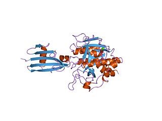
In molecular biology, the carboxypeptidase A inhibitor family is a family of proteins which is represented by the well-characterised metallocarboxypeptidase A inhibitor (MCPI) from potatoes, which belongs to the MEROPS inhibitor family I37, clan IE. It inhibits metallopeptidases belonging to MEROPS peptidase family M14, carboxypeptidase A. In Russet Burbank potatoes, it is a mixture of approximately equal amounts of two polypeptide chains containing 38 or 39 amino acid residues. The chains differ in their amino terminal sequence only and are resistant to fragmentation by proteases. The structure of the complex between bovine carboxypeptidase A and the 39-amino-acid carboxypeptidase A inhibitor from potatoes has been determined at 2.5-Angstrom resolution.

<span class="mw-page-title-main">Trappin protein transglutaminase binding domain</span>

In molecular biology, the trappin protein transglutaminase binding domain or cementoin is a protein domain found at the N-terminus of Whey Acidic Protein (WAP) domain-containing protease inhibitors such as trappin-2. This N-terminal domain enables it to become cross-linked to extracellular matrix proteins by transglutaminase. This domain contains several repeated motifs with the consensus sequence Gly-Gln-Asp-Pro-Val-Lys, and these together can anchor the whole molecule to extracellular matrix proteins, such as laminin, fibronectin, beta-crystallin, collagen IV, fibrinogen, and elastin, by transglutaminase-catalysed cross-links. The whole domain is rich in glutamine and lysine, thus allowing transglutaminase(s) to catalyse the formation of an intermolecular epsilon-(gamma-glutamyl)lysine isopeptide bond.

<span class="mw-page-title-main">Dipeptidyl-peptidase IV family</span>

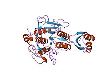
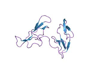
In molecular biology, the dipeptidyl-peptidase IV family is a family of serine peptidases which belong to MEROPS peptidase family S9, subfamily S9B. The protein fold of the peptidase domain for members of this family resembles that of serine carboxypeptidase D, the type example of clan SC. The type example of this family is Dipeptidyl peptidase-4.

<span class="mw-page-title-main">Ecotin</span>

In molecular biology, ecotin is a protease inhibitor which belongs to MEROPS inhibitor family I11, clan IN. Ecotins are dimeric periplasmic proteins from Escherichia coli and related Gram-negative bacteria that have been shown to be potent inhibitors of many trypsin-fold serine proteases of widely varying substrate specificity, which belong to MEROPS peptidase family S1. Phylogenetic analysis suggested that ecotin has an exogenous target, possibly neutrophil elastase. Ecotin from E. coli, Yersinia pestis, and Pseudomonas aeruginosa, all species that encounter the mammalian immune system, inhibit neutrophil elastase strongly while ecotin from the plant pathogen Pantoea citrea inhibits neutrophil elastase 1000-fold less potently. Ecotins all potently inhibit pancreatic digestive peptidases trypsin and chymotrypsin, while showing more variable inhibition of the blood peptidases Factor Xa, thrombin, and urokinase-type plasminogen activator.

<span class="mw-page-title-main">Haemadin</span>

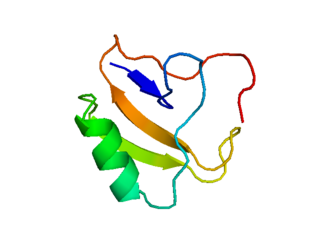
In molecular biology, haemadin is an anticoagulant peptide synthesised by the Indian leech, Haemadipsa sylvestris. It adopts a secondary structure consisting of five short beta-strands (beta1-beta5), which are arranged in two antiparallel distorted sheets formed by strands beta1-beta4-beta5 and beta2-beta3 facing each other. This beta-sandwich is stabilised by six enclosed cysteines arranged in a [1-2, 3-5, 4-6] disulfide pairing resulting in a disulfide-rich hydrophobic core that is largely inaccessible to bulk solvent. The close proximity of disulfide bonds [3-5] and [4-6] organises haemadin into four distinct loops. The N-terminal segment of this domain binds to the active site of thrombin, inhibiting it.

<span class="mw-page-title-main">Cyanobacterial clock proteins</span> Proteins that regulate circadian rhythms

In molecular biology, the cyanobacterial clock proteins are the main circadian regulator in cyanobacteria. The cyanobacterial clock proteins comprise three proteins: KaiA, KaiB and KaiC. The kaiABC complex may act as a promoter-nonspecific transcription regulator that represses transcription, possibly by acting on the state of chromosome compaction. This complex is expressed from a KaiABC operon.

<span class="mw-page-title-main">3C-like protease</span> Class of enzymes

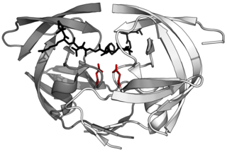
The 3C-like protease (3CLpro) or main protease (Mpro), formally known as C30 endopeptidase or 3-chymotrypsin-like protease, is the main protease found in coronaviruses. It cleaves the coronavirus polyprotein at eleven conserved sites. It is a cysteine protease and a member of the PA clan of proteases. It has a cysteine-histidine catalytic dyad at its active site and cleaves a Gln–(Ser/Ala/Gly) peptide bond.

<span class="mw-page-title-main">Pacifastin</span>

Pacifastin is a family of serine proteinase inhibitors found in arthropods. Pacifastin inhibits the serine peptidases trypsin and chymotrypsin.

References

  1. "antiprotease". The Free Dictionary.
  2. Roy, Mrinalini; Rawat, Aadish; Kaushik, Sanket; Jyoti, Anupam; Srivastava, Vijay Kumar (2022-08-01). "Endogenous cysteine protease inhibitors in upmost pathogenic parasitic protozoa". Microbiological Research. 261: 127061. doi:10.1016/j.micres.2022.127061. ISSN   0944-5013. PMID   35605309. S2CID   248741177.
  3. OMIM - PROTEASE INHIBITOR 1; PI
  4. Rawlings ND, Tolle DP, Barrett AJ (March 2004). "Evolutionary families of peptidase inhibitors". Biochem. J. 378 (Pt 3): 705–16. doi:10.1042/BJ20031825. PMC   1224039 . PMID   14705960.
  5. Tangrea MA, Bryan PN, Sari N, Orban J (July 2002). "Solution structure of the pro-hormone convertase 1 pro-domain from Mus musculus". J. Mol. Biol. 320 (4): 801–12. doi:10.1016/S0022-2836(02)00543-0. PMID   12095256.
  6. Jain SC, Shinde U, Li Y, Inouye M, Berman HM (November 1998). "The crystal structure of an autoprocessed Ser221Cys-subtilisin E-propeptide complex at 2.0 A resolution". J. Mol. Biol. 284 (1): 137–44. doi:10.1006/jmbi.1998.2161. PMID   9811547.
  7. Groves MR, Taylor MA, Scott M, Cummings NJ, Pickersgill RW, Jenkins JA (October 1996). "The prosequence of procaricain forms an alpha-helical domain that prevents access to the substrate-binding cleft". Structure. 4 (10): 1193–203. doi: 10.1016/s0969-2126(96)00127-x . PMID   8939744.
  8. Olonen A, Kalkkinen N, Paulin L (July 2003). "A new type of cysteine proteinase inhibitor--the salarin gene from Atlantic salmon (Salmo salar L.) and Arctic charr (Salvelinus alpinus)". Biochimie. 85 (7): 677–81. doi:10.1016/S0300-9084(03)00128-7. PMID   14505823.
  9. Green TB, Ganesh O, Perry K, Smith L, Phylip LH, Logan TM, Hagen SJ, Dunn BM, Edison AS (April 2004). "IA3, an aspartic proteinase inhibitor from Saccharomyces cerevisiae, is intrinsically unstructured in solution". Biochemistry. 43 (14): 4071–81. doi:10.1021/bi034823n. PMID   15065849.
  10. Tanaka K, Aoki H, Oda K, Murao S, Saito H, Takahashi H (November 1990). "Nucleotide sequence of the gene for a metalloproteinase inhibitor of Streptomyces nigrescens (SMPI)". Nucleic Acids Res. 18 (21): 6433. doi:10.1093/nar/18.21.6433. PMC   332545 . PMID   2243793.
  11. Murai H, Hara S, Ikenaka T, Oda K, Murao S (January 1985). "Amino acid sequence of Streptomyces metallo-proteinase inhibitor from Streptomyces nigrescens TK-23". J. Biochem. 97 (1): 173–80. doi:10.1093/oxfordjournals.jbchem.a135041. PMID   3888972.
  12. Ohno A, Tate S, Seeram SS, Hiraga K, Swindells MB, Oda K, Kainosho M (September 1998). "NMR structure of the Streptomyces metalloproteinase inhibitor, SMPI, isolated from Streptomyces nigrescens TK-23: another example of an ancestral beta gamma-crystallin precursor structure". J. Mol. Biol. 282 (2): 421–33. doi:10.1006/jmbi.1998.2022. PMID   9735297.
  13. Monteiro AC, Abrahamson M, Lima AP, Vannier-Santos MA, Scharfstein J (November 2001). "Identification, characterization and localization of chagasin, a tight-binding cysteine protease inhibitor in Trypanosoma cruzi". J. Cell Sci. 114 (Pt 21): 3933–42. doi:10.1242/jcs.114.21.3933. PMID   11719560.
  14. Figueiredo da Silva AA; de Carvalho Vieira L; Krieger MA; Goldenberg S; Zanchin NI; Guimarães BG (February 2007). "Crystal structure of chagasin, the endogenous cysteine-protease inhibitor from Trypanosoma cruzi". J. Struct. Biol. 157 (2): 416–23. doi:10.1016/j.jsb.2006.07.017. PMID   17011790.
  15. Wang SX, Pandey KC, Scharfstein J, Whisstock J, Huang RK, Jacobelli J, Fletterick RJ, Rosenthal PJ, Abrahamson M, Brinen LS, Rossi A, Sali A, McKerrow JH (May 2007). "The structure of chagasin in complex with a cysteine protease clarifies the binding mode and evolution of an inhibitor family". Structure. 15 (5): 535–43. doi: 10.1016/j.str.2007.03.012 . PMID   17502099.
  16. Brzin J, Rogelj B, Popovic T, Strukelj B, Ritonja A (June 2000). "Clitocypin, a new type of cysteine proteinase inhibitor from fruit bodies of mushroom clitocybe nebularis". J. Biol. Chem. 275 (26): 20104–9. doi: 10.1074/jbc.M001392200 . PMID   10748021.
  17. Iwanaga S, Okada M, Isawa H, Morita A, Yuda M, Chinzei Y (May 2003). "Identification and characterization of novel salivary thrombin inhibitors from the ixodidae tick, Haemaphysalis longicornis". Eur. J. Biochem. 270 (9): 1926–34. doi: 10.1046/j.1432-1033.2003.03560.x . PMID   12709051.
This article incorporates text from the public domain Pfam and InterPro: IPR022217
This article incorporates text from the public domain Pfam and InterPro: IPR019506
This article incorporates text from the public domain Pfam and InterPro: IPR013201
This article incorporates text from the public domain Pfam and InterPro: IPR019507
This article incorporates text from the public domain Pfam and InterPro: IPR007141
This article incorporates text from the public domain Pfam and InterPro: IPR018990
This article incorporates text from the public domain Pfam and InterPro: IPR019508
This article incorporates text from the public domain Pfam and InterPro: IPR021716
This article incorporates text from the public domain Pfam and InterPro: IPR022713
This article incorporates text from the public domain Pfam and InterPro: IPR019509
This article incorporates text from the public domain Pfam and InterPro: IPR021719
This article incorporates text from the public domain Pfam and InterPro: IPR010259