Alphatorquevirus

Last updated
Alphatorquevirus
Virus classification OOjs UI icon edit-ltr.svg
(unranked): Virus
Family: Anelloviridae
Genus:Alphatorquevirus

Alphatorquevirus is a genus of viruses in the family Anelloviridae , in group II in the Baltimore classification. It encompasses numerous species of the virus that was formerly known as TTV (transfusion-transmitted virus), torque teno virus, SENV (senior virus), SANBAN, and various others. [1] The genus contains 26 species.

Contents

Taxonomy

The genus contains the following species: [2]

History

TTV was first reported in a Japanese patient in 1997 by the research scientist T. Nishizawa. [3] The virus is extremely common, even in healthy individualsas much as 100% prevalent in some countries, and in approximately 10% of blood donors in the UK and the US. Although it does not appear to cause symptoms of hepatitis on its own, it is often found in patients with liver disease. [4] For the most part, TTV infection is believed to be asymptomatic.

Initially found in Japanese patients with hepatitis of unknown cause, TTV was detected in various populations without proven pathology, including blood donors. This new virus was initially discovered in 1997 by means of representational difference analysis (RDA) in the plasma of a Japanese patient (initials T.T.) with posttransfusion hepatitis. A sequence (N22) of 500 nucleotides (nt) was first characterized and further extended to about 3700 nt (TA278 clone). [5] At that time, sequence analysis suggested that TTV was related to the Parvoviridae family. At the end of 1998, two independent studies demonstrated the presence of an additional GC-rich region of about 120 nt which led to the discovery of the circular nature of the TTV genome (~3800 nt). This finding established the relationship of TTV with the Circoviridae family. [6]

Etymology

Initially the virus was named TTV after a patient with T.T. initials. Later the name torque (necklace) teno (from Latin tenuis - "thin") virus was adopted as it preserved the original abbreviation. [7]

Viral spread

The large number of epidemiological studies permitted to clearly point out the global distribution of the virus (Africa, North and South America, Asia, Europe, Oceania) in rural and urban populations. Despite that the link between TTV infection and a given pathology has not been shown, the hypothesis of a relation between viral load and the immune status of the host was suggested. Moreover, although initially suspected to be transmitted only by blood transfusion, [8] the global dispersion of the virus in populations and its detection in various biologic samples (plasma, saliva, feces, etc...) suggest combined modes of diffusion, and in particular the spread by saliva droplets. [9] Sexual transmission has also been proposed. [10]

Related viruses have been found in chimpanzees, apes, African monkeys, tupaias, chickens, pigs, cows, sheep and dogs. [11]

Vertical transmission has been reported in pigs. [12] [13]

Genome

TTV's genome is a negative sense, circular single-stranded piece of DNA, approximately 3.8 kb in length; it is a non-enveloped virus with a virion of about 40 nm in diameter. While bearing some similarity to members of the group Circoviridae , it lacks sequence homology with any known viruses. It is classified under the family Anelloviridae .

Its genome contains 2 large open reading frames, encoding 770 and 202 amino acids, as well as several smaller ORFs. The genomic region -154/-76 contains a critical promoter.

Isolates have been classified into five main clades numbered 1 to 5. TTV genogroup 3 also includes the 8 virus strains known as SENV-A to H.

Clinical

These viruses are not currently believed to cause disease in humans. Infection with these viruses tends to lead to lifelong viraemia and their possible association with disease remains under investigation. Higher than usual viral loads have been associated with severe idiopathic inflammatory myopathies, cancer and lupus.

Examination of faecal samples in 135 Brazilians with gastroenteritis showed evidence of the virus in 121 (91%). [14] The presence of multiple genotypes was common.

The presence of this virus in acute lung injury and exacerbations of idiopathic lung fibrosis has been reported. [15]

Increased viral loads in cases of congenital mannose-binding lectin deficiencies have been reported. [16]

A possible case of aplastic anaemia with hepatitis has been reported. [17]

One case of post-transplant hepatitis has been reported. [18]

An association with head/neck cancer has been proposed. [19]

TTV viral loads have been shown to increase in patients with immunosuppression. Increased levels of TTV have been observed, for example, in sepsis. [20]

Since TTV is ubiquitous, and viral replication correlates with immune status, TTV has been studied as a promising marker to assess global functional immune competence in transplant recipients. [21]

Replication

Not much is known about TTV's replication, however based on animal circoviral studies, a double strand replication structure appears necessary. Some studies have described the presence of double strand TTV DNA in various tissues and organs suggesting an active replication in these localizations. [22] These findings also minimize the hypothetic implication of TTV in hepatic disorders. No other data are at the present time available for TLMV (TTV-like Mini Virus – the strain infecting humans).

Related Research Articles

<span class="mw-page-title-main">Hepatitis C</span> Human viral infection

Hepatitis C is an infectious disease caused by the hepatitis C virus (HCV) that primarily affects the liver; it is a type of viral hepatitis. During the initial infection period, people often have mild or no symptoms. Early symptoms can include fever, dark urine, abdominal pain, and yellow tinged skin. The virus persists in the liver, becoming chronic, in about 70% of those initially infected. Early on, chronic infection typically has no symptoms. Over many years however, it often leads to liver disease and occasionally cirrhosis. In some cases, those with cirrhosis will develop serious complications such as liver failure, liver cancer, or dilated blood vessels in the esophagus and stomach.

Hepatitis D is a type of viral hepatitis caused by the hepatitis delta virus (HDV). HDV is one of five known hepatitis viruses: A, B, C, D, and E. HDV is considered to be a satellite because it can propagate only in the presence of the hepatitis B virus (HBV). Transmission of HDV can occur either via simultaneous infection with HBV (coinfection) or superimposed on chronic hepatitis B or hepatitis B carrier state (superinfection).

<span class="mw-page-title-main">Hepatitis A</span> Acute infectious disease of the liver

Hepatitis A is an infectious disease of the liver caused by Hepatovirus A (HAV); it is a type of viral hepatitis. Many cases have few or no symptoms, especially in the young. The time between infection and symptoms, in those who develop them, is 2–6 weeks. When symptoms occur, they typically last 8 weeks and may include nausea, vomiting, diarrhea, jaundice, fever, and abdominal pain. Around 10–15% of people experience a recurrence of symptoms during the 6 months after the initial infection. Acute liver failure may rarely occur, with this being more common in the elderly.

<span class="mw-page-title-main">Hepatitis E</span> Human disease caused by Orthohepevirus A

Hepatitis E is inflammation of the liver caused by infection with the hepatitis E virus (HEV); it is a type of viral hepatitis. Hepatitis E has mainly a fecal-oral transmission route that is similar to hepatitis A, although the viruses are unrelated. In retrospect, the earliest known epidemic of hepatitis E occurred in 1955 in New Delhi, but the virus was not isolated until 1983 by Russian scientists investigating an outbreak in Afghanistan. HEV is a positive-sense, single-stranded, nonenveloped, RNA icosahedral virus and one of five known human hepatitis viruses: A, B, C, D, and E.

<span class="mw-page-title-main">Hepatitis C virus</span> Species of virus

The hepatitis C virus (HCV) is a small, enveloped, positive-sense single-stranded RNA virus of the family Flaviviridae. The hepatitis C virus is the cause of hepatitis C and some cancers such as liver cancer and lymphomas in humans.

<i>Orthohepevirus A</i> Species of virus

The hepatitis E virus (HEV) is the causative agent of hepatitis E. It is of the species Orthohepevirus A.

GB virus C (GBV-C), formerly known as hepatitis G virus (HGV) and also known as human pegivirus – HPgV is a virus in the family Flaviviridae and a member of the Pegivirus, is known to infect humans, but is not known to cause human disease. Reportedly, HIV patients coinfected with GBV-C can survive longer than those without GBV-C, but the patients may be different in other ways. Research is active into the virus' effects on the immune system in patients coinfected with GBV-C and HIV.

Human Immunodeficiency Virus (HIV) and Hepatitis C Virus (HCV) co-infection is a multi-faceted, chronic condition that significantly impacts public health. According to the World Health Organization (WHO), 2 to 15% of those infected with HIV are also affected by HCV, increasing their risk of morbidity and mortality due to accelerated liver disease. The burden of co-infection is especially high in certain high-risk groups, such as intravenous drug users and men who have sex with men. These individuals who are HIV-positive are commonly co-infected with HCV due to shared routes of transmission including, but not limited to, exposure to HIV-positive blood, sexual intercourse, and passage of the Hepatitis C virus from mother to infant during childbirth.

Anelloviridae is a family of viruses. They are classified as vertebrate viruses and have a non-enveloped capsid, which is round with isometric, icosahedral symmetry and has a triangulation number of 3.

<span class="mw-page-title-main">Hepatitis B</span> Human viral infection

Hepatitis B is an infectious disease caused by the Hepatitis B virus (HBV) that affects the liver; it is a type of viral hepatitis. It can cause both acute and chronic infection.

<i>Hepatitis B virus</i> Species of the genus Orthohepadnavirus

Hepatitis B virus (HBV) is a partially double-stranded DNA virus, a species of the genus Orthohepadnavirus and a member of the Hepadnaviridae family of viruses. This virus causes the disease hepatitis B.

Betatorquevirus is a genus of viruses in the family Anelloviridae, in group II in the Baltimore classification. The genus Betatorquevirus includes all "torque teno mini viruses" (TTMV), numbered from 1 to 38 as 38 species.

Gammatorquevirus is a genus of viruses in the family Anelloviridae, in group II in the Baltimore classification. It contains 15 species. The fifteen species are all named "torque teno midi virus" (TTMDV), number 1–15.

Iotatorquevirus is a genus of viruses in the family Anelloviridae, in group II in the Baltimore classification. It includes one species: Iotatorquevirus suida1a.

<span class="mw-page-title-main">Interferon Lambda 3</span> Protein-coding gene in the species Homo sapiens

Interferon lambda 3 encodes the IFNL3 protein. IFNL3 was formerly named IL28B, but the Human Genome Organization Gene Nomenclature Committee renamed this gene in 2013 while assigning a name to the then newly discovered IFNL4 gene. Together with IFNL1 and IFNL2, these genes lie in a cluster on chromosomal region 19q13. IFNL3 shares ~96% amino-acid identity with IFNL2, ~80% identity with IFNL1 and ~30% identity with IFNL4.

<span class="mw-page-title-main">Ledipasvir</span> Hepatitis C drug

Ledipasvir is a drug for the treatment of hepatitis C that was developed by Gilead Sciences. After completing Phase III clinical trials, on February 10, 2014, Gilead filed for U.S. approval of a ledipasvir/sofosbuvir fixed-dose combination tablet for genotype 1 hepatitis C. The ledipasvir/sofosbuvir combination is a direct-acting antiviral agent that interferes with HCV replication and can be used to treat patients with genotypes 1a or 1b without PEG-interferon or ribavirin.

Infections of the hepatitis C virus (HCV) in children and pregnant women are less understood than those in other adults. Worldwide, the prevalence of HCV infection in pregnant women and children has been estimated to 1-8% and 0.05-5% respectively. The vertical transmission rate has been estimated to be 3-5% and there is a high rate of spontaneous clearance (25-50%) in the children. Higher rates have been reported for both vertical transmission. and prevalence in children (15%).
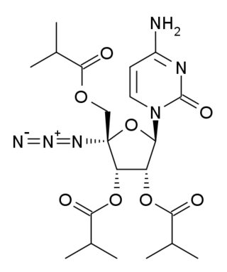
<span class="mw-page-title-main">Balapiravir</span> Chemical compound

Balapiravir is an experimental antiviral drug which acts as a polymerase inhibitor. There were efforts to develop it as a potential treatment for hepatitis C, and it was subsequently also studied in Dengue fever, but was not found to be useful. Lower doses failed to produce measurable reductions in viral load, while higher doses produced serious side effects such as lymphopenia which precluded further development of the drug. Subsequent research found that excess cytokine production triggered by Dengue virus infection prevented the conversion of the balapiravir prodrug to its active form, thereby blocking the activity of the drug.

Torque teno sus virus, belonging to the family Anelloviridae, is a group of virus strains that are non-enveloped, with a single-stranded circular DNA genome ranging from 2.6 to 2.8 kb in size. These swine infecting anelloviruses are divided into two genera: Iotatorquevirus and Kappatorquevirus. Torque teno sus virus has been found in pigs worldwide. TTSuVs are mainly transmitted by fecal-oral route. The prevalence of these viruses is relatively high. For now, there is not known disease caused exclusively by TTSuV. There is the possibility that TTSuV may worsen the progression of other diseases and therefore increase the economic losses for pig industry.

<span class="mw-page-title-main">Interferon Lambda 4</span> Protein-coding gene in the species Homo sapiens

Interferon lambda 4 is one of the most recently discovered human genes and the newest addition to the interferon lambda protein family. This gene encodes the IFNL4 protein, which is involved in immune response to viral infection.

References

  1. Biagini P (2009). "Classification of TTV and Related Viruses (Anelloviruses)". TT Viruses. Current Topics in Microbiology and Immunology. Vol. 331. pp. 21–33. doi:10.1007/978-3-540-70972-5_2. ISBN   978-3-540-70971-8. PMID   19230555.
  2. "Virus Taxonomy: 2020 Release". International Committee on Taxonomy of Viruses (ICTV). March 2021. Retrieved 23 May 2021.
  3. Nishizawa, T; Okamoto, H; Konishi, K; Yoshizawa, H; Miyakawa, Y; Mayumi, M (8 December 1997). "A novel DNA virus (TTV) associated with elevated transaminase levels in posttransfusion hepatitis of unknown etiology". Biochemical and Biophysical Research Communications. 241 (1): 92–7. doi:10.1006/bbrc.1997.7765. PMID   9405239.
  4. Biagini, P: "Human circoviruses.", pages 95-101. Veterinary Microbiology, 2004.
  5. Nishizawa, T.; Okamoto, H.; Konishi, K.; Yoshizawa, H.; Miyakawa, Y.; Mayumi, M. (1997). "A novel DNA virus (TTV) associated with elevated transaminase levels in posttransfusion hepatitis of unknown etiology". Biochem. Biophys. Res. Commun. 241 (1): 92–97. doi:10.1006/bbrc.1997.7765. PMID   9405239.
  6. Miyata, H.; Tsunoda, H.; Kazi, A.; Yamada, A.; Khan, M.A.; Yeung, B.J.; Murakami, J.; Kamahora, T.; Shiraki, K.; Hino, S. (1999). "Identification of a novel GC-rich 113-nucleotide region to complete the circular, single-stranded DNA genome of TT virus, the first human circovirus". J. Virol. 73 (5): 3582–3586. doi:10.1128/JVI.73.5.3582-3586.1999. PMC   104131 . PMID   10196248.
  7. "AABB website" (PDF). Retrieved December 25, 2017.
  8. Biagini, P.; Gallian, P.; Cantaloube, J.F.; De Micco, P.; de Lamballerie, X. (1998). "Presence of TT virus in French blood donors and intravenous drug users". J. Hepatol. 29 (4): 684–685. doi:10.1016/s0168-8278(98)80167-0. PMID   9824281.
  9. Gallian, P.; Biagini, P.; Zhong, S.; Louinssi, M.; Yeo, W.; Cantaloube, J.F.; Attoui, H.; de Micco, P.; Johnson, P.J.; de Lamballerie, X. (2000). "TT virus: a study of molecular epidemiology and transmission of genotypes 1, 2 and 3". J. Clin. Virol. 17 (1): 43–49. doi:10.1016/s1386-6532(00)00066-4. PMID   10814938.
  10. Zheng, MY; Lin, Y; Li, DJ; Ruan, HB; Chen, Y; Wu, TT (2010). "TTV and HPV co-infection in cervical smears of patients with cervical lesions in littoral of Zhejiang province". Zhonghua Shi Yan He Lin Chuang Bing du Xue Za Zhi. 24 (2): 110–112. PMID   21110428.
  11. Hino, S; Miyata, H (2007). "Torque teno virus (TTV): current status". Rev Med Virol. 17 (1): 45–57. doi:10.1002/rmv.524. PMID   17146841. S2CID   25965110.
  12. Martínez-Guinó, L; Kekarainen, T; Segalés, J (2009). "Evidence of Torque teno virus (TTV) vertical transmission in swine". Theriogenology. 71 (9): 1390–1395. doi:10.1016/j.theriogenology.2009.01.010. PMID   19249089.
  13. Pozzuto, T; Mueller, B; Meehan, B; Ringler, SS; McIntosh, KA; Ellis, JA; Mankertz, A; Krakowka, S (2009). "In utero transmission of porcine torque teno viruses". Vet Microbiol. 137 (3–4): 375–379. doi:10.1016/j.vetmic.2009.02.001. PMID   19282113.
  14. Pinho-Nascimento, CA; Leite, JP; Niel, C; Diniz-Mendes, L (2011). "Torque teno virus in fecal samples of patients with gastroenteritis: Prevalence, genogroups distribution, and viral load". J Med Virol. 83 (6): 1107–11. doi:10.1002/jmv.22024. PMID   21503927. S2CID   2296541.
  15. Wootton SC, Kim DS, Kondoh Y, Chen E, Lee JS, Song JW, Huh JW, Taniguchi H, Chiu C, Boushey H, Lancaster LH, Wolters PJ, Derisi J, Ganem D, Collard HR (2011) Viral infection in acute exacerbation of idiopathic pulmonary fibrosis. Am J Respir Crit Care Med.
  16. Maggi F, Pifferi M, Michelucci A, Albani M, Sbranti S, Lanini L, Simi P, Macchia P, Pistello M, Bendinelli M (2011) Torque teno virus viremia load size in patients with selected congenital defects of innate immunity. Clin Vaccine Immunol. 18(4):692–694.
  17. Ishimura M, Ohga S, Ichiyama M, Kusuhara K, Takada H, Hara T, Takahashi M, Okamoto H (2010) Hepatitis-associated aplastic anemia during a primary infection of genotype 1a torque teno virus. Eur J Pediatr. 2010 Jul;169(7):899–902
  18. Piaggio F, Dodi F, Bottino G, Andorno E, Gentile R, Ferrari C, Barabino G, Giannone A, Immordino G, Miggino M, Magoni Rossi A, Moraglia E, Gasloli G, Gelli M, Ferrante R, Morelli N, Casaccia M, Valente U (2009) Torque Teno Virus—cause of viral liver disease following liver transplantation: a case report. Transplant Proc. 41(4):1378–1379
  19. Hettmann, A; Demcsák, A; Bach, Á; Decsi, G; Dencs, Á; Pálinkó, D; Rovó, L; Nagy, K; Minarovits, J; Takács, M (2016). "Detection and phylogenetic analysis of Torque Teno Virus in salivary and tumor biopsy samples from head and neck carcinoma patients". Intervirology. 59 (2): 123–129. doi: 10.1159/000452974 . PMID   27924796.
  20. Walton, Andrew H.; Muenzer, Jared T.; Rasche, David; Boomer, Jonathan S.; Sato, Bryan; Brownstein, Bernard H.; Pachot, Alexandre; Brooks, Terrence L.; Deych, Elena; Shannon, William D.; Green, Jonathan M.; Storch, Gregory A.; Hotchkiss, Richard S. (11 June 2014). "Reactivation of Multiple Viruses in Patients with Sepsis". PLOS ONE. 9 (6): e98819. Bibcode:2014PLoSO...998819W. doi: 10.1371/journal.pone.0098819 . PMC   4053360 . PMID   24919177.
  21. Jaksch, Peter; Kundi, Michael; Görzer, Irene; Muraközy, Gabriella; Lambers, Christopher; Benazzo, Alberto; Hoetzenecker, Konrad; Klepetko, Walter; Puchhammer-Stöckl, Elisabeth (5 November 2018). "Torque Teno Virus as a Novel Biomarker Targeting the Efficacy of Immunosuppression After Lung Transplantation". The Journal of Infectious Diseases. 218 (12): 1922–1928. doi: 10.1093/infdis/jiy452 . PMID   30053048.
  22. Okamoto, H.; Nishizawa, T.; Takahashi, M.; Asabe, S.; Tsuda, F.; Yoshikawa, A. (2001a). "Heterogeneous distribution of tt virus of distinct genotypes in multiple tissues from infected humans". Virology. 288 (2): 358–368. doi: 10.1006/viro.2001.1097 . PMID   11601907.