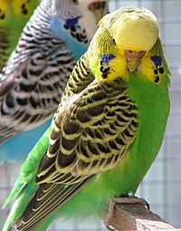
The budgerigar gets its yellow color from a psittacofulvin pigment and its green color from a combination of the same yellow pigment and blue structural color. The blue and white bird in the background lacks the yellow pigment. The dark markings on both birds are due to the black pigment eumelanin.
Biological pigments, also known simply as pigments or biochromes,[1] are substances produced by living organisms that have a color resulting from selective color absorption. Biological pigments include plant pigments and flower pigments. Many biological structures, such as skin, eyes, feathers, fur and hair contain pigments such as melanin in specialized cells called chromatophores. In some species, pigments accrue over very long periods during an individual's lifespan.[2]
Pigment color differs from structural color in that it is the same for all viewing angles, whereas structural color is the result of selective reflection or iridescence, usually because of multilayer structures. For example, butterfly wings typically contain structural color, although many butterflies have cells that contain pigment as well.[3]
Biological pigments
See conjugated systems for electron bond chemistry that causes these molecules to have pigment.
The primary function of pigments in plants is photosynthesis, which uses the green pigment chlorophyll and several colorful pigments that absorb as much light energy as possible.[4][5] Pigments are also known to play a role in pollination where pigment accumulation or loss can lead to floral color change, signaling to pollinators which flowers are rewarding and contain more pollen and nectar.[6]
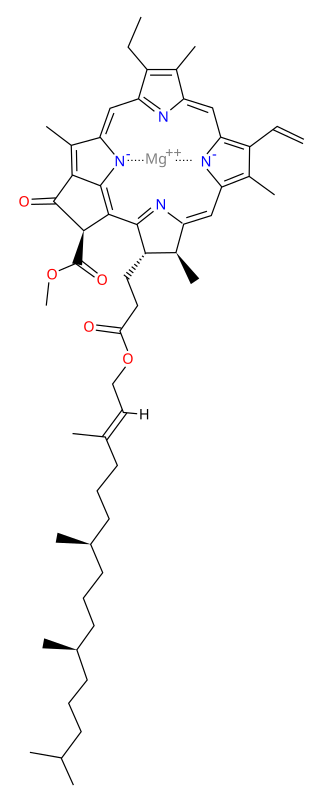
Chlorophyll is the primary pigment in plants; it is a chlorin that absorbs blue and red wavelengths of light while reflecting a majority of green. It is the presence and relative abundance of chlorophyll that gives plants their green color. All land plants and green algae possess two forms of this pigment: chlorophyll a and chlorophyll b. Kelps, diatoms, and other photosynthetic heterokonts contain chlorophyll c instead of b, while red algae possess only chlorophyll a. All chlorophylls serve as the primary means plants use to intercept light in order to fuel photosynthesis.
Carotenoids are red, orange, or yellow tetraterpenoids. During the process of photosynthesis, they have functions in light-harvesting (as accessory pigments), in photoprotection (energy dissipation via non-photochemical quenching as well as singlet oxygen scavenging for prevention of photooxidative damage), and also serve as protein structural elements. In higher plants, they also serve as precursors to the plant hormone abscisic acid.
Betalains are red or yellow pigments. Like anthocyanins they are water-soluble, but unlike anthocyanins they are synthesized from tyrosine. This class of pigments is found only in the Caryophyllales (including cactus and amaranth), and never co-occur in plants with anthocyanins.[5] Betalains are responsible for the deep red color of beets.
Anthocyanins (literally "flower blue") are water-solubleflavonoidpigments that appear red to blue, according to pH. They occur in all tissues of higher plants, providing color in leaves, plant stem, roots, flowers, and fruits, though not always in sufficient quantities to be noticeable. Anthocyanins are most visible in the petals of flowers of many species.[5]
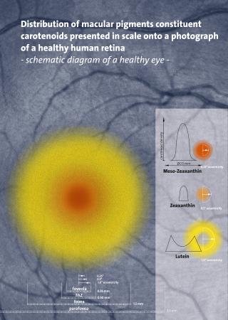
Plants, in general, contain six ubiquitous carotenoids: neoxanthin, violaxanthin, antheraxanthin, zeaxanthin, lutein and β-carotene.[7] Lutein is a yellow pigment found in fruits and vegetables and is the most abundant carotenoid in plants. Lycopene is the red pigment responsible for the color of tomatoes. Other less common carotenoids in plants include lutein epoxide (in many woody species), lactucaxanthin (found in lettuce), and alpha carotene (found in carrots).[8]
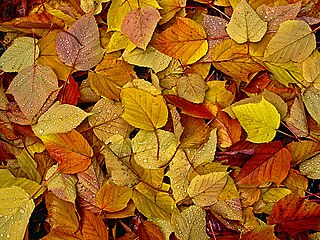
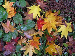
A particularly noticeable manifestation of pigmentation in plants is seen with autumn leaf color, a phenomenon that affects the normally green leaves of many deciduoustrees and shrubs whereby they take on, during a few weeks in the autumn season, various shades of red, yellow, purple, and brown.[9]
Chlorophylls degrade into colorless tetrapyrroles known as nonfluorescent chlorophyll catabolites (NCCs).[10] As the predominant chlorophylls degrade, the hidden pigments of yellow xanthophylls and orange beta-carotene are revealed. These pigments are present throughout the year, but the red pigments, the anthocyanins, are synthesized de novo once roughly half of chlorophyll has been degraded. The amino acids released from degradation of light harvesting complexes are stored all winter in the tree's roots, branches, stems, and trunk until next spring when they are recycled to re‑leaf the tree.
Pigments in algae
Algae are very diverse photosynthetic organisms, which differ from plants in that they are aquatic organisms, they do not present vascular tissue and do not generate an embryo. However, both types of organisms share the possession of photosynthetic pigments, which absorb and release energy that is later used by the cell. These pigments in addition to chlorophylls, are phycobiliproteins, fucoxanthins, xanthophylls and carotenes, which serve to trap the energy of light and lead it to the primary pigment, which is responsible for initiating oxygenic photosynthesis reactions.
Pigmentation is used by many animals for protection, by means of camouflage, mimicry, or warning coloration. Some animals including fish, amphibians and cephalopods use pigmented chromatophores to provide camouflage that varies to match the background.
However, some biological pigments in animals, such as heme groups that help to carry oxygen in the blood, are colored as a result of happenstance. Their color does not have a protective or signalling function.
A variety of diseases and abnormal conditions that involve pigmentation are in humans and animals, either from absence of or loss of pigmentation or pigment cells, or from the excess production of pigment.
Albinism is an inherited disorder characterized by total or partial loss of melanin. Humans and animals that suffer from albinism are called "albinistic" (the term "albino" is also sometimes used, but may be considered offensive when applied to people).
Lamellar ichthyosis, also called "fish scale disease", is an inherited condition in which one symptom is excess production of melanin. The skin is darker than normal, and is characterized by darkened, scaly, dry patches.
Melasma is a condition in which dark brown patches of pigment appear on the face, influenced by hormonal changes. When it occurs during a pregnancy, this condition is called the mask of pregnancy.
ocular pigmentation is an accumulation of pigment in the eye, and may be caused by latanoprost medication.[17]
Vitiligo is a condition in which there is a loss of pigment-producing cells called melanocytes in patches of skin.
Pigments in marine animals
Carotenoids and carotenoproteins
Carotenoids are the most common group of pigments found in nature.[18] Over 600 different kinds of carotenoids are found in animals, plants, and microorganisms.
Marine animals are incapable of making their own carotenoids and thus rely on plants for these pigments. Carotenoproteins are especially common among marine animals. These complexes are responsible for the various colors (red, purple, blue, green, etc.) to these marine invertebrates for mating rituals and camouflage. There are two main types of carotenoproteins: Type A and Type B. Type A has carotenoids (chromogen) which are stoichiometrically associated with a simple protein (glycoprotein). The second type, Type B, has carotenoids which are associated with a lipo protein and is usually less stable. While Type A is commonly found in the surface (shells and skins) of marine invertebrates, Type B is usually in eggs, ovaries, and blood. The colors and characteristic absorption of these carotenoprotein complexes are based upon the chemical binding of the chromogen and the protein subunits.
For example, the blue carotenoprotein, linckiacyanin has about 100-200 carotenoid molecules per every complex.[19] In addition, the functions of these pigment-protein complexes also change their chemical structure as well. Carotenoproteins that are within the photosynthetic structure are more common, but complicated. Pigment-protein complexes that are outside of the photosynthetic system are less common, but have a simpler structure. For example, there are only two of these blue astaxanthin-proteins in the jellyfish, Velella velella, contains only about 100 carotenoids per complex.[citation needed]
A common carotenoid in animals is astaxanthin, which gives off a purple-blue and green pigment. Astaxanthin's color is formed by creating complexes with proteins in a certain order. For example, the crustochrin has approximately 20 astaxanthin molecules bonded with protein. When the complexes interact by exciton-exciton interaction, it lowers the absorbance maximum, changing the different color pigments.
In lobsters, there are various types of astaxanthin-protein complexes present. The first one is crustacyanin (max 632nm), a slate-blue pigment found in the lobster's carapace. The second one is crustochrin (max 409), a yellow pigment which is found on the outer layer of the carapace. Lastly, the lipoglycoprotein and ovoverdin forms a bright green pigment that is usually present in the outer layers of the carapace and the lobster eggs.[20][21]
Tetrapyrroles
Tetrapyrroles are the next most common group of pigments.[citation needed] They have four pyrrole rings, each ring consisting of C4H4NH. The main role of the tetrapyrroles is their connection in the biological oxidation process. Tetrapyrroles have a major role in electron transport and act as a replacement for many enzymes. They also have a role in the pigmentation of the marine organism's tissues.
Melanin
Melanin[22] is a class of compounds that serves as a pigment with different structures responsible for dark, tan, yellowish / reddish pigments in marine animals. It is produced as the amino acid tyrosine is converted into melanin, which is found in the skin, hair, and eyes. Derived from aerobic oxidation of phenols, they are polymers.
There are several different types of melanins considering that they are an aggregate of smaller component molecules, such as nitrogen containing melanins. There are two classes of pigments: black and brown insoluble eumelanins, which are derived from aerobic oxidation of tyrosine in the presence of tyrosinase, and the alkali-soluble phaeomelanins which range from a yellow to red brown color, arising from the deviation of the eumelanin pathway through the intervention of cysteine and/or glutathione. Eumelanins are usually found in the skin and eyes. Several different melanins include melanoprotein (dark brown melanin that is stored in high concentrations in the ink sac of the cuttlefish Sepia Officianalis), echinoidea (found in sand dollars, and the hearts of sea urchins), holothuroidea (found in sea cucumbers), and ophiuroidea (found in brittle and snake stars). These melanins are possibly polymers which arise from the repeated coupling of simple bi-polyfunctional monomeric intermediates, or of high molecular weights. The compounds benzothiazole and tetrahydroisoquinoline ring systems act as UV-absorbing compounds.
Bioluminescence
The only light source in the deep sea, marine animals give off visible light energy called bioluminescence,[23] a subset of chemiluminescence. This is the chemical reaction in which chemical energy is converted to light energy. It is estimated that 90% of deep-sea animals produce some sort of bioluminescence. Considering that a large proportion of the visible light spectrum is absorbed before reaching the deep sea, most of the emitted light from the sea-animals is blue and green. However, some species may emit a red and infrared light, and there has even been a genus that is found to emit yellow bioluminescence. The organ that is responsible for the emission of bioluminescence is known as photophores. This type is only present in squid and fish, and is used to illuminate their ventral surfaces, which disguise their silhouettes from predators. The uses of the photophores in the sea-animals differ, such as lenses for controlling intensity of color, and the intensity of the light produced. Squids have both photophores and chromatophores which controls both of these intensities. Another thing that is responsible for the emission of bioluminescence, which is evident in the bursts of light that jellyfish emit, start with a luciferin (a photogen) and ends with the light emitter (a photagogikon.) Luciferin, luciferase, salt, and oxygen react and combine to create a single unit called photo-proteins, which can produce light when reacted with another molecule such as Ca+. Jellyfish use this as a defense mechanism; when a smaller predator is attempting to devour a jellyfish, it will flash its lights, which would therefore lure a larger predator and chase the smaller predator away. It is also used as mating behavior.
In reef-building coral and sea anemones, they fluoresce; light is absorbed at one wavelength, and re-emitted at another. These pigments may act as natural sunscreens, aid in photosynthesis, serve as warning coloration, attract mates, warn rivals, or confuse predators.
Chromatophores
Chromatophores are color pigment changing cells that are directly stimulated by central motor neurons. They are primarily used for quick environmental adaptation for camouflaging. The process of changing the color pigment of their skin relies on a single highly developed chromatophore cell and many muscles, nerves, glial and sheath cells. Chromatophores contract and contain vesicles that stores three different liquid pigments. Each color is indicated by the three types of chromatophore cells: erythrophores, melanophores, and xanthophores. The first type is the erythrophores, which contains reddish pigments such as carotenoids and pteridines. The second type is the melanophores, which contains black and brown pigments such as the melanins. The third type is the xanthophores which contains yellow pigments in the forms of carotenoids. The various colors are made by the combination of the different layers of the chromatophores. These cells are usually located beneath the skin or scale the animals. There are two categories of colors generated by the cell – biochromes and schematochromes. Biochromes are colors chemically formed microscopic, natural pigments. Their chemical composition is created to take in some color of light and reflect the rest. In contrast, schematochromes (structural colors) are colors created by light reflections from a colorless surface and refractions by tissues. Schematochromes act like prisms, refracting and dispersing visible light to the surroundings, which will eventually reflect a specific combination of colors. These categories are determined by the movement of pigments within the chromatophores. The physiological color changes are short-term and fast, found in fishes, and are a result from an animal's response to a change in the environment. In contrast, the morphological color changes are long-term changes, occurs in different stages of the animal, and are due to the change of numbers of chromatophores. To change the color pigments, transparency, or opacity, the cells alter in form and size, and stretch or contract their outer covering.
Photo-protective pigments
Due to damage from UV-A and UV-B, marine animals have evolved to have compounds that absorb UV light and act as sunscreen. Mycosporine-like amino acids (MAAs) can absorb UV rays at 310-360nm. Melanin is another well-known UV-protector. Carotenoids and photopigments both indirectly act as photo-protective pigments, as they quench oxygen free-radicals. They also supplement photosynthetic pigments that absorb light energy in the blue region.
Defensive role of pigments
It's known that animals use their color patterns to warn off predators, however it has been observed that a sponge pigment mimicked a chemical which involved the regulation of moulting of an amphipod that was known to prey on sponges. So whenever that amphipod eats the sponge, the chemical pigments prevents the moulting, and the amphipod eventually dies.
Environmental influence on color
Coloration in invertebrates varies based on the depth, water temperature, food source, currents, geographic location, light exposure, and sedimentation. For example, the amount of carotenoid a certain sea anemone decreases as we go deeper into the ocean. Thus, the marine life that resides on deeper waters is less brilliant than the organisms that live in well-lit areas due to the reduction of pigments. In the colonies of the colonial ascidian-cyanophyte symbiosis Trididemnum solidum, their colors are different depending on the light regime in which they live. The colonies that are exposed to full sunlight are heavily calcified, thicker, and are white. In contrast the colonies that live in shaded areas have more phycoerythrin (pigment that absorbs green) in comparison to phycocyanin (pigment that absorbs red), thinner, and are purple. The purple color in the shaded colonies are mainly due to the phycobilin pigment of the algae, meaning the variation of exposure in light changes the colors of these colonies.
Adaptive coloration
Aposematism is the warning coloration to signal potential predators to stay away. In many chromodorid nudibranchs, they take in distasteful and toxic chemicals emitted from sponges and store them in their repugnatorial glands (located around the mantle edge). Predators of nudibranchs have learned to avoid these certain nudibranchs based on their bright color patterns. Preys also protect themselves by their toxic compounds ranging from a variety of organic and inorganic compounds.
Physiological activities
Pigments of marine animals serve several different purposes, other than defensive roles. Some pigments are known to protect against UV (see photo-protective pigments.) In the nudibranch Nembrotha Kubaryana, tetrapyrrole pigment 13 has been found to be a potent antimicrobial agent. Also in this creature, tamjamines A, B, C, E, and F has shown antimicrobial, antitumor, and immunosuppressive activities.
Sesquiterpenoids are recognized for their blue and purple colors, but it has also been reported to exhibit various bioactivities such as antibacterial, immunoregulating, antimicrobial, and cytotoxic, as well as the inhibitory activity against cell division in the fertilized sea urchin and ascidian eggs. Several other pigments have been shown to be cytotoxic. In fact, two new carotenoids that were isolated from a sponge called Phakellia stelliderma showed mild cytotoxicity against mouse leukemia cells. Other pigments with medical involvements include scytonemin, topsentins, and debromohymenialdisine have several lead compounds in the field of inflammation, rheumatoid arthritis and osteoarthritis respectively. There's evidence that topsentins are potent mediators of immunogenic inflation, and topsentin and scytonemin are potent inhibitors of neurogenic inflammation.
Chlorophyll is any of several related green pigments found in cyanobacteria and in the chloroplasts of algae and plants. Its name is derived from the Greek words χλωρός, khloros and φύλλον, phyllon ("leaf"). Chlorophyll allow plants to absorb energy from light.
Photosynthesis is a system of biological processes by which photosynthetic organisms, such as most plants, algae, and cyanobacteria, convert light energy, typically from sunlight, into the chemical energy necessary to fuel their activities. Photosynthetic organisms use intracellular organic compounds to store the chemical energy they produce in photosynthesis within organic compounds like sugars, glycogen, cellulose and starches. Photosynthesis is usually used to refer to oxygenic photosynthesis, a process that produces oxygen. To use this stored chemical energy, the organisms' cells metabolize the organic compounds through another process called cellular respiration. Photosynthesis plays a critical role in producing and maintaining the oxygen content of the Earth's atmosphere, and it supplies most of the biological energy necessary for complex life on Earth.
Chromatophores are cells that produce color, of which many types are pigment-containing cells, or groups of cells, found in a wide range of animals including amphibians, fish, reptiles, crustaceans and cephalopods. Mammals and birds, in contrast, have a class of cells called melanocytes for coloration.
Carotenoids are yellow, orange, and red organic pigments that are produced by plants and algae, as well as several bacteria, archaea, and fungi. Carotenoids give the characteristic color to pumpkins, carrots, parsnips, corn, tomatoes, canaries, flamingos, salmon, lobster, shrimp, and daffodils. Over 1,100 identified carotenoids can be further categorized into two classes – xanthophylls and carotenes.
Chromoplasts are plastids, heterogeneous organelles responsible for pigment synthesis and storage in specific photosynthetic eukaryotes. It is thought that like all other plastids including chloroplasts and leucoplasts they are descended from symbiotic prokaryotes.
Photobiology is the scientific study of the beneficial and harmful interactions of light in living organisms. The field includes the study of photophysics, photochemistry, photosynthesis, photomorphogenesis, visual processing, circadian rhythms, photomovement, bioluminescence, and ultraviolet radiation effects.
Xanthophylls are yellow pigments that occur widely in nature and form one of two major divisions of the carotenoid group; the other division is formed by the carotenes. The name is from Greek: xanthos (ξανθός), meaning "yellow", and phyllon (φύλλον), meaning "leaf"), due to their formation of the yellow band seen in early chromatography of leaf pigments.
Fucoxanthin is a xanthophyll, with formula C42H58O6. It is found as an accessory pigment in the chloroplasts of brown algae and most other heterokonts, giving them a brown or olive-green color. Fucoxanthin absorbs light primarily in the blue-green to yellow-green part of the visible spectrum, peaking at around 510-525 nm by various estimates and absorbing significantly in the range of 450 to 540 nm.
Plant physiology is a subdiscipline of botany concerned with the functioning, or physiology, of plants.
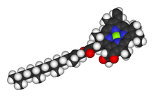
Astaxanthin is a keto-carotenoid within a group of chemical compounds known as terpenes. Astaxanthin is a metabolite of zeaxanthin and canthaxanthin, containing both hydroxyl and ketone functional groups. It is a lipid-soluble pigment with red coloring properties, which result from the extended chain of conjugated double bonds at the center of the compound. The presence of the hydroxyl functional groups and the hydrophobic hydrocarbons render the molecule amphiphilic.
Photosystems are functional and structural units of protein complexes involved in photosynthesis. Together they carry out the primary photochemistry of photosynthesis: the absorption of light and the transfer of energy and electrons. Photosystems are found in the thylakoid membranes of plants, algae, and cyanobacteria. These membranes are located inside the chloroplasts of plants and algae, and in the cytoplasmic membrane of photosynthetic bacteria. There are two kinds of photosystems: PSI and PSII.
Chlorophyll a is a specific form of chlorophyll used in oxygenic photosynthesis. It absorbs most energy from wavelengths of violet-blue and orange-red light, and it is a poor absorber of green and near-green portions of the spectrum. Chlorophyll does not reflect light but chlorophyll-containing tissues appear green because green light is diffusively reflected by structures like cell walls. This photosynthetic pigment is essential for photosynthesis in eukaryotes, cyanobacteria and prochlorophytes because of its role as primary electron donor in the electron transport chain. Chlorophyll a also transfers resonance energy in the antenna complex, ending in the reaction center where specific chlorophylls P680 and P700 are located.
A chromophore is a molecule which absorbs light at a particular wavelength and emits color as a result. Chromophores are commonly referred to as colored molecules for this reason. The word is derived from Ancient Greek χρῶμᾰ (chroma) 'color', and -φόρος (phoros) 'carrier of'. Many molecules in nature are chromophores, including chlorophyll, the molecule responsible for the green colors of leaves. The color that is seen by our eyes is that of the light not absorbed by the reflecting object within a certain wavelength spectrum of visible light. The chromophore indicates a region in the molecule where the energy difference between two separate molecular orbitals falls within the range of the visible spectrum. Visible light that hits the chromophore can thus be absorbed by exciting an electron from its ground state into an excited state. In biological molecules that serve to capture or detect light energy, the chromophore is the moiety that causes a conformational change in the molecule when hit by light.
Zeaxanthin is one of the most common carotenoids in nature, and is used in the xanthophyll cycle. Synthesized in plants and some micro-organisms, it is the pigment that gives paprika, corn, saffron, goji (wolfberries), and many other plants and microbes their characteristic color.
A light-harvesting complex consists of a number of chromophores which are complex subunit proteins that may be part of a larger super complex of a photosystem, the functional unit in photosynthesis. It is used by plants and photosynthetic bacteria to collect more of the incoming light than would be captured by the photosynthetic reaction center alone. The light which is captured by the chromophores is capable of exciting molecules from their ground state to a higher energy state, known as the excited state. This excited state does not last very long and is known to be short-lived.
Photoprotection is the biochemical process that helps organisms cope with molecular damage caused by sunlight. Plants and other oxygenic phototrophs have developed a suite of photoprotective mechanisms to prevent photoinhibition and oxidative stress caused by excess or fluctuating light conditions. Humans and other animals have also developed photoprotective mechanisms to avoid UV photodamage to the skin, prevent DNA damage, and minimize the downstream effects of oxidative stress.
Autumn leaf color is a phenomenon that affects the normally green leaves of many deciduous trees and shrubs by which they take on, during a few weeks in the autumn season, various shades of yellow, orange, red, purple, and brown. The phenomenon is commonly called autumn colours or autumn foliage in British English and fall colors, fall foliage, or simply foliage in American English.
Non-photochemical quenching (NPQ) is a mechanism employed by plants and algae to protect themselves from the adverse effects of high light intensity. It involves the quenching of singlet excited state chlorophylls (Chl) via enhanced internal conversion to the ground state, thus harmlessly dissipating excess excitation energy as heat through molecular vibrations. NPQ occurs in almost all photosynthetic eukaryotes, and helps to regulate and protect photosynthesis in environments where light energy absorption exceeds the capacity for light utilization in photosynthesis.
Amelanism is a pigmentation abnormality characterized by the lack of pigments called melanins, commonly associated with a genetic loss of tyrosinase function. Amelanism can affect fish, amphibians, reptiles, birds, and mammals including humans. The appearance of an amelanistic animal depends on the remaining non-melanin pigments. The opposite of amelanism is melanism, a higher percentage of melanin.
Albinism is the congenital absence of melanin in an animal or plant resulting in white hair, feathers, scales and skin and reddish pink or blue eyes. Individuals with the condition are referred to as albinos.
↑ Young AJ, Phillip D, Savill J (1997). "Carotenoids in higher plant photosynthesis.". In Pessaraki M (ed.). Handbook of Photosynthesis. New York: Taylor and Francis. pp.575–596.
↑ García-Plazaola JI, Matsubara S, Osmond CB (September 2007). "The lutein epoxide cycle in higher plants: its relationships to other xanthophyll cycles and possible functions". Functional Plant Biology. 34 (9): 759–773. doi:10.1071/FP07095. PMID32689404.
↑ Milicua JC, Barandiaran A, Macarulla JM, Garate AM, Gomez R (November 1985). "Structural characteristics of the carotenoids binding to the blue carotenoprotein from Procambarus clarkii". Experientia. 41 (11): 1485–6. doi:10.1007/BF01950050. S2CID37966773.
↑ Bandaranayake WM (April 2006). "The nature and role of pigments of marine invertebrates". Natural Product Reports. 23 (2): 223–55. doi:10.1039/b307612c. PMID16572229.
This page is based on this Wikipedia article Text is available under the CC BY-SA 4.0 license; additional terms may apply. Images, videos and audio are available under their respective licenses.