Cell fusion

Last updated

Cell fusion is an important cellular process in which several uninucleate cells (cells with a single nucleus) combine to form a multinucleate cell, known as a syncytium. Cell fusion occurs during differentiation of myoblasts, osteoclasts and trophoblasts, during embryogenesis, and morphogenesis. [1] Cell fusion is a necessary event in the maturation of cells so that they maintain their specific functions throughout growth.

Contents

History

In 1847 Theodore Schwann expanded upon the theory that all living organisms are composed of cells when he added that discrete cells are the basis of life. Schwann observed that in certain cells the walls and cavities of the cells coalesce together. This observation provided the first hint that cells fuse. It was not until 1960 that cell biologists deliberately fused cells for the first time. To fuse the cells, biologists combined isolated mouse cells and induced fusion of their outer membrane using the Sendai virus (a respiratory virus in mice). Each of the fused hybrid cells contained a single nucleus with chromosomes from both fusion partners. Synkaryon became the name of this type of cell combined with a nucleus. In the late 1960s biologists successfully fused cells of different types and from different species. The hybrid products of these fusions, heterokaryon, were hybrids that maintained two or more separate nuclei. This work was headed by Henry Harris at the University of Oxford and Nils Ringertz from Sweden's Karolinska Institute. These two men are responsible for reviving the interest of cell fusion. The hybrid cells interested biologists in the area of how different kinds of cytoplasm affect different kinds of nuclei. The work conducted by Henry and Nils showed that proteins from one gene fusion affect gene expression in the other partner's nucleus, and vice versa. These hybrid cells that were created were considered forced exceptions to normal cellular integrity and it was not until 2002 that the possibility of cell fusion between cells of different types may have a real function in mammals. [2]

Types of cell fusion

a Cells of the same lineage fuse to form a cell with multiple nuclei, known as a syncytium. The fused cell can have an altered phenotype and new functions such as barrier formation.
b Cells of different lineage fuse to form a cell with multiple nuclei, known as a heterokaryon. The fused cells might have undergone a reversion of phenotype or show transdifferentiation.
c Cells of different lineage or the same lineage fuse to form a cell with a single nucleus, known as a synkaryon. New functions of the fused cell can include a reversion of phenotype, transdifferentiation and proliferation. If nuclear fusion occurs, the fused nucleus initially contains the complete chromosomal content of both fusion partners (4N), but ultimately chromosomes are lost and/or re-sorted (see arrows). If nuclear fusion does not occur, a heterokaryon (or syncytium) can become a synkaryon by shedding an entire nucleus. CellFusionTypes.jpg
a Cells of the same lineage fuse to form a cell with multiple nuclei, known as a syncytium. The fused cell can have an altered phenotype and new functions such as barrier formation.
b Cells of different lineage fuse to form a cell with multiple nuclei, known as a heterokaryon. The fused cells might have undergone a reversion of phenotype or show transdifferentiation.
c Cells of different lineage or the same lineage fuse to form a cell with a single nucleus, known as a synkaryon. New functions of the fused cell can include a reversion of phenotype, transdifferentiation and proliferation. If nuclear fusion occurs, the fused nucleus initially contains the complete chromosomal content of both fusion partners (4N), but ultimately chromosomes are lost and/or re-sorted (see arrows). If nuclear fusion does not occur, a heterokaryon (or syncytium) can become a synkaryon by shedding an entire nucleus.

Homotypic cell fusion occurs between cells of the same type. An example of this would be osteoclasts or myofibers fusing together with their respective type of cells. When the two nuclei merge a synkaryon is produced. Cell fusion normally occurs with nuclear fusion, however in the absence of nuclear fusion, the cell would be described as a binucleated heterokaryon. A heterokaryon is the melding of two or more cells into one and it may reproduce itself for several generations. [3] If two of the same type of cells fuse but their nuclei do not fuse, then the resulting cell is called a syncytium. [4]

Heterotypic cell fusion occurs between cells of different types. The result of this fusion is also a synkaryon produced by the merging of the nuclei, and a binucleated heterokaryon in the absence of nuclear fusion. An example of this would be Bone Marrow Derived Cells (BMDCs) being fused with parenchymatous organs. [5]

Methods of cell fusion

There are four methods that cell biologists and biophysicists use to fuse cells. These four ways include electrical cell fusion, polyethylene glycol cell fusion, and sendai virus induced cell fusion and a newly developed method termed optically controlled thermoplasmonics.

BTX ECM 2001 Electrofusion generator cell fusion applications manufactured by BTX Harvard Apparatus, Holliston MA USA ECM 2001 Hybridoma System.jpg
BTX ECM 2001 Electrofusion generator cell fusion applications manufactured by BTX Harvard Apparatus, Holliston MA USA

Electrical cell fusion is an essential step in some of the most innovative methods in modern biology. This method begins when two cells are brought into contact by dielectrophoresis. Dielectrophoresis uses a high frequency alternating current, unlike electrophoresis in which a direct current is applied. Once the cells are brought together, a pulsed voltage is applied. The pulse voltage causes the cell membrane to permeate and subsequent combining of the membranes and the cells then fuse. After this, alternative voltage is applied for a brief period of time to stabilize the process. The result of this is that the cytoplasm has mixed together and the cell membrane has completely fused. All that remains separate is the nuclei, which will fuse at a later time within the cell, making the result a heterokaryon cell. [6]

Polyethylene glycol cell fusion is the simplest, but most toxic, way to fuse cells. In this type of cell fusion polyethylene glycol, PEG, acts as a dehydrating agent and fuses not only plasma membranes but also intracellular membranes. This leads to cell fusion since PEG induces cell agglutination and cell-to-cell contact. Though this type of cell fusion is the most widely used, it still has downfalls. Oftentimes PEG can cause uncontrollable fusion of multiple cells, leading to the appearance of giant polykaryons. Also, standard PEG cell fusion is poorly reproducible and different types of cells have various fusion susceptibilities. This type of cell fusion is widely used for the production of somatic cell hybrids and for nuclear transfer in mammalian cloning. [7]

Sendai virus induced cell fusion occurs in four different temperature stages. During the first stage, which lasts no longer than 10 minutes, viral adsorption takes place and the adsorbed virus can be inhibited by viral antibodies. The second stage, which is 20 minutes, is pH dependent and an addition of viral antiserum can still inhibit ultimate fusion. In the third, antibody-refractory stage, viral envelope constituents remain detectable on the surface of cells. During the fourth stage, cell fusion becomes evident and HA neuraminidase and fusion factor begin to disappear. The first and second stages are the only two that are pH dependent. [8]

Thermoplasmonics induced cell fusion Thermoplasmonics is based on a near infrared (NIR) laser and a plasmonic nanoparticle. The laser which typically acts as an optical trap, is used to heat the nanoscopic plasmonic particle to very high and extremely locally elevated temperatures. Optical trapping of such a nanoheater at the interface between two membrane vesicles, or two cells, leads to immediate fusion of the two verified by both content and lipid mixing. Advantages include full flexibility of which cells to fuse and fusion can be performed in any buffer condition unlike electroformation which is affected by salt.

In human therapy

Alternative forms of restoring organ function and replacing damaged cells are needed with donor organs and tissue for transplantation being so scarce. It is because of the scarcity that biologists have begun considering the potential for therapeutic cell fusion. Biologists have been discussing implications of the observation that cell fusion can occur with restorative effects following tissue damage or cell transplantation. Though using cell fusion for this is being talked about and worked on, there are still many challenges those who wish to implement cell fusion as a therapeutic tool face. These challenges include choosing the best cells to use for the reparative fusion, determining the best way to introduce the chosen cells into the desired tissue, discovering methods to increase the incidence of cell fusion, and ensuring that the resulting fusion products will function properly. If these challenges can be overcome then cell fusion may have therapeutic potential. [9]

Role in plant cells

In plants, cell fusion happens far less frequently compared to eukaryotic cells, however it does occur in some situations. Plant cells have evolved unique methods to fuse cells, largely in part due to the cell wall that surrounds plant cells. The cell wall in a plant cell will become altered prior to fusion, usually becoming thinner or even forming a bridge between cells that are about to fuse. Gamete fusion can also occur in plants. [10]

Role in cancer progression

Cell fusion has become an area of focus for research in cancer progression in humans. When multiple types of differentiated cells fuse, the resulting cell could potentially be polyploid. Polyploid cells can be unstable due to their different genetic combinations which can often result in the cell becoming diseased. Polyploid cells can also result in unscheduled endoreplication, a process when DNA is replicated within the cell without the cell dividing, which has been linked to cancer development because of the increase in genetic instability within the cell. Metastasis, the spreading of cancer cells to different areas of the body and one of the leading causes of cancer related death, is a process that is linked to cell fusion. Cells derived from bone marrow fuse with malignant tumor cells, creating cells that have traits of each parent cell. These fused, cancerous cells have migration capabilities inherited from the bone marrow derived cell (BMDC) that allow it to travel throughout the body. [11]

Microorganisms

Fungi

Plasmogamy is the stage of the sexual cycle of fungi in which two cells fuse together to share a common cytoplasm while bringing haploid nuclei from both partners together in the same cell.

Amoebozoa

Cell fusion (plasmogamy or syngamy) is a stage in the Amoebozoa sexual cycle. [12]

Bacteria

In Escherichia coli spontaneous zygogenesis (Z-mating) involves cell fusion, and appears to be a form of true sexuality in prokaryotes. Bacteria that perform Z-mating are called Szp+. [13]

Other uses

See also

Related Research Articles

<span class="mw-page-title-main">Apoptosis</span> Programmed cell death in multicellular organisms

Apoptosis is a form of programmed cell death that occurs in multicellular organisms and in some eukaryotic, single-celled microorganisms such as yeast. Biochemical events lead to characteristic cell changes (morphology) and death. These changes include blebbing, cell shrinkage, nuclear fragmentation, chromatin condensation, DNA fragmentation, and mRNA decay. The average adult human loses between 50 and 70 billion cells each day due to apoptosis. For an average human child between eight and fourteen years old, each day the approximate lost is 20 to 30 billion cells.

<span class="mw-page-title-main">Chimera (genetics)</span> Single organism composed of two or more different populations of genetically distinct cells

A genetic chimerism or chimera is a single organism composed of cells with more than one distinct genotype. In animals and human chimeras, this means an individual derived from two or more zygotes, which can include possessing blood cells of different blood types, and subtle variations in form (phenotype). Animal chimeras are produced by the merger of two embryos. In plant chimeras, however, the distinct types of tissue may originate from the same zygote, and the difference is often due to mutation during ordinary cell division. Normally, genetic chimerism is not visible on casual inspection; however, it has been detected in the course of proving parentage. In contrast, an individual where each cell contains genetic material from two organisms of different breeds, varieties, species or genera is called a hybrid.

A syncytium or symplasm is a multinucleate cell that can result from multiple cell fusions of uninuclear cells, in contrast to a coenocyte, which can result from multiple nuclear divisions without accompanying cytokinesis. The muscle cell that makes up animal skeletal muscle is a classic example of a syncytium cell. The term may also refer to cells interconnected by specialized membranes with gap junctions, as seen in the heart muscle cells and certain smooth muscle cells, which are synchronized electrically in an action potential.

<span class="mw-page-title-main">Karyogamy</span> Fusion of the nuclei of two haploid eukaryotic cells

Karyogamy is the final step in the process of fusing together two haploid eukaryotic cells, and refers specifically to the fusion of the two nuclei. Before karyogamy, each haploid cell has one complete copy of the organism's genome. In order for karyogamy to occur, the cell membrane and cytoplasm of each cell must fuse with the other in a process known as plasmogamy. Once within the joined cell membrane, the nuclei are referred to as pronuclei. Once the cell membranes, cytoplasm, and pronuclei fuse, the resulting single cell is diploid, containing two copies of the genome. This diploid cell, called a zygote or zygospore can then enter meiosis, or continue to divide by mitosis. Mammalian fertilization uses a comparable process to combine haploid sperm and egg cells (gametes) to create a diploid fertilized egg.

<span class="mw-page-title-main">Heterokaryon</span>

A heterokaryon is a multinucleate cell that contains genetically different nuclei. Heterokaryotic and heterokaryosis are derived terms. This is a special type of syncytium. This can occur naturally, such as in the mycelium of fungi during sexual reproduction, or artificially as formed by the experimental fusion of two genetically different cells, as e.g., in hybridoma technology.

<span class="mw-page-title-main">Phagosome</span>

In cell biology, a phagosome is a vesicle formed around a particle engulfed by a phagocyte via phagocytosis. Professional phagocytes include macrophages, neutrophils, and dendritic cells (DCs).

<span class="mw-page-title-main">Viral envelope</span> Outermost layer of many types of the infectious agent

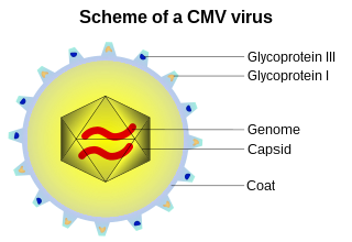
A viral envelope is the outermost layer of many types of viruses. It protects the genetic material in their life cycle when traveling between host cells. Not all viruses have envelopes. A viral envelope protein or E protein is a protein in the envelope, which may be acquired by the capsid from an infected host cell.

<i>Murine respirovirus</i> Sendai virus, virus of rodents

Murine respirovirus, formerly Sendai virus (SeV) and previously also known as murine parainfluenza virus type 1 or hemagglutinating virus of Japan (HVJ), is an enveloped, 150-200 nm–diameter, negative sense, single-stranded RNA virus of the family Paramyxoviridae. It typically infects rodents and it is not pathogenic for humans or domestic animals

<span class="mw-page-title-main">Viral entry</span> Earliest stage of infection in the viral life cycle

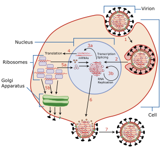
Viral entry is the earliest stage of infection in the viral life cycle, as the virus comes into contact with the host cell and introduces viral material into the cell. The major steps involved in viral entry are shown below. Despite the variation among viruses, there are several shared generalities concerning viral entry.

<span class="mw-page-title-main">Cytopathic effect</span>

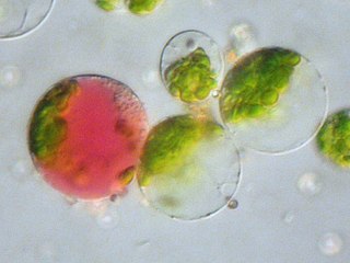
Cytopathic effect or cytopathogenic effect refers to structural changes in host cells that are caused by viral invasion. The infecting virus causes lysis of the host cell or when the cell dies without lysis due to an inability to replicate. Both of these effects occur due to CPEs. If a virus causes these morphological changes in the host cell, it is said to be cytopathogenic. Common examples of CPE include rounding of the infected cell, fusion with adjacent cells to form syncytia, and the appearance of nuclear or cytoplasmic inclusion bodies.

A fusion mechanism is any mechanism by which cell fusion or virus–cell fusion takes place, as well as the machinery that facilitates these processes. Cell fusion is the formation of a hybrid cell from two separate cells. There are three major actions taken in both virus–cell fusion and cell–cell fusion: the dehydration of polar head groups, the promotion of a hemifusion stalk, and the opening and expansion of pores between fusing cells. Virus–cell fusions occur during infections of several viruses that are health concerns relevant today. Some of these include HIV, Ebola, and influenza. For example, HIV infects by fusing with the membranes of immune system cells. In order for HIV to fuse with a cell, it must be able to bind to the receptors CD4, CCR5, and CXCR4. Cell fusion also occurs in a multitude of mammalian cells including gametes and myoblasts.

The parasexual cycle, a process restricted to fungi and single-celled organisms, is a nonsexual mechanism of parasexuality for transferring genetic material without meiosis or the development of sexual structures. It was first described by Italian geneticist Guido Pontecorvo in 1956 during studies on Aspergillus nidulans. A parasexual cycle is initiated by the fusion of hyphae (anastomosis) during which nuclei and other cytoplasmic components occupy the same cell. Fusion of the unlike nuclei in the cell of the heterokaryon results in formation of a diploid nucleus (karyogamy), which is believed to be unstable and can produce segregants by recombination involving mitotic crossing-over and haploidization. Mitotic crossing-over can lead to the exchange of genes on chromosomes; while haploidization probably involves mitotic nondisjunctions which randomly reassort the chromosomes and result in the production of aneuploid and haploid cells. Like a sexual cycle, parasexuality gives the species the opportunity to recombine the genome and produce new genotypes in their offspring. Unlike a sexual cycle, the process lacks coordination and is exclusively mitotic.

Multinucleate cells are eukaryotic cells that have more than one nucleus per cell, i.e., multiple nuclei share one common cytoplasm. Mitosis in multinucleate cells can occur either in a coordinated, synchronous manner where all nuclei divide simultaneously or asynchronously where individual nuclei divide independently in time and space. Certain organisms may have a multinuclear stage of their life cycle. For example, slime molds have a vegetative, multinucleate life stage called a plasmodium.

<span class="mw-page-title-main">IFITM1</span> Protein-coding gene in the species Homo sapiens

Interferon-induced transmembrane protein 1 is a protein that in humans is encoded by the IFITM1 gene. IFITM1 has also recently been designated CD225. This protein has several additional names: fragilis, IFI17 [interferon-induced protein 17], 9-27 [Interferon-inducible protein 9-27] and Leu13.

<span class="mw-page-title-main">Somatic fusion</span> Genetic modification fusing plants into a hybrid

Somatic fusion, also called protoplast fusion, is a type of genetic modification in plants by which two distinct species of plants are fused together to form a new hybrid plant with the characteristics of both, a somatic hybrid. Hybrids have been produced either between different varieties of the same species or between two different species.

<span class="mw-page-title-main">Lipid bilayer fusion</span>

In membrane biology, fusion is the process by which two initially distinct lipid bilayers merge their hydrophobic cores, resulting in one interconnected structure. If this fusion proceeds completely through both leaflets of both bilayers, an aqueous bridge is formed and the internal contents of the two structures can mix. Alternatively, if only one leaflet from each bilayer is involved in the fusion process, the bilayers are said to be hemifused. In hemifusion, the lipid constituents of the outer leaflet of the two bilayers can mix, but the inner leaflets remain distinct. The aqueous contents enclosed by each bilayer also remain separated.

Microcell Mediated Chromosome Transfer (or MMCT) is a technique used in cell biology and genetics to transfer a chromosome from a defined donor cell line into a recipient cell line. MMCT has been in use since the 1970s and has contributed to a multitude of discoveries including tumor, metastasis and telomerase suppressor genes as well as information about epigenetics, x-inactivation, mitochondrial function and aneuploidy. MMCT follows the basic procedure where donor cells (i.e. cells providing one or more chromosomes or fragments to a recipient cell) are induced to multinucleate their chromosomes. These nuclei are then forced through the cell membrane to create microcells, which can be fused to a recipient cell line.

Cell–cell fusogens are glycoproteins that facilitate the fusion of cell to cell membranes. Cell–cell fusion is critical for the merging of gamete genomes and the development of organs in multicellular organisms. Cell-cell fusion occurs when both actin cytoskeleton and fusogenic proteins properly rearrange across the cell membrane. This process is led by actin-propelled membrane protrusions.

<span class="mw-page-title-main">Intracellular delivery</span> Scientific research area

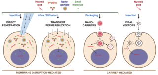
Intracellular delivery is the process of introducing external materials into living cells. Materials that are delivered into cells include nucleic acids, proteins, peptides, impermeable small molecules, synthetic nanomaterials, organelles, and micron-scale tracers, devices and objects. Such molecules and materials can be used to investigate cellular behavior, engineer cell operations or correct a pathological function.

A hybrid cell line is a fusion of cells from two different cell types. When the membrane of two cells merge, the nuclei combine to form a polykaryote. These fusions can happen spontaneously as in the case of tumor hybrid cells, or may be induced by a variety of laboratory techniques.

References

  1. "6.3. Cell fusion". Herkules.oulu.fi. Retrieved 2013-08-16.
  2. Ogle, Brenda M.; Platt, Jeffrey L. (1 January 2004). "The Biology of Cell Fusion: Cells of different types and from different species can fuse, potentially transferring disease, repairing tissues and taking part in development". American Scientist. 92 (5): 420–427. doi:10.1511/2004.49.943. JSTOR   27858450.
  3. "Definition of Cell fusion".
  4. Ogle, B. M.; Cascalho, M.; Platt, J. L. (2005). "Cells derived by fusion". Nature Reviews Molecular Cell Biology. 6 (7): 567–575. doi:10.1038/nrm1678. PMID   15957005. S2CID   22584685.
  5. Singec, Ilyas; Snyder, Evan Y. (2008). "Inflammation as a matchmaker: Revisiting cell fusion". Nature Cell Biology. 10 (5): 503–505. doi:10.1038/ncb0508-503. PMID   18454127. S2CID   30269058.
  6. "Principles And Applications Of Electrical Cell Fusion".
  7. Pedrazzoli, Filippo; Chrysantzas, Iraklis; Dezzani, Luca; Rosti, Vittorio; Vincitorio, Massimo; Sitar, Giammaria (1 January 2011). "Cell fusion in tumor progression: the isolation of cell fusion products by physical methods". Cancer Cell International. 11: 32. doi: 10.1186/1475-2867-11-32 . PMC   3187729 . PMID   21933375.
  8. Wainberg, M. A.; Howe, C. (1 October 1973). "Factors Affecting Cell Fusion Induced by Sendai Virus". J Virol. 12 (4): 937–939. doi:10.1128/JVI.12.4.937-939.1973. PMC   356713 . PMID   4359961.
  9. Sullivan, Stephen; Eggan, Kevin (1 January 2006). "The potential of cell fusion for human therapy". Stem Cell Rev. 2 (4): 341–349. doi:10.1007/BF02698061. PMID   17848721. S2CID   11446688.
  10. Maruyama, Daisuke; Ohtsu, Mina; Higashiyama, Tetsuya (2016-12-01). "Cell fusion and nuclear fusion in plants". Seminars in Cell & Developmental Biology. The Rhomboid Superfamily in Development and Disease. 60: 127–135. doi:10.1016/j.semcdb.2016.07.024. ISSN   1084-9521.
  11. Bastida-Ruiz, Daniel; Van Hoesen, Kylie; Cohen, Marie (April 2016). "The Dark Side of Cell Fusion". International Journal of Molecular Sciences. 17 (5): 638. doi: 10.3390/ijms17050638 . ISSN   1422-0067. PMC   4881464 . PMID   27136533.
  12. Hofstatter PG, Brown MW, Lahr DJG (November 2018). "Comparative Genomics Supports Sex and Meiosis in Diverse Amoebozoa". Genome Biol Evol. 10 (11): 3118–3128. doi:10.1093/gbe/evy241. PMC   6263441 . PMID   30380054.
  13. Gratia JP, Thiry M (September 2003). "Spontaneous zygogenesis in Escherichia coli, a form of true sexuality in prokaryotes". Microbiology (Reading, Engl.). 149 (Pt 9): 2571–84. doi: 10.1099/mic.0.26348-0 . PMID   12949181.
  14. Gammal, Roseann; Baker, Krista; Heilman, Destin (2011). "Heterokaryon Technique for Analysis of Cell Type-specific Localization". Journal of Visualized Experiments (49): 2488. doi:10.3791/2488. ISSN   1940-087X. PMC   3197295 . PMID   21445034.

Further reading