Chromosome conformation capture

Last updated
Chromosome conformation capture technologies Chromosome Conformation Capture Technology (en).svg
Chromosome conformation capture technologies

Chromosome conformation capture techniques (often abbreviated to 3C technologies or 3C-based methods [1] ) are a set of molecular biology methods used to analyze the spatial organization of chromatin in a cell. These methods quantify the number of interactions between genomic loci that are nearby in 3-D space, but may be separated by many nucleotides in the linear genome. [2] Such interactions may result from biological functions, such as promoter-enhancer interactions, or from random polymer looping, where undirected physical motion of chromatin causes loci to collide. [3] Interaction frequencies may be analyzed directly, [4] or they may be converted to distances and used to reconstruct 3-D structures. [5]

Contents

The chief difference between 3C-based methods is their scope. For example, when using PCR to detect interaction in a 3C experiment, the interactions between two specific fragments are quantified. In contrast, Hi-C quantifies interactions between all possible pairs of fragments simultaneously. Deep sequencing of material produced by 3C also produces genome-wide interactions maps.

History

Historically, microscopy was the primary method of investigating nuclear organization, [6] which can be dated back to 1590. [7]

Experimental methods

All 3C methods start with a similar set of steps, performed on a sample of cells.

Chromosome conformation techniques.jpg

First, the cell genomes are cross-linked with formaldehyde, [28] which introduces bonds that "freeze" interactions between genomic loci. Treatment of cells with 1-3% formaldehyde, for 10-30min at room temperature is most common, however, standardization for preventing high protein-DNA cross linking is necessary, as this may negatively affect the efficiency of restriction digestion in the subsequent step. [29] The genome is then cut into fragments with a restriction endonuclease. The size of restriction fragments determines the resolution of interaction mapping. Restriction enzymes (REs) that make cuts on 6bp recognition sequences, such as EcoR1 or HindIII, are used for this purpose, as they cut the genome once every 4000bp, giving ~ 1 million fragments in the human genome. [29] [30] For more precise interaction mapping, a 4bp recognizing RE may also be used. The next step is, proximity based ligation. This takes place at low DNA concentrations or within intact, permeabilized nuclei [26] in the presence of T4 DNA ligase, [31] such that ligation between cross-linked interacting fragments is favored over ligation between fragments that are not cross-linked. Subsequently, interacting loci are quantified by amplifying ligated junctions by PCR methods. [29] [31]

Original methods

3C (one-vs-one)

The chromosome conformation capture (3C) experiment quantifies interactions between a single pair of genomic loci. For example, 3C can be used to test a candidate promoter-enhancer interaction. Ligated fragments are detected using PCR with known primers. [2] [17] That is why this technique requires the prior knowledge of the interacting regions.

4C (one-vs-all)

Chromosome conformation capture-on-chip (4C) (also known as circular chromosome conformation capture) captures interactions between one locus and all other genomic loci. It involves a second ligation step, to create self-circularized DNA fragments, which are used to perform inverse PCR. Inverse PCR allows the known sequence to be used to amplify the unknown sequence ligated to it. [32] [2] [19] In contrast to 3C and 5C, the 4C technique does not require the prior knowledge of both interacting chromosomal regions. Results obtained using 4C are highly reproducible with most of the interactions that are detected between regions proximal to one another. On a single microarray, approximately a million interactions can be analyzed. [ citation needed ]

5C (many-vs-many)

Chromosome conformation capture carbon copy (5C) detects interactions between all restriction fragments within a given region, with this region's size typically no greater than a megabase. [2] [20] This is done by ligating universal primers to all fragments. However, 5C has relatively low coverage. The 5C technique overcomes the junctional problems at the intramolecular ligation step and is useful for constructing complex interactions of specific loci of interest. This approach is unsuitable for conducting genome-wide complex interactions since that will require millions of 5C primers to be used.[ citation needed ]

Hi-C (all-vs-all)

Hi-C uses high-throughput sequencing to find the nucleotide sequence of fragments [2] [22] and uses paired end sequencing, which retrieves a short sequence from each end of each ligated fragment. As such, for a given ligated fragment, the two sequences obtained should represent two different restriction fragments that were ligated together in the proximity based ligation step. The pair of sequences are individually aligned to the genome, thus determining the fragments involved in that ligation event. Hence, all possible pairwise interactions between fragments are tested.

Sequence capture-based methods

A number of methods use oligonucleotide capture to enrich 3C and Hi-C libraries for specific loci of interest. [33] [34] These methods include Capture-C, [35] NG Capture-C, [36] Capture-3C, [35] HiCap, [33] [37] Capture Hi-C. [38] and Micro Capture-C. [39] These methods are able to produce higher resolution and sensitivity than 4C based methods, [40] Micro Capture-C provides the highest resolution of the available 3C techniques and it is possible to generate base pair resolution data. [39]

Single-cell methods

Single-cell adaptations of these methods, such as ChIP-seq and Hi-C can be used to investigate the interactions occurring in individual cells. [41] [42]

Multi-interaction methods

A number of methods sequence multiple ligation junctions simultaneously to detect higher-order structures where multiple regions of chromatin may be interacting. These methods include Tri-C, [43] 3way 4C/C-walks, [44] and multi-contact 4C (MC-4C). [45]

Immunoprecipitation-based methods

ChIP-loop

ChIP-loop combines 3C with ChIP-seq to detect interactions between two loci of interest mediated by a protein of interest. [2] [46] The ChIP-loop may be useful in identifying long-range cis-interactions and trans interaction mediated through proteins since frequent DNA collisions will not occur. [ citation needed ]

Genome wide methods

ChIA-PET combines Hi-C with ChIP-seq to detect all interactions mediated by a protein of interest. [2] [23] HiChIP was designed to allow similar analysis as ChIA-PET with less input material. [47]

Biological impact

3C methods have led to a number of biological insights, including the discovery of new structural features of chromosomes, the cataloguing of chromatin loops, and increased understanding of transcriptional regulation mechanisms (the disruption of which can lead to disease). [6]

3C methods have demonstrated the importance of spatial proximity of regulatory elements to the genes that they regulate. For example, in tissues that express globin genes, the β-globin locus control region forms a loop with these genes. This loop is not found in tissues where the gene is not expressed. [48] This technology has further aided the genetic and epigenetic study of chromosomes both in model organisms and in humans.[ not verified in body ]

These methods have revealed large-scale organization of the genome into topologically associating domains (TADs), which correlate with epigenetic markers. Some TADs are transcriptionally active, while others are repressed. [49] Many TADs have been found in D. melanogaster, mouse and human. [50] Moreover, CTCF and cohesin play important roles in determining TADs and enhancer-promoter interactions. The result shows that the orientation of CTCF binding motifs in an enhancer-promoter loop should be facing to each other in order for the enhancer to find its correct target. [51]

Human disease

There are several diseases caused by defects in promoter-enhancer interactions, which are reviewed in this paper. [52]

Beta thalassemia is a certain type of blood disorder caused by a deletion of LCR enhancer element. [53] [54]

Holoprosencephaly is cephalic disorder caused by a mutation in the SBE2 enhancer element, which in turn weakened the production of SHH gene. [55]

PPD2 (polydactyly of a triphalangeal thumb) is caused by a mutation of ZRS enhancer, which in turn strengthened the production of SHH gene. [56] [57]

Adenocarcinoma of the lung can be caused by a duplication of enhancer element for MYC gene. [58]

T-cell acute lymphoblastic leukemia is caused by an introduction of a new enhancer. [59]

Data analysis

Heat map and circular plot visualization of Hi-C data. a. Hi-C interactions among all chromosomes from G401 human kidney cells, as plotted by the my5C software. b. Heat map visualization illustrating the bipartite structure of the mouse X chromosome, as plotted by Hi-Browse. c. Heat map visualization of a 3 Mbp locus (chr4:18000000-21000000), produced by Juicebox, using in-situ Hi-C data from the GM12878 cell line. d. Circular plot of the bipartite mouse X chromosome, generated by the Epigenome Browser. Image from Hi c visualization.gif
Heat map and circular plot visualization of Hi-C data. a. Hi-C interactions among all chromosomes from G401 human kidney cells, as plotted by the my5C software. b. Heat map visualization illustrating the bipartite structure of the mouse X chromosome, as plotted by Hi-Browse. c. Heat map visualization of a 3 Mbp locus (chr4:18000000-21000000), produced by Juicebox, using in-situ Hi-C data from the GM12878 cell line. d. Circular plot of the bipartite mouse X chromosome, generated by the Epigenome Browser. Image from

The different 3C-style experiments produce data with very different structures and statistical properties. As such, specific analysis packages exist for each experiment type. [34]

Hi-C data is often used to analyze genome-wide chromatin organization, such as topologically associating domains (TADs), linearly contiguous regions of the genome that are associated in 3-D space. [49] Several algorithms have been developed to identify TADs from Hi-C data. [4] [64]

Hi-C and its subsequent analyses are evolving. Fit-Hi-C [3] is a method based on a discrete binning approach with modifications of adding distance of interaction (initial spline fitting, aka spline-1) and refining the null model (spline-2). The result of Fit-Hi-C is a list of pairwise intra-chromosomal interactions with their p-values and q-values. [63]

The 3-D organization of the genome can also be analyzed via eigendecomposition of the contact matrix. Each eigenvector corresponds to a set of loci, which are not necessarily linearly contiguous, that share structural features. [65]

A significant confounding factor in 3C technologies is the frequent non-specific interactions between genomic loci that occur due to random polymer behavior. An interaction between two loci must be confirmed as specific through statistical significance testing. [3]

Normalization of Hi-C contact map

There are two major ways of normalizing raw Hi-C contact heat maps. The first way is to assume equal visibility, meaning there is an equal chance for each chromosomal position to have an interaction. Therefore, the true signal of a Hi-C contact map should be a balanced matrix (Balanced matrix has constant row sums and column sums). An example of algorithms that assumes equal visibility is Sinkhorn-Knopp algorithm, which scales the raw Hi-C contact map into a balanced matrix.

The other way is to assume there is a bias associated with each chromosomal position. The contact map value at each coordinate will be the true signal at that position times bias associated with the two contact positions. An example of algorithms that aim to solve this model of bias is iterative correction, which iteratively regressed out row and column bias from the raw Hi-C contact map. There are a number of software tools available for analysis of Hi-C data. [66]

DNA motif analysis

DNA motifs are specific short DNA sequences, often 8-20 nucleotides in length [67] which are statistically overrepresented in a set of sequences with a common biological function. Currently, regulatory motifs on the long-range chromatin interactions have not been studied extensively. Several studies have focused on elucidating the impact of DNA motifs in promoter-enhancer interactions.

Bailey et al. has identified that ZNF143 motif in the promoter regions provides sequence specificity for promoter-enhancer interactions. [68] Mutation of ZNF143 motif decreased the frequency of promoter-enhancer interactions suggesting that ZNF143 is a novel chromatin-looping factor.

For genome-scale motif analysis, in 2016, Wong et al. reported a list of 19,491 DNA motif pairs for K562 cell line on the promoter-enhancer interactions. [69] As a result, they proposed that motif pairing multiplicity (number of motifs that are paired with a given motif) is linked to interaction distance and regulatory region type. In the next year, Wong published another article reporting 18,879 motif pairs in 6 human cell lines. [70] A novel contribution of this work is MotifHyades, a motif discovery tool that can be directly applied to paired sequences.

Cancer genome analysis

The 3C-based techniques can provide insights into the chromosomal rearrangements in the cancer genomes. [71] Moreover, they can show changes of spatial proximity for regulatory elements and their target genes, which bring deeper understanding of the structural and functional basis of the genome. [72]

Related Research Articles

Chromatin is a complex of DNA and protein found in eukaryotic cells. The primary function is to package long DNA molecules into more compact, denser structures. This prevents the strands from becoming tangled and also plays important roles in reinforcing the DNA during cell division, preventing DNA damage, and regulating gene expression and DNA replication. During mitosis and meiosis, chromatin facilitates proper segregation of the chromosomes in anaphase; the characteristic shapes of chromosomes visible during this stage are the result of DNA being coiled into highly condensed chromatin.

In molecular biology and genetics, transcriptional regulation is the means by which a cell regulates the conversion of DNA to RNA (transcription), thereby orchestrating gene activity. A single gene can be regulated in a range of ways, from altering the number of copies of RNA that are transcribed, to the temporal control of when the gene is transcribed. This control allows the cell or organism to respond to a variety of intra- and extracellular signals and thus mount a response. Some examples of this include producing the mRNA that encode enzymes to adapt to a change in a food source, producing the gene products involved in cell cycle specific activities, and producing the gene products responsible for cellular differentiation in multicellular eukaryotes, as studied in evolutionary developmental biology.

DNA footprinting is a method of investigating the sequence specificity of DNA-binding proteins in vitro. This technique can be used to study protein-DNA interactions both outside and within cells.

<span class="mw-page-title-main">CTCF</span> Transcription factor

Transcriptional repressor CTCF also known as 11-zinc finger protein or CCCTC-binding factor is a transcription factor that in humans is encoded by the CTCF gene. CTCF is involved in many cellular processes, including transcriptional regulation, insulator activity, V(D)J recombination and regulation of chromatin architecture.

ChIP-sequencing, also known as ChIP-seq, is a method used to analyze protein interactions with DNA. ChIP-seq combines chromatin immunoprecipitation (ChIP) with massively parallel DNA sequencing to identify the binding sites of DNA-associated proteins. It can be used to map global binding sites precisely for any protein of interest. Previously, ChIP-on-chip was the most common technique utilized to study these protein–DNA relations.

DNA adenine methyltransferase identification, often abbreviated DamID, is a molecular biology protocol used to map the binding sites of DNA- and chromatin-binding proteins in eukaryotes. DamID identifies binding sites by expressing the proposed DNA-binding protein as a fusion protein with DNA methyltransferase. Binding of the protein of interest to DNA localizes the methyltransferase in the region of the binding site. Adenine methylation does not occur naturally in eukaryotes and therefore adenine methylation in any region can be concluded to have been caused by the fusion protein, implying the region is located near a binding site. DamID is an alternate method to ChIP-on-chip or ChIP-seq.

Paired-end tags (PET) are the short sequences at the 5’ and 3' ends of a DNA fragment which are unique enough that they (theoretically) exist together only once in a genome, therefore making the sequence of the DNA in between them available upon search or upon further sequencing. Paired-end tags (PET) exist in PET libraries with the intervening DNA absent, that is, a PET "represents" a larger fragment of genomic or cDNA by consisting of a short 5' linker sequence, a short 5' sequence tag, a short 3' sequence tag, and a short 3' linker sequence. It was shown conceptually that 13 base pairs are sufficient to map tags uniquely. However, longer sequences are more practical for mapping reads uniquely. The endonucleases used to produce PETs give longer tags but sequences of 50–100 base pairs would be optimal for both mapping and cost efficiency. After extracting the PETs from many DNA fragments, they are linked (concatenated) together for efficient sequencing. On average, 20–30 tags could be sequenced with the Sanger method, which has a longer read length. Since the tag sequences are short, individual PETs are well suited for next-generation sequencing that has short read lengths and higher throughput. The main advantages of PET sequencing are its reduced cost by sequencing only short fragments, detection of structural variants in the genome, and increased specificity when aligning back to the genome compared to single tags, which involves only one end of the DNA fragment.

Chromatin Interaction Analysis by Paired-End Tag Sequencing is a technique that incorporates chromatin immunoprecipitation (ChIP)-based enrichment, chromatin proximity ligation, Paired-End Tags, and High-throughput sequencing to determine de novo long-range chromatin interactions genome-wide.

<span class="mw-page-title-main">Cas9</span> Microbial protein found in Streptococcus pyogenes M1 GAS

Cas9 is a 160 kilodalton protein which plays a vital role in the immunological defense of certain bacteria against DNA viruses and plasmids, and is heavily utilized in genetic engineering applications. Its main function is to cut DNA and thereby alter a cell's genome. The CRISPR-Cas9 genome editing technique was a significant contributor to the Nobel Prize in Chemistry in 2020 being awarded to Emmanuelle Charpentier and Jennifer Doudna.

<span class="mw-page-title-main">STARR-seq</span>

STARR-seq is a method to assay enhancer activity for millions of candidates from arbitrary sources of DNA. It is used to identify the sequences that act as transcriptional enhancers in a direct, quantitative, and genome-wide manner.

<span class="mw-page-title-main">Topologically associating domain</span> Self-interacting genomic region

A topologically associating domain (TAD) is a self-interacting genomic region, meaning that DNA sequences within a TAD physically interact with each other more frequently than with sequences outside the TAD. The median size of a TAD in mouse cells is 880 kb, and they have similar sizes in non-mammalian species. Boundaries at both side of these domains are conserved between different mammalian cell types and even across species and are highly enriched with CCCTC-binding factor (CTCF) and cohesin. In addition, some types of genes appear near TAD boundaries more often than would be expected by chance.

<span class="mw-page-title-main">Nuclear organization</span> Spatial distribution of chromatin within a cell nucleus

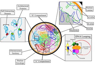
Nuclear organization refers to the spatial distribution of chromatin within a cell nucleus. There are many different levels and scales of nuclear organisation. Chromatin is a higher order structure of DNA.

<span class="mw-page-title-main">Insulated neighborhood</span>

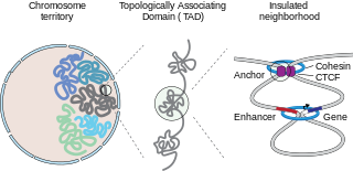
In mammalian biology, insulated neighborhoods are chromosomal loop structures formed by the physical interaction of two DNA loci bound by the transcription factor CTCF and co-occupied by cohesin. Insulated neighborhoods are thought to be structural and functional units of gene control because their integrity is important for normal gene regulation. Current evidence suggests that these structures form the mechanistic underpinnings of higher-order chromosome structures, including topologically associating domains (TADs). Insulated neighborhoods are functionally important in understanding gene regulation in normal cells and dysregulated gene expression in disease.

<span class="mw-page-title-main">Single cell epigenomics</span> Study of epigenomics in individual cells by single cell sequencing

Single cell epigenomics is the study of epigenomics in individual cells by single cell sequencing. Since 2013, methods have been created including whole-genome single-cell bisulfite sequencing to measure DNA methylation, whole-genome ChIP-sequencing to measure histone modifications, whole-genome ATAC-seq to measure chromatin accessibility and chromosome conformation capture.

<span class="mw-page-title-main">Genome architecture mapping</span>

In molecular biology, genome architecture mapping (GAM) is a cryosectioning method to map colocalized DNA regions in a ligation independent manner. It overcomes some limitations of Chromosome conformation capture (3C), as these methods have a reliance on digestion and ligation to capture interacting DNA segments. GAM is the first genome-wide method for capturing three-dimensional proximities between any number of genomic loci without ligation.

DXZ4 is a variable number tandemly repeated DNA sequence. In humans it is composed of 3kb monomers containing a highly conserved CTCF binding site. CTCF is a transcription factor protein and the main insulator responsible for partitioning of chromatin domains in the vertebrate genome.

Human epigenome is the complete set of structural modifications of chromatin and chemical modifications of histones and nucleotides. These modifications affect according to cellular type and development status. Various studies show that epigenome depends on exogenous factors.

<span class="mw-page-title-main">Hi-C (genomic analysis technique)</span> Genomic analysis technique

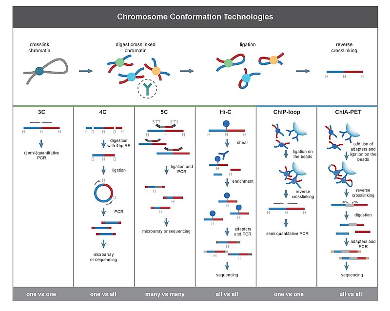
Hi-C is a high-throughput genomic and epigenomic technique first described in 2009 by Lieberman-Aiden et al. to capture chromatin conformation. In general, Hi-C is considered as a derivative of a series of chromosome conformation capture technologies, including but not limited to 3C, 4C, and 5C. Hi-C comprehensively detects genome-wide chromatin interactions in the cell nucleus by combining 3C and next-generation sequencing (NGS) approaches and has been considered as a qualitative leap in C-technology development and the beginning of 3D genomics.

<span class="mw-page-title-main">PLAC-Seq</span> Proximity ligation assisted chip-seq technology

Proximity ligation-assisted chromatin immunoprecipitation sequencing (PLAC-seq) is a chromatin conformation capture(3C)-based technique to detect and quantify genomic chromatin structure from a protein-centric approach. PLAC-seq combines in situ Hi-C and chromatin immunoprecipitation (ChIP), which allows for the identification of long-range chromatin interactions at a high resolution with low sequencing costs. Mapping long-range 3-dimensional(3D) chromatin interactions is important in identifying transcription enhancers and non-coding variants that can be linked to human diseases.

Pore-C is an emerging genomic technique which utilizes chromatin conformation capture (3C) and Oxford Nanopore Technologies' (ONT) long-read sequencing to characterize three-dimensional (3D) chromatin structure. To characterize concatemers, the originators of Pore-C developed an algorithm to identify alignments that are assigned to a restriction fragment; concatemers with greater than two associated fragments are deemed high order. Pore-C attempts to improve on previous 3C technologies, such as Hi-C and SPRITE, by not requiring DNA amplification prior to sequencing. This technology was developed as a simpler and more easily scalable method of capturing higher-order chromatin structure and mapping regions of chromatin contact. In addition, Pore-C can be used to visualize epigenomic interactions due to the capability of ONT long-read sequencing to detect DNA methylation. Applications of this technology include analysis of combinatorial chromatin interactions, the generation of de novo chromosome scale assemblies, visualization of regions associated with multi-locus histone bodies, and detection and resolution of structural variants.

References

  1. de Wit E, de Laat W (January 2012). "A decade of 3C technologies: insights into nuclear organization". Genes & Development. 26 (1): 11–24. doi:10.1101/gad.179804.111. PMC   3258961 . PMID   22215806.
  2. 1 2 3 4 5 6 7 Hakim O, Misteli T (March 2012). "SnapShot: Chromosome confirmation capture". Cell. 148 (5): 1068.e1–2. doi:10.1016/j.cell.2012.02.019. PMC   6374129 . PMID   22385969.
  3. 1 2 3 Ay F, Bailey TL, Noble WS (June 2014). "Statistical confidence estimation for Hi-C data reveals regulatory chromatin contacts". Genome Research. 24 (6): 999–1011. doi:10.1101/gr.160374.113. PMC   4032863 . PMID   24501021.
  4. 1 2 3 Rao SS, Huntley MH, Durand NC, Stamenova EK, Bochkov ID, Robinson JT, Sanborn AL, Machol I, Omer AD, Lander ES, Aiden EL (December 2014). "A 3D map of the human genome at kilobase resolution reveals principles of chromatin looping". Cell. 159 (7): 1665–80. doi:10.1016/j.cell.2014.11.021. PMC   5635824 . PMID   25497547.
  5. Varoquaux N, Ay F, Noble WS, Vert JP (June 2014). "A statistical approach for inferring the 3D structure of the genome". Bioinformatics. 30 (12): i26–33. doi:10.1093/bioinformatics/btu268. PMC   4229903 . PMID   24931992.
  6. 1 2 Denker A, de Laat W (June 2016). "The second decade of 3C technologies: detailed insights into nuclear organization". Genes & Development. 30 (12): 1357–82. doi:10.1101/gad.281964.116. PMC   4926860 . PMID   27340173.
  7. "Who invented the microscope? A complete Microscope History". Vision Engineering Ltd. Archived from the original on 22 April 2018.
  8. Martins LA (1999). "Did Sutton and Boveri propose the so-called Sutton-Boveri chromosome hypothesis?". Genet. Mol. Biol. 22 (2): 261–272. doi: 10.1590/S1415-47571999000200022 .
  9. "Genes and genetics: The language of scientific discovery". Oxford English Dictionary. Oxford University Press. 2012-08-16.
  10. Harris M (2015-02-05). "Heterochromatin and euchromatin mains".
  11. 1 2 3 Deichmann U (August 2016). "Epigenetics: The origins and evolution of a fashionable topic". Developmental Biology. 416 (1): 249–254. doi: 10.1016/j.ydbio.2016.06.005 . PMID   27291929.
  12. Lu H, Liu X, Deng Y, Qing H (December 2013). "DNA methylation, a hand behind neurodegenerative diseases". Frontiers in Aging Neuroscience. 5: 85. doi: 10.3389/fnagi.2013.00085 . PMC   3851782 . PMID   24367332.
  13. "The Francis Crick Papers: The Discovery of the Double Helix, 1951–1953".
  14. "Francis Crick, Rosalind Franklin, James Watson, and Maurice Wilkins". Science History Institute. 2016-06-01. Retrieved 2023-02-28.
  15. Cremer T, Cremer M (March 2010). "Chromosome territories". Cold Spring Harbor Perspectives in Biology. 2 (3): a003889. doi:10.1101/cshperspect.a003889. PMC   2829961 . PMID   20300217.
  16. Cullen KE, Kladde MP, Seyfred MA (July 1993). "Interaction between transcription regulatory regions of prolactin chromatin". Science. 261 (5118): 203–6. Bibcode:1993Sci...261..203C. doi:10.1126/science.8327891. PMID   8327891.
  17. 1 2 Dekker J, Rippe K, Dekker M, Kleckner N (February 2002). "Capturing chromosome conformation". Science. 295 (5558): 1306–11. Bibcode:2002Sci...295.1306D. doi:10.1126/science.1067799. PMID   11847345. S2CID   3561891.
  18. Osborne CS, Ewels PA, Young AN (January 2011). "Meet the neighbours: tools to dissect nuclear structure and function". Briefings in Functional Genomics. 10 (1): 11–7. doi:10.1093/bfgp/elq034. PMC   3080762 . PMID   21258046.
  19. 1 2 Simonis M, Klous P, Splinter E, Moshkin Y, Willemsen R, de Wit E, van Steensel B, de Laat W (November 2006). "Nuclear organization of active and inactive chromatin domains uncovered by chromosome conformation capture-on-chip (4C)". Nature Genetics. 38 (11): 1348–54. doi:10.1038/ng1896. PMID   17033623. S2CID   22787572.
  20. 1 2 Dostie J, Richmond TA, Arnaout RA, Selzer RR, Lee WL, Honan TA, et al. (October 2006). "Chromosome Conformation Capture Carbon Copy (5C): a massively parallel solution for mapping interactions between genomic elements". Genome Research. 16 (10): 1299–309. doi:10.1101/gr.5571506. PMC   1581439 . PMID   16954542.
  21. Albert I, Mavrich TN, Tomsho LP, Qi J, Zanton SJ, Schuster SC, Pugh BF (March 2007). "Translational and rotational settings of H2A.Z nucleosomes across the Saccharomyces cerevisiae genome". Nature. 446 (7135): 572–6. Bibcode:2007Natur.446..572A. doi:10.1038/nature05632. PMID   17392789. S2CID   4416890.
  22. 1 2 Lieberman-Aiden E, van Berkum NL, Williams L, Imakaev M, Ragoczy T, Telling A, et al. (October 2009). "Comprehensive mapping of long-range interactions reveals folding principles of the human genome". Science. 326 (5950): 289–93. Bibcode:2009Sci...326..289L. doi:10.1126/science.1181369. PMC   2858594 . PMID   19815776.
  23. 1 2 Fullwood MJ, Liu MH, Pan YF, Liu J, Xu H, Mohamed YB, et al. (November 2009). "An oestrogen-receptor-alpha-bound human chromatin interactome". Nature. 462 (7269): 58–64. Bibcode:2009Natur.462...58F. doi:10.1038/nature08497. PMC   2774924 . PMID   19890323.
  24. Dixon JR, Selvaraj S, Yue F, Kim A, Li Y, Shen Y, Hu M, Liu JS, Ren B (April 2012). "Topological domains in mammalian genomes identified by analysis of chromatin interactions". Nature. 485 (7398): 376–80. Bibcode:2012Natur.485..376D. doi:10.1038/nature11082. PMC   3356448 . PMID   22495300.
  25. Nora EP, Lajoie BR, Schulz EG, Giorgetti L, Okamoto I, Servant N, Piolot T, van Berkum NL, Meisig J, Sedat J, Gribnau J, Barillot E, Blüthgen N, Dekker J, Heard E (April 2012). "Spatial partitioning of the regulatory landscape of the X-inactivation centre". Nature. 485 (7398): 381–5. Bibcode:2012Natur.485..381N. doi:10.1038/nature11049. PMC   3555144 . PMID   22495304.
  26. 1 2 Nagano, Takashi; Lubling, Yaniv; Stevens, Tim J.; Schoenfelder, Stefan; Yaffe, Eitan; Dean, Wendy; Laue, Ernest D.; Tanay, Amos; Fraser, Peter (October 2013). "Single-cell Hi-C reveals cell-to-cell variability in chromosome structure". Nature. 502 (7469): 59–64. Bibcode:2013Natur.502...59N. doi:10.1038/nature12593. PMC   3869051 . PMID   24067610.
  27. Rao, Suhas; Huntley, Miriam (December 2014). "A 3D map of the human genome at kilobase resolution reveals principles of chromatin looping". Cell. 159 (7): 1665–1680. doi:10.1016/j.cell.2014.11.021. PMC   5635824 . PMID   25497547.
  28. Gavrilov A, Eivazova E, Priozhkova I, Lipinski M, Razin S, Vassetzky Y (2009). "Chromosome Conformation Capture (From 3C to 5C) and Its ChIP-Based Modification". Chromatin Immunoprecipitation Assays. review. Methods in Molecular Biology. Vol. 567. pp. 171–88. doi:10.1007/978-1-60327-414-2_12. ISBN   978-1-60327-413-5. PMID   19588093.
  29. 1 2 3 Naumova N, Smith EM, Zhan Y, Dekker J (November 2012). "Analysis of long-range chromatin interactions using Chromosome Conformation Capture". Methods. 58 (3): 192–203. doi:10.1016/j.ymeth.2012.07.022. PMC   3874837 . PMID   22903059.
  30. Belton JM, Dekker J (June 2015). "Chromosome Conformation Capture (3C) in Budding Yeast". Cold Spring Harbor Protocols. 2015 (6): 580–6. doi: 10.1101/pdb.prot085175 . PMID   26034304.
  31. 1 2 Gavrilov AA, Golov AK, Razin SV (2013-03-26). "Actual ligation frequencies in the chromosome conformation capture procedure". PLOS ONE. 8 (3): e60403. Bibcode:2013PLoSO...860403G. doi: 10.1371/journal.pone.0060403 . PMC   3608588 . PMID   23555968.
  32. Zhao, Zhihu; Tavoosidana, Gholamreza; Sjolinder, Mikael; Gondor, Anita; Mariano, Piero; Wang, Sha; Kanduri, Chandrasekhar; Lezcano, Magda; Sandhu, Kuljeet Singh; Singh, Umashankar; Pant, Vinod; Tiwari, Vijay; Kurukuti, Sreenivasulu; Ohlsson, Rolf (2006). "Circular chromosome conformation capture (4C) uncovers extensive networks of epigenetically regulated intra- and interchromosomal interactions". Nature Genetics. 38 (11): 1341–7. doi:10.1038/ng1891. PMID   17033624. S2CID   2660843.
  33. 1 2 USpatent 10287621
  34. 1 2 Schmitt AD, Hu M, Ren B (December 2016). "Genome-wide mapping and analysis of chromosome architecture". Nature Reviews Molecular Cell Biology. 17 (12): 743–755. doi:10.1038/nrm.2016.104. PMC   5763923 . PMID   27580841.
  35. 1 2 Hughes JR, Roberts N, McGowan S, Hay D, Giannoulatou E, Lynch M, et al. (February 2014). "Analysis of hundreds of cis-regulatory landscapes at high resolution in a single, high-throughput experiment". Nature Genetics. 46 (2): 205–12. doi:10.1038/ng.2871. hdl: 2318/144575 . PMID   24413732. S2CID   205348099.
  36. Davies JO, Telenius JM, McGowan SJ, Roberts NA, Taylor S, Higgs DR, Hughes JR (January 2016). "Multiplexed analysis of chromosome conformation at vastly improved sensitivity". Nature Methods. 13 (1): 74–80. doi:10.1038/nmeth.3664. PMC   4724891 . PMID   26595209.
  37. Sahlén, Pelin; Abdullayev, Ilgar; Ramsköld, Daniel; Matskova, Liudmila; Rilakovic, Nemanja; Lötstedt, Britta; Albert, Thomas J.; Lundeberg, Joakim; Sandberg, Rickard (2015-08-03). "Genome-wide mapping of promoter-anchored interactions with close to single-enhancer resolution". Genome Biology. 16 (1): 156. doi: 10.1186/s13059-015-0727-9 . ISSN   1474-760X. PMC   4557751 . PMID   26313521.
  38. Jäger R, Migliorini G, Henrion M, Kandaswamy R, Speedy HE, Heindl A, Whiffin N, Carnicer MJ, Broome L, Dryden N, Nagano T, Schoenfelder S, Enge M, Yuan Y, Taipale J, Fraser P, Fletcher O, Houlston RS (February 2015). "Capture Hi-C identifies the chromatin interactome of colorectal cancer risk loci". Nature Communications. 6: 6178. Bibcode:2015NatCo...6.6178J. doi:10.1038/ncomms7178. PMC   4346635 . PMID   25695508.
  39. 1 2 Hua P, Badat M, Hanssen L, Hentges L, Crump N, Downes D, Jeziorska DM, Oudelaar AM, Schwessinger R, Taylor S, Milne TA, Hughes JR, Higgs DR, Davies, JO (June 2021). "Defining genome architecture at base-pair resolution". Nature. 595 (7865): 125–129. Bibcode:2021Natur.595..125H. doi:10.1038/s41586-021-03639-4. PMID   34108683. S2CID   235394147.
  40. Davies JO, Oudelaar AM, Higgs DR, Hughes JR (January 2017). "How best to identify chromosomal interactions: a comparison of approaches". Nature Methods. 14 (2): 125–134. doi:10.1038/nmeth.4146. PMID   28139673. S2CID   4136037.
  41. Nagano T, Lubling Y, Stevens TJ, Schoenfelder S, Yaffe E, Dean W, et al. (October 2013). "Single-cell Hi-C reveals cell-to-cell variability in chromosome structure". Nature. 502 (7469): 59–64. Bibcode:2013Natur.502...59N. doi:10.1038/nature12593. PMC   3869051 . PMID   24067610.
  42. Schwartzman O, Tanay A (December 2015). "Single-cell epigenomics: techniques and emerging applications". Nature Reviews Genetics. 16 (12): 716–26. doi:10.1038/nrg3980. PMID   26460349. S2CID   10326803.
  43. Oudelaar, A. Marieke; Davies, James O. J.; Hanssen, Lars L. P.; Telenius, Jelena M.; Schwessinger, Ron; Liu, Yu; Brown, Jill M.; Downes, Damien J.; Chiariello, Andrea M.; Bianco, Simona; Nicodemi, Mario (2018). "Single-allele chromatin interactions identify regulatory hubs in dynamic compartmentalized domains". Nature Genetics. 50 (12): 1744–1751. doi:10.1038/s41588-018-0253-2. ISSN   1546-1718. PMC   6265079 . PMID   30374068.
  44. Olivares-Chauvet, Pedro; Mukamel, Zohar; Lifshitz, Aviezer; Schwartzman, Omer; Elkayam, Noa Oded; Lubling, Yaniv; Deikus, Gintaras; Sebra, Robert P.; Tanay, Amos (2016). "Capturing pairwise and multi-way chromosomal conformations using chromosomal walks". Nature. 540 (7632): 296–300. Bibcode:2016Natur.540..296O. doi:10.1038/nature20158. ISSN   1476-4687. PMID   27919068. S2CID   786054.
  45. Allahyar, Amin; Vermeulen, Carlo; Bouwman, Britta A. M.; Krijger, Peter H. L.; Verstegen, Marjon J. A. M.; Geeven, Geert; van Kranenburg, Melissa; Pieterse, Mark; Straver, Roy; Haarhuis, Judith H. I.; Jalink, Kees (2018). "Enhancer hubs and loop collisions identified from single-allele topologies". Nature Genetics. 50 (8): 1151–1160. doi:10.1038/s41588-018-0161-5. ISSN   1546-1718. PMID   29988121. S2CID   49667747.
  46. Horike S, Cai S, Miyano M, Cheng JF, Kohwi-Shigematsu T (January 2005). "Loss of silent-chromatin looping and impaired imprinting of DLX5 in Rett syndrome". Nature Genetics. 37 (1): 31–40. doi:10.1038/ng1491. PMID   15608638. S2CID   2884412.
  47. Mumbach MR, Rubin AJ, Flynn RA, Dai C, Khavari PA, Greenleaf WJ, Chang HY (November 2016). "HiChIP: efficient and sensitive analysis of protein-directed genome architecture". Nature Methods. 13 (11): 919–922. doi:10.1038/nmeth.3999. PMC   5501173 . PMID   27643841.
  48. Tolhuis B, Palstra RJ, Splinter E, Grosveld F, de Laat W (December 2002). "Looping and interaction between hypersensitive sites in the active beta-globin locus". Molecular Cell. 10 (6): 1453–65. doi: 10.1016/S1097-2765(02)00781-5 . PMID   12504019.
  49. 1 2 Cavalli G, Misteli T (March 2013). "Functional implications of genome topology". Nature Structural & Molecular Biology. 20 (3): 290–9. doi:10.1038/nsmb.2474. PMC   6320674 . PMID   23463314.
  50. Dekker J, Marti-Renom MA, Mirny LA (June 2013). "Exploring the three-dimensional organization of genomes: interpreting chromatin interaction data". Nature Reviews Genetics. 14 (6): 390–403. doi:10.1038/nrg3454. PMC   3874835 . PMID   23657480.
  51. Guo Y, Xu Q, Canzio D, Shou J, Li J, Gorkin DU, et al. (August 2015). "CRISPR Inversion of CTCF Sites Alters Genome Topology and Enhancer/Promoter Function". Cell. 162 (4): 900–10. doi:10.1016/j.cell.2015.07.038. PMC   4642453 . PMID   26276636.
  52. Krijger PH, de Laat W (December 2016). "Regulation of disease-associated gene expression in the 3D genome". Nature Reviews Molecular Cell Biology. 17 (12): 771–782. doi:10.1038/nrm.2016.138. PMID   27826147. S2CID   11484886.
  53. Fritsch EF, Lawn RM, Maniatis T (June 1979). "Characterisation of deletions which affect the expression of fetal globin genes in man". Nature. 279 (5714): 598–603. Bibcode:1979Natur.279..598F. doi:10.1038/279598a0. PMID   450109. S2CID   4243029.
  54. Van der Ploeg LH, Konings A, Oort M, Roos D, Bernini L, Flavell RA (February 1980). "gamma-beta-Thalassaemia studies showing that deletion of the gamma- and delta-genes influences beta-globin gene expression in man". Nature. 283 (5748): 637–42. Bibcode:1980Natur.283..637V. doi:10.1038/283637a0. PMID   6153459. S2CID   4371542.
  55. Jeong Y, El-Jaick K, Roessler E, Muenke M, Epstein DJ (February 2006). "A functional screen for sonic hedgehog regulatory elements across a 1 Mb interval identifies long-range ventral forebrain enhancers". Development. 133 (4): 761–72. doi: 10.1242/dev.02239 . PMID   16407397.
  56. Lettice LA, Heaney SJ, Purdie LA, Li L, de Beer P, Oostra BA, et al. (July 2003). "A long-range Shh enhancer regulates expression in the developing limb and fin and is associated with preaxial polydactyly". Human Molecular Genetics. 12 (14): 1725–35. doi: 10.1093/hmg/ddg180 . PMID   12837695.
  57. Wieczorek D, Pawlik B, Li Y, Akarsu NA, Caliebe A, May KJ, et al. (January 2010). "A specific mutation in the distant sonic hedgehog (SHH) cis-regulator (ZRS) causes Werner mesomelic syndrome (WMS) while complete ZRS duplications underlie Haas type polysyndactyly and preaxial polydactyly (PPD) with or without triphalangeal thumb". Human Mutation. 31 (1): 81–9. doi:10.1002/humu.21142. PMID   19847792. S2CID   1715146.
  58. Zhang X, Choi PS, Francis JM, Imielinski M, Watanabe H, Cherniack AD, Meyerson M (February 2016). "Identification of focally amplified lineage-specific super-enhancers in human epithelial cancers". Nature Genetics. 48 (2): 176–82. doi:10.1038/ng.3470. PMC   4857881 . PMID   26656844.
  59. Mansour MR, Abraham BJ, Anders L, Berezovskaya A, Gutierrez A, Durbin AD, et al. (December 2014). "Oncogene regulation. An oncogenic super-enhancer formed through somatic mutation of a noncoding intergenic element". Science. 346 (6215): 1373–7. doi:10.1126/science.1259037. PMC   4720521 . PMID   25394790.
  60. Lajoie BR, van Berkum NL, Sanyal A, Dekker J (October 2009). "My5C: web tools for chromosome conformation capture studies". Nature Methods. 6 (10): 690–1. doi:10.1038/nmeth1009-690. PMC   2859197 . PMID   19789528.
  61. Deng X, Ma W, Ramani V, Hill A, Yang F, Ay F, et al. (August 2015). "Bipartite structure of the inactive mouse X chromosome". Genome Biology. 16 (1): 152. doi: 10.1186/s13059-015-0728-8 . PMC   4539712 . PMID   26248554.
  62. Zhou X, Lowdon RF, Li D, Lawson HA, Madden PA, Costello JF, Wang T (May 2013). "Exploring long-range genome interactions using the WashU Epigenome Browser". Nature Methods. 10 (5): 375–6. doi:10.1038/nmeth.2440. PMC   3820286 . PMID   23629413.
  63. 1 2 Yardımcı GG, Noble WS (February 2017). "Software tools for visualizing Hi-C data". Genome Biology. 18 (1): 26. doi: 10.1186/s13059-017-1161-y . PMC   5290626 . PMID   28159004.
  64. Dixon JR, Selvaraj S, Yue F, Kim A, Li Y, Shen Y, et al. (April 2012). "Topological domains in mammalian genomes identified by analysis of chromatin interactions". Nature. 485 (7398): 376–80. Bibcode:2012Natur.485..376D. doi:10.1038/nature11082. PMC   3356448 . PMID   22495300.
  65. Imakaev M, Fudenberg G, McCord RP, Naumova N, Goloborodko A, Lajoie BR, et al. (October 2012). "Iterative correction of Hi-C data reveals hallmarks of chromosome organization". Nature Methods. 9 (10): 999–1003. doi:10.1038/nmeth.2148. PMC   3816492 . PMID   22941365.
  66. Imakaev M, Fudenberg G, McCord RP, Naumova N, Goloborodko A, Lajoie BR, Dekker J, Mirny LA (October 2012). "Iterative correction of Hi-C data reveals hallmarks of chromosome organization". Nature Methods. 9 (10): 999–1003. doi:10.1038/nmeth.2148. PMC   3816492 . PMID   22941365.
  67. Zambelli F, Pesole G, Pavesi G (March 2013). "Motif discovery and transcription factor binding sites before and after the next-generation sequencing era". Briefings in Bioinformatics. 14 (2): 225–37. doi:10.1093/bib/bbs016. PMC   3603212 . PMID   22517426.
  68. Bailey, S. D., Zhang, X., Desai, K., Aid, M., Corradin, O., Cowper-Sal·lari, R., … Lupien, M. (2015). ZNF143 provides sequence specificity to secure chromatin interactions at gene promoters. Nature Communications, 2, 6186. Retrieved from https://doi.org/10.1038/ncomms7186
  69. K. Wong, Y. Li, and C. Peng, “Identification of coupling DNA motif pairs on long-range chromatin interactions in human,” vol. 32, no. September 2015, pp. 321–324, 2016.
  70. Ka-Chun Wong; MotifHyades: expectation maximization for de novo DNA motif pair discovery on paired sequences, Bioinformatics, Volume 33, Issue 19, 1 October 2017, Pages 3028–3035, https://doi.org/10.1093/bioinformatics/btx381
  71. Harewood L, Kishore K, Eldridge MD, Wingett S, Pearson D, Schoenfelder S, Collins VP, Fraser P (June 2017). "Hi-C as a tool for precise detection and characterisation of chromosomal rearrangements and copy number variation in human tumours". Genome Biology. 18 (1): 125. doi: 10.1186/s13059-017-1253-8 . PMC   5488307 . PMID   28655341.
  72. Taberlay PC, Achinger-Kawecka J, Lun AT, Buske FA, Sabir K, Gould CM, et al. (June 2016). "Three-dimensional disorganization of the cancer genome occurs coincident with long-range genetic and epigenetic alterations". Genome Research. 26 (6): 719–31. doi:10.1101/gr.201517.115. PMC   4889976 . PMID   27053337.

Further reading

See also