
Eumycetoma, also known as Madura foot, is a persistent fungal infection of the skin and the tissues just under the skin, affecting most commonly the feet, although it can occur in hands and other body parts. It starts as a painless wet nodule, which may be present for years before ulceration, swelling, grainy discharge and weeping from sinuses and fistulae, followed by bone deformity.

Cochliobolus lunatus is a fungal plant pathogen that can cause disease in humans and other animals. The anamorph of this fungus is known as Curvularia lunata, while C. lunatus denotes the teleomorph or sexual stage. They are, however, the same biological entity. C. lunatus is the most commonly reported species in clinical cases of reported Cochliobolus infection.

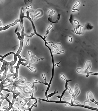
Setosphaeria rostrata is a heat tolerant fungus with an asexual reproductive form (anamorph) known as Exserohilum rostratum. This fungus is a common plant pathogen, causing leaf spots as well as crown rot and root rot in grasses. It is also found in soils and on textiles in subtropical and tropical regions. Exserohilum rostratum is one of the 35 Exserohilum species implicated uncommonly as opportunistic pathogens of humans where it is an etiologic agent of sinusitis, keratitis, skin lesions and an often fatal meningoencephalitis. Infections caused by this species are most often seen in regions with hot climates like Israel, India and the southern USA.
Exophiala jeanselmei is a saprotrophic fungus in the family Herpotrichiellaceae. Four varieties have been discovered: Exophiala jeanselmei var. heteromorpha, E. jeanselmei var. lecanii-corni, E. jeanselmei var. jeanselmei, and E. jeanselmei var. castellanii. Other species in the genus Exophiala such as E. dermatitidis and E. spinifera have been reported to have similar annellidic conidiogenesis and may therefore be difficult to differentiate.

Pseudallescheria boydii is a species of fungus classified in the Ascomycota. It is associated with some forms of eumycetoma/maduromycosis and is the causative agent of pseudallescheriasis. Typically found in stagnant and polluted water, it has been implicated in the infection of immunocompromised and near-drowned pneumonia patients. Treatment of infections with P. boydii is complicated by resistance to many of the standard antifungal agents normally used to treat infections by filamentous fungi.

Exophiala dermatitidis is a thermophilic black yeast, and a member of the Herpotrichiellaceae. While the species is only found at low abundance in nature, metabolically active strains are commonly isolated in saunas, steam baths, and dish washers. Exophiala dermatitidis only rarely causes infection in humans, however cases have been reported around the world. In East Asia, the species has caused lethal brain infections in young and otherwise healthy individuals. The fungus has been known to cause cutaneous and subcutaneous phaeohyphomycosis, and as a lung colonist in people with cystic fibrosis in Europe. In 2002, an outbreak of systemic E. dermatitidis infection occurred in women who had received contaminated steroid injections at North Carolina hospitals.
Ochroconis gallopava, also called Dactylaria gallopava or Dactylaria constricta var. gallopava, is a member of genus Dactylaria. Ochroconis gallopava is a thermotolerant, darkly pigmented fungus that causes various infections in fowls, turkeys, poults, and immunocompromised humans first reported in 1986. Since then, the fungus has been increasingly reported as an agent of human disease especially in recipients of solid organ transplants. Ochroconis gallopava infection has a long onset and can involve a variety of body sites. Treatment of infection often involves a combination of antifungal drug therapy and surgical excision.
Phaeohyphomycosis is a diverse group of fungal infections, caused by dematiaceous fungi whose morphologic characteristics in tissue include hyphae, yeast-like cells, or a combination of these. It can be associated with an array of melanistic filamentous fungi including Alternaria species, Exophiala jeanselmei, and Rhinocladiella mackenziei.
Thielavia subthermophila is a ubiquitous, filamentous fungus that is a member of the phylum Ascomycota and order Sordariales. Known to be found on plants of arid environments, it is an endophyte with thermophilic properties, and possesses dense, pigmented mycelium. Thielavia subthermophila has rarely been identified as a human pathogen, with a small number of clinical cases including ocular and brain infections. For treatment, antifungal drugs such as amphotericin B have been used topically or intravenously, depending upon the condition.
Scedosporiosis is the general name for any mycosis – i.e., fungal infection – caused by a fungus from the genus Scedosporium. Current population-based studies suggest Scedosporium prolificans and Scedosporium apiospermum to be among the most common infecting agents from the genus, although infections caused by other members thereof are not unheard of. The latter is an asexual form (anamorph) of another fungus, Pseudallescheria boydii. The former is a "black yeast", currently not characterized as well, although both of them have been described as saprophytes.

Exophiala phaeomuriformis is thermophilic fungus belonging to the genus Exophiala and the family Herpotrichiellaceae. it is a member of the group of fungi known as black yeasts, and is typically found in hot and humid locations, such as saunas, bathrooms, and dishwashers. This species can cause skin infections and is typically classified as a Biosafety Risk Group 2 agent.

Cladosporium cladosporioides is a darkly pigmented mold that occurs world-wide on a wide range of materials both outdoors and indoors. It is one of the most common fungi in outdoor air where its spores are important in seasonal allergic disease. While this species rarely causes invasive disease in animals, it is an important agent of plant disease, attacking both the leaves and fruits of many plants. This species produces asexual spores in delicate, branched chains that break apart readily and drift in the air. It is able to grow under low water conditions and at very low temperatures.
Fonsecaea compacta is a saprophytic fungal species found in the family Herpotrichiellaceae. It is a rare etiological agent of chromoblastomycosis, with low rates of correspondence observed from reports. The main active components of F. compacta are glycolipids, yet very little is known about its composition. F. compacta is widely regarded as a dysplastic variety of Fonsecaea pedrosoi, its morphological precursor. The genus Fonsecaea presently contains two species, F. pedrosoi and F. compacta. Over 100 strains of F. pedrosoi have been isolated but only two of F. compacta.

Rhinocladiella mackenziei is a deeply pigmented mold that is a common cause of human cerebral phaeohyphomycosis. Rhinocladiella mackenziei was believed to be endemic solely to the Middle East, due to the first cases of infection being limited to the region. However, cases of R. mackenziei infection are increasingly reported from regions outside the Middle East. This pathogen is unique in that the majority of cases have been reported from immunologically normal people.
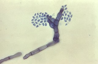
Cladophialophora carrionii is a melanized fungus in the genus Cladophialophora that is associated with decaying plant material like cacti and wood. It is one of the most frequent species of Cladophialophora implicated in human disease. Cladophialophora carrionii is a causative agent of chromoblastomycosis, a subcutaneous infection that occurs in sub-tropical areas such as Madagascar, Australia and northwestern Venezuela. Transmission occurs through traumatic implantation of plant material colonized by C. carrionii, mainly infecting rural workers. When C. carrionii infects its host, it transforms from a mycelial state to a muriform state to better tolerate the extreme conditions in the host's body.

Cladosporium sphaerospermum is a radiotrophic fungus belonging to the genus Cladosporium and was described in 1886 by Albert Julius Otto Penzig from the decaying leaves and branches of Citrus. It is a dematiaceous (darkly-pigmented) fungus characterized by slow growth and largely asexual reproduction. Cladosporium sphaerospermum consists of a complex of poorly morphologically differentiated, "cryptic" species that share many physiological and ecological attributes. In older literature, all of these sibling species were classified as C. sphaerospermum despite their unique nature. Accordingly, there is confusion in older literature reports on the physiological and habitat regularities of C. sphaerospermum in the strict sense. This fungus is most phylogenetically similar to C. fusiforme. According to modern phylogenetic analyses, the previously synonymized species, Cladosporium langeroni, is a distinct species.
Cladosporium oxysporum is an airborne fungus that is commonly found outdoors and is distributed throughout the tropical and subtropical region, it is mostly located In Asia and Africa. It spreads through airborne spores and is often extremely abundant in outdoor air during the spring and summer seasons. It mainly feeds on decomposing organic matter in warmer climates, but can also be parasitic and feed on living plants. The airborne spores can occasionally cause cutaneous infections in humans, and the high prevalence of C. oxysporum in outdoor air during warm seasons contributes to its importance as an etiological agent of allergic disease and possibly human cutaneous phaeohyphomycosis in tropical regions.
Curvularia pallescens is a soil fungus, that commonly grows on crops found in tropical regions. The conidia of the fungus are distinguishable from those of related species due to their lack of curvature. C. pallescens has been reported to cause infection in plants, and in immunocompetent individuals. This species is the anamorph of Cochliobolus pallescens.
Cladophialophora arxii is a black yeast shaped dematiaceous fungus that is able to cause serious phaeohyphomycotic infections. C. arxii was first discovered in 1995 in Germany from a 22-year-old female patient suffering multiple granulomatous tracheal tumours. It is a clinical strain that is typically found in humans and is also capable of acting as an opportunistic fungus of other vertebrates Human cases caused by C. arxii have been reported from all parts of the world such as Germany and Australia.
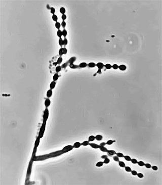
Cladophialophora is a genus of fungi in the family Herpotrichiellaceae. It has 35 species. The genus contains black yeast-like fungi, some of which are species of important medical significance. Cladophialophora bantiana causes the rare brain disease cerebral phaeohyphomycosis. Cladophialophora carrionii is a common cause of chromoblastomycosis in semi-arid climates. Some of the species are endophytes–associating with plants. For example, Cladophialophora yegresii is a cactus endophyte, which is sometimes introduced into humans via cactus spines.
