Colletotrichum lindemuthianum

Last updated

Colletotrichum lindemuthianum
Colletotrichum lindemuthianum.jpg
Colletotrichum lindemuthianum damage to bean pods.
Scientific classification OOjs UI icon edit-ltr.svg
Domain: Eukaryota
Kingdom: Fungi
Division: Ascomycota
Class: Sordariomycetes
Order: Glomerellales
Family: Glomerellaceae
Genus: Colletotrichum
Species:
C. lindemuthianum
Binomial name
Colletotrichum lindemuthianum
(Sacc. & Magnus) Briosi & Cavara, (1889)
Synonyms

Gloeosporium lindemuthianumSacc. & Magnus, (1878)
Glomerella cingulata f.sp. phaseoli
Glomerella lindemuthianaShear [as 'lindemuthianum'], (1913)

Contents

Colletotrichum lindemuthianum is a fungus which causes anthracnose, or black spot disease, of the common bean plant ( Phaseolus vulgaris ). It is considered a hemibiotrophic pathogen because it spends part of its infection cycle as a biotroph, living off of the host but not harming it, and the other part as a necrotroph, killing and obtaining nutrients from the host tissues.

History

The anthracnose of common bean was first identified in 1875 in the fruit and vegetable garden of the Agricultural Institute of Popplesdorf, Germany by Lindemuth. [1] By 1878, Saccardo and Magnus had made many observations on the cause of the anthracnose disease, recording their results in Michelia I:129. [2] They concluded that it was caused by a fungus, which they named Gloeosporium lindemuthianum after Lindemuth himself. [1] Several years later, Briosi and Cavara discovered the presence of setae on the fungus, reclassifying it from the genus Gloeosporium to Colletotrichum, where it remains today. [1] [2] Recognizing the devastating effect the fungus was having on common bean populations worldwide, it quickly became a heavily studied subject among scientists, who principally investigated means of controlling its spread. In 1911, Barrus reported the discovery of multiple fungal strains, each of which differed in its ability to infect certain varieties of bean plants, which initiated the work of Edgerton and Moreland, who found eleven different strains of the pathogen, but theorized more may exist. [1] Since then, numerous strains have been identified, each targeting specific varieties of bean plants. During the early part of the 20th century, the various races were identified by use of the Greek alphabet, paired with numbers, but at the turn of the 21st century a naming system using binary code was adopted. [3] Under the binary naming system, each plant cultivar is given a binary number, and the code for a particular race of the pathogen is determined by the sum of the binary numbers of the cultivars which it infects. [3]

Life cycle

Infection

An illustration of the biotrophic primary hyphae post-inoculation. Colletotrichum Infection.png
An illustration of the biotrophic primary hyphae post-inoculation.

The spores of C. lindemuthianum are dispersed by rain splash and must quickly attach to the aerial parts of the plant in order to infect the host. [4] A heavy rainfall may spread the spores as far as 4.5 meters from the host plant. [3] The spore will then germinate on the new host and form a short germ tube which develops an appressorium, or 'pressing' organ. [5] As the germ tube grows, it pulls the spore and the appressorium together, causing an indentation to occur in the cell wall. An infection peg is then able to protrude from the appressorium and penetrate through the cell wall. Once through the cell wall, an infection hypha grows and develops into an infection vesicle. [1] [6]

Biotrophic phase

The first stage after infection is known as the biotrophic phase, and consists of a broad primary hyphae, which develops out of the infection vesicle. The primary hyphae occasionally penetrates through additional cell walls by use of mechanical force, but usually will not grow very far from the infection vesicle. It always stays along a wall, such that half of the hyphae's circumference is in contact with the cell wall at all times. The primary hyphae do not penetrate the host cell's plasma membranes, but instead grows between it and the cell wall. As such, these hyphae do not intentionally kill any cells. [1] [7] In these early stages of infection, proteins are released by the infection vesicle which suppress the host's defense responses. One such protein is formed by the nitrogen starvation-induced gene CgDN3. The proteins suppress any hypersensitivity responses from the host to allow unhindered growth and development of the fungus. During the biotrophic phase, the pathogen gains nutrients by transferring hexoses and amino acids from the living host cell to the fungus by use of monosaccharide-H+ symporters. [7]

Necrotrophic phase

About 48–72 hours after inoculation, depending on the environment, the biotrophic phase ends and the necrotrophic phase begins. It is marked by the development of many thin hyphae, known as secondary or necrotrophic hyphae, which develop off of the primary hyphae and the infection hypha. Unlike the much larger primary hyphae, these secondary hyphae move freely through the host in all directions, penetrating cell walls and membranes alike. [7] In order to more easily ramify throughout the host tissue, the hyphae release enzymes which break down host-specific proteins. [6] Some such enzymes include cutinase, proteases, pectin and pectate lyases. One enzyme, endopolygalacturonase, is a highly specialized cell wall-degrading enzyme which is critical for the growth of the mycelium. Endopolygalacturonase is normally produced by fruiting plants and induces ripening of fruit by degrading polygalacturonan present in the cell walls. C. lindemuthianum releases large amounts of this enzyme, which not only weakens the cell wall by removing the polygalacturonan, ng the area available for cell-wall degradation by other enzymes. [8] All of this cell degradation is not allowed to go to waste, however, as the breaking down of the cell wall releases many oligo and monosaccharides which are then free to be taken up by the fungus. This is a much broader availability of sugars than was available in the biotrophic phase, which consisted mostly of glucose and fructose derived from sucrose. Because of how rapidly the fungus is killing the host, it no longer releases any defense response prevention proteins, relying purely on the rapid death of the host and growth of the pathogen. [7] Additionally, since the mycelium must rapidly spread during the necrotrophic stage in order to supply nutrients to the fungus, it will more easily thrive in younger bean plants, which have softer tissues than their older counterparts. [1] While there is very little discoloration of the host cells before secondary hyphae development, discoloration becomes rapidly apparent about 100 hours post infection. [1] Black spots begin to develop on the surface of the plant, and grows radially outward: the manifestation of anthracnose disease. These are the first visual symptoms of an infected plant, and usually occur along leaf veins on the underside of the leaf. As the lesions grow, they become indented in the center, where conidia begin to develop. These conidia are colorless at first, but develop into light pink, flesh-colored pustules ready to be spread to new hosts by rain. In the event of infection of a bean pod, the conidia may develop in the seeds themselves, where they can remain dormant until the seed begins to germinate and grow, at which point the fungus will begin to grow on the young host. [9]

Conidial anastomosis tubes

Conidial anastomosis tubes as found in C. lindemuthianum. Image (f): fusion point, (b) birth scar, (c) conidia, (a) CATs Conidial Anastomosis Tubes.png
Conidial anastomosis tubes as found in C. lindemuthianum. Image (f): fusion point, (b) birth scar, (c) conidia, (a) CATs

Maturing colonies Colletotrichum lindemuthianum, produce conidial anastomosis tubes (CATs) between conidia before leaving the host and before germination, [10] in contrast to the genetic model fungus Neurospora crassa (bread mould) that produces conidial anastomosis tubes from conidia and conidial germ tubes during germination. [11] Other Colletotrichum species produce conidial anastomosis tubes as well. [10] The initial characterization of CATs was made in this plant pathogen in 2003 by Roca et al. in Brazil. [10] [12] CATs first form 15 days after the start of fruiting body development and grow from the conidia. As they grow, they may either fuse with other CATs, as evidenced by fusion points, or make direct contact with another conidium. Because each conidium creates CATs, the result is a network of conidia all connected together. The CAT network is developed very quickly, often within 1 hour of the first CAT connection. The exact purpose of this connection network is unclear, but it permits free flow of cytoplasm, proteins, organelles and even nuclei between conidia. It is unclear whether these CATs can play a role in genetic recombination, as no sexual stages for C. lindemuthianum has been found in nature. [10]

Economic effects

Green Bean Crop, Storeton Benkid77 Green Bean Crop, Storeton 200709.JPG
Green Bean Crop, Storeton

Dry bean is an ancient crop, first domesticated over 7000 years ago and is still a staple crop globally. [3] Today, the largest bean producing nations are Brazil, India, China, Mexico, The United States of America, Myanmar, Canada, and Argentina. In all of these nations, anthracnose is considered one of the most invasive and destructive dry bean diseases, capable of destroying up to 95% of a plantation's yield while also threatening growth and development rates. As such, it is very important for producers to have disease-free certified seeds, which are generally produced in arid regions such as Idaho, where the relative humidity does not surpass the necessary 92% for spore germination. Further precaution is often taken by producers, who apply fungicides such as Azoxystrobin, fluodiooxonil or metalaxyl-m. Applied at a concentration of 125 grams per hectare of dry bean crop, azoxystrobin has been shown to increase bean yields by up to 20%. [3]

Growing in culture

It has been known for some time that the fungus can be grown in a culture. The culture is generally made of a nutrient rich agar, as the fungus generally has a hard time germinating in water due to the water solubility of the appressorium. For optimal growth, the culture should be kept at 22 °C and at a pH of 8, although it can be grown in temperatures between 0–34 °C and pH levels between 3–11. [1]

Diseases on fungus

In 1975, British mycologist Rawlinson published findings of many isometric, uniformly sized particles that he identified as double-stranded RNA viruses in the extract of the α5 race of fungus. The α5 race is noted for its unusually poor sporulation in cultures and its weak pathogenicity. Rawlinson hypothesized these features were caused by the observed viral particles. [13] However, the particles proved to be inseparable from the fungus, making an effective control difficult to acquire. Instead, he compared growth, pathogenicity and morphology of the α5 race with other races of C. lindemuthianum which were not infected. No significant differences were observed between the infected and non-infected races of the pathogen, although all α races proved to be highly nonpathogenic, regularly scoring under 1 on the 5 point pathogenicity scale, whereas other races, such as the δ races, had pathogenicity ratings of 5. While the virus has no observable negative effects on the fungus, its purpose or how it infected the fungus is still unknown. [13]

Related Research Articles

<span class="mw-page-title-main">Conidium</span> Asexual, non-motile spore of a fungus

A conidium, sometimes termed an asexual chlamydospore or chlamydoconidium, is an asexual, non-motile spore of a fungus. The word conidium comes from the Ancient Greek word for dust, κόνις (kónis). They are also called mitospores due to the way they are generated through the cellular process of mitosis. They are produced exogenously. The two new haploid cells are genetically identical to the haploid parent, and can develop into new organisms if conditions are favorable, and serve in biological dispersal.

<span class="mw-page-title-main">Appressorium</span> Structure produced by some fungi

An appressorium is a specialized cell typical of many fungal plant pathogens that is used to infect host plants. It is a flattened, hyphal "pressing" organ, from which a minute infection peg grows and enters the host, using turgor pressure capable of punching through even Mylar.

<i>Rhizoctonia solani</i> Species of fungus

Rhizoctonia solani is a species of fungus in the order Cantharellales. Basidiocarps are thin, effused, and web-like, but the fungus is more typically encountered in its anamorphic state, as hyphae and sclerotia. The name Rhizoctonia solani is currently applied to a complex of related species that await further research. In its wide sense, Rhizoctonia solani is a facultative plant pathogen with a wide host range and worldwide distribution. It causes various plant diseases such as root rot, damping off, and wire stem. It can also form mycorrhizal associations with orchids.

Glomerella graminicola is an economically important crop parasite affecting both wheat and maize where it causes the plant disease Anthracnose Leaf Blight.

<i>Thielaviopsis basicola</i> Species of fungus

Thielaviopsis basicola is the plant-pathogen fungus responsible for black root rot disease. This particular disease has a large host range, affecting woody ornamentals, herbaceous ornamentals, agronomic crops, and even vegetable crops. Examples of susceptible hosts include petunia, pansy, poinsettia, tobacco, cotton, carrot, lettuce, tomato, and others. Symptoms of this disease resemble nutrient deficiency but are truly a result of the decaying root systems of plants. Common symptoms include chlorotic lower foliage, yellowing of plant, stunting or wilting, and black lesions along the roots. The lesions along the roots may appear red at first, getting darker and turning black as the disease progresses. Black root lesions that begin in the middle of a root can also spread further along the roots in either direction. Due to the nature of the pathogen, the disease can easily be identified by the black lesions along the roots, especially when compared to healthy roots. The black lesions that appear along the roots are a result of the formation of chlamydospores, resting spores of the fungus that contribute to its pathogenicity. The chlamydospores are a dark brown-black color and cause the "discoloration" of the roots when they are produced in large amounts.

<i>Colletotrichum kahawae</i> Species of fungus

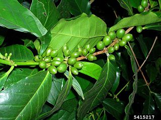
Colletotrichum kahawae is a fungal plant pathogen that causes coffee berry disease (CBD) on Coffea arabica crops. The pathogen is an ascomycete that reproduces asexually. The asexual spores (conidia) are stored within acervuli. This disease is considered to be one of the major factors hampering C.arabica production in the African continent, which represents the current geographic range of the fungus. Coffee berry disease causes dark necrosis in spots and causes the green berries of the coffee to drop prematurely. High humidity, relatively warm temperatures, and high altitude are ideal for disease formation. Given the severity of the disease and the lack of effective control measures, there is great concern that the fungus may spread to other coffee producing continents, such as South America, which could have catastrophic consequences.

<i>Colletotrichum gossypii</i> Species of fungus

Colletotrichum gossypii is a plant pathogen. This fungus is affiliated with cotton plants where it causes anthracnose. Its reproduction in the plants is asexual. The conidia have only one nucleus. Before conidia germination fusion by mean of conidial anastomosis tube could happen. The conidia could germinate in media plates.

Alternaria carthami is a necrotrophic plant pathogen of safflower. The fungus is in the order Pleosporales and family Pleosporaceae. It was first isolated in India, has spread globally and can have devastating effects on safflower yield, and resultant oilseed production. A. carthami is known to be seed-borne and appears as irregular brown lesions on safflower leaves and stems.

<i>Phomopsis obscurans</i> Species of fungus

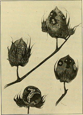
Phomopsis obscurans is a common fungus found in strawberry plants, which causes the disease of leaf blight. Common symptoms caused by the pathogen begin as small circular reddish-purple spots and enlarge to form V-shaped lesions that follow the vasculature of the plant's leaves. Although the fungus infects leaves early in the growing season when the plants are beginning to develop, leaf blight symptoms are most apparent on older plants towards the end of the growing season. The disease can weaken strawberry plants through the destruction of foliage, which results in reduced yields. In years highly favorable for disease development, leaf blight can ultimately lead to the death of the strawberry plants. A favorable environment for the growth and development of the Phomopsis obscurans pathogen is that of high temperature, high inoculum density, a long period of exposure to moisture, and immature host tissue. In the case of disease management, a conjunction of cultural practices is the most effective way of reducing the infection.

Colletotrichum capsici is a species of fungus and plant pathogen which causes leaf blight on Chlorophytum borivilianum, basil, chickpea and pepper as well as dieback in pigeonpea and anthracnose in poinsettia.

<i>Colletotrichum coccodes</i> Pathogenic fungus

Colletotrichum coccodes is a plant pathogen, which causes anthracnose on tomato and black dot disease of potato. Fungi survive on crop debris and disease emergence is favored by warm temperatures and wet weather.

<i>Glomerella cingulata</i> Species of fungus

Glomerella cingulata is a fungal plant pathogen, being the name of the sexual stage (teleomorph) while the more commonly referred to asexual stage (anamorph) is called Colletotrichum gloeosporioides. For most of this article the pathogen will be referred to as C. gloeosporioides. This pathogen is a significant problem worldwide, causing anthracnose and fruit rotting diseases on hundreds of economically important hosts.

Microdochium panattonianum is a fungal plant pathogen. This pathogen causes anthracnose of lettuce, a disease which produces necrotic lesions in cultivated lettuce. In extended periods of wet weather, M. panattonianum can cause significant crop-losses. The impact of this pathogen is exacerbated by farming lettuce without crop rotation, and by planting of susceptible lettuce varieties, such as Romaine lettuce.

Colletotrichum fragariae is a fungal plant pathogen infecting strawberries. It is not a well known fungus, and there are many similar fungi that are related to it. It is part of the Colletotrichum genus. It is a pathogen that occurs in strawberries. It leads to the disease known as anthracnose. This is typically at the crown of the strawberry, which is why it is often called crown rot. It is also known as the Anthracnose Crown rot. The fungus also infects leaves and is known as leaf spot, which is common among all Colletotrichum. This is not as common in the fragariae, as it is more common in the crown. This fungus is also better at infecting younger strawberries/seedlings. The most common way to control this disease is fungicides that are harmful to the environment. There have been studies done to see if the fungus infects other hosts but other than some weeds, it is very specific to Strawberries.

<i>Botrytis fabae</i> Species of fungus

Botrytis fabae is a plant pathogen, a fungus that causes chocolate spot disease of broad or fava bean plants, Vicia faba. It was described scientifically by Mexican-born Galician microbiologist Juan Rodríguez Sardiña in 1929.

Conidial anastomosis tubes (CATs) are cells formed from the conidia of many filamentous fungi. These cells have a tubular shape and form an anastomosis (bridge) that allows fusion between conidia.

Entomophaga grylli is a fungal pathogen which infects and kills grasshoppers. It is the causal agent of one of the most widespread diseases affecting grasshoppers. This is sometimes known as summit disease because infected insects climb to the upper part of a plant and grip the tip of the stem as they die; this ensures widespread dispersal of the fungal spores. The fungus is a species complex with several different pathotypes, each one of which seems to be host-specific to different subfamilies of grasshoppers. The pathogen is being investigated for its possible use in biological pest control of grasshoppers.

<i>Alternaria brassicicola</i> Species of fungus

Alternaria brassicicola is a fungal necrotrophic plant pathogen that causes black spot disease on a wide range of hosts, particularly in the genus of Brassica, including a number of economically important crops such as cabbage, Chinese cabbage, cauliflower, oilseeds, broccoli and canola. Although mainly known as a significant plant pathogen, it also contributes to various respiratory allergic conditions such as asthma and rhinoconjunctivitis. Despite the presence of mating genes, no sexual reproductive stage has been reported for this fungus. In terms of geography, it is most likely to be found in tropical and sub-tropical regions, but also in places with high rain and humidity such as Poland. It has also been found in Taiwan and Israel. Its main mode of propagation is vegetative. The resulting conidia reside in the soil, air and water. These spores are extremely resilient and can overwinter on crop debris and overwintering herbaceous plants.

Hemibiotrophs are the spectrum of plant pathogens, including bacteria, oomycete and a group of plant pathogenic fungi that keep its host alive while establishing itself within the host tissue, taking up the nutrients with brief biotrophic-like phase. It then, in later stages of infection switches to a necrotrophic life-style, where it rampantly kills the host cells, deriving its nutrients from the dead tissues.

<i>Colletotrichum fioriniae</i> Fungal species Colletotrichum fioriniae

Colletotrichum fioriniae is a fungal plant pathogen and endophyte of fruits and foliage of many broadleaved plants worldwide. It causes diseases on agriculturally important crops, including anthracnose of strawberry, ripe rot of grapes, bitter rot of apple, anthracnose of peach, and anthracnose of blueberry. Its ecological role in the natural environment is less well understood, other than it is a common leaf endophyte of many temperate trees and shrubs and in some cases may function as an entomopathogen.

References

  1. 1 2 3 4 5 6 7 8 9 Leach, Julian Gilbert. 1922.The parasitism of colletotrichum lindemuthianum. University of Minnesota. Retrieved 30 March 2014
  2. 1 2 Stoneman, Bertha. 1898. A comparative study of the development of some anthracnoses. Botanical Gazette 26 (2) (August, 1898).
  3. 1 2 3 4 5 Pynenburg, Gerard Martin. 2010. Argonomic and economic assessment of intensive pest management of dry bean. M.Sc., University of Guelph. Retrieved 4 April 2014
  4. Mercure, E.W.; H. Kunoh; R.L. Nicholson (December 1994). "Adhesion of Colletotrichum graminicola conidia to corn leaves, a requirement for disease development". Physiological and Molecular Plant Pathology. 45 (6): 407. doi:10.1016/S0885-5765(05)80039-6.
  5. Bailey, J.A. (1992). Colletotrichum: biology, pathology and control. p. 88. Archived from the original on 2014-04-13. Retrieved 2013-05-08.
  6. 1 2 Dean, Ralph; VanKan, Jan A. L.; Pretorius, Zacharias A.; Hammond-Kosack, Kim E.; Di Pietro, Antonio; Spanu, Pietro De.; Rudd, Jason J.; Dickman, Marty; Kahmann, Regine; Ellis, Jeff; Foster, Gary D. (2012). The Top 10 fungal pathogens in molecular plant pathology. Molecular Plant Pathology. p. 414. Accessed 31 March 2014.
  7. 1 2 3 4 Munch, Steffen, Ulrike Lingner, Daniela S. Floss, Nancy Ludwig, Norbert Sauer, and Holger B. Deising. 2008. The hemibiotrophic lifestyle of colletotrichum species. Elsevier(165): 41.
  8. Acosta-rodríguez, Ismael, Carlos Piñón-escobedo, Ma Guadalupe Zavala-páramo, Everardo López-romero, and Horacio Cano-camacho. 2005. Degradation of cellulose by the bean-pathogenic fungus colletotrichum lindemuthianum. production of extracellular cellulolytic enzymes by cellulose induction. Antonie van Leeuwenhoek 87, (4) (05): 301-10
  9. Saettler, A. February 01, 1983. Bean anthracnose Seed-transmitted disease caused by the fungus Colletotrichum lindemuthianum.. Extension bulletin E - Cooperative Extension Service, Michigan State University. no. 1671, (accessed March 27, 2014).
  10. 1 2 3 4 Roca M., M.G.; Davide, L.C.; Mendes-Costa, M.C.; Wheals, A. (2003). "Conidial anastomoses tubes in Colletotrichum". Fungal Genetics and Biology. 40 (2): 138–145. doi:10.1016/S1087-1845(03)00088-4. PMID   14516766.
  11. Roca, M.G.; Arlt, J.; Jeffree, C.E.; Read, N.D. (2005). "Cell biology of conidial anastomosis tubes in Neurospora crassa". Eukaryotic Cell. 4 (5): 911–919. doi:10.1128/EC.4.5.911-919.2005. PMC   1140100 . PMID   15879525.
  12. Glass, N.L.; Fleissner, A. (2006). "Re-wiring the network: understanding the mechanism of function of anastomosis in filamentous fungi.". In Kues, U.; Fisher, R. (eds.). Growth, Differentiation and Sexuality (The Mycota. Mycota. Vol. 1. Springer. pp. 123–139. ISBN   978-3-540-28134-4.
  13. 1 2 RAWLINSON, CJ. 1975. DOUBLE-STRANDED-RNA VIRUS IN COLLETOTRICHUM-LINDEMUTHIANUM. Transactions of the British Mycological Society 65, no. oct, (accessed April 01, 2014).

Further reading

Roca M., M. Gabriela; Davide, Lisete C.; Mendes-Costa, Maria C. Cytogenetics of Colletotrichum lindemuthianum (Glomerella cingulata f. sp. phaseoli) Fitopatologia brasileira, vol. 28 no. 4 Brasília July/Aug. 2003