Colorectal polyp

Last updated
Colon polyps
Polyp-2.jpeg
Polyp of sigmoid colon as revealed by colonoscopy. Approximately 1 cm in diameter. The polyp was removed by snare cautery.
Specialty Gastroenterology   OOjs UI icon edit-ltr-progressive.svg

A colorectal polyp is a polyp (fleshy growth) occurring on the lining of the colon or rectum. [1] Untreated colorectal polyps can develop into colorectal cancer. [2]

Contents

Colorectal polyps are often classified by their behaviour (i.e. benign vs. malignant) or cause (e.g. as a consequence of inflammatory bowel disease). They may be benign (e.g. hyperplastic polyp), pre-malignant (e.g. tubular adenoma) or malignant (e.g. colorectal adenocarcinoma).

Signs and symptoms

Colorectal polyps are not usually associated with symptoms. [2] When they occur, symptoms include bloody stools; changes in frequency or consistency of stools (such as a week or more of constipation or diarrhoea); [3] and fatigue arising from blood loss. [2] Anemia arising from iron deficiency can also present due to chronic blood loss, even in the absence of bloody stools. [3] [4] Another symptom may be an increased mucus production especially those involving villous adenomas. [4] Copious production of mucus causes loss of potassium that can occasionally result in symptomatic hypokalemia. [4] Occasionally, if a polyp is big enough to cause a bowel obstruction, there may be nausea, vomiting and severe constipation. [3]

Structure

Polyps are either pedunculated (attached to the intestinal wall by a stalk) or sessile (grow directly from the wall). [5] [6] :1342 In addition to the gross appearance categorization, they are further divided by their histologic appearance as tubular adenoma which are tubular glands, villous adenoma which are long finger like projections on the surface, and tubulovillous adenoma which has features of both. [6] :1342

Genetics

Hereditary syndromes causing increased colorectal polyp formation include:

Several genes have been associated with polyposis, such as GREM1, MSH3, MLH3, NTHL1, RNF43 and RPS20. [8]

Familial adenomatous polyposis

Familial adenomatous polyposis (FAP) is a form of hereditary cancer syndrome involving the APC gene located on chromosome q521. [9] The syndrome was first described in 1863 by Virchow on a 15-year-old boy with multiple polyps in his colon. [9] The syndrome involves development of multiple polyps at an early age and those left untreated will all eventually develop cancer. [9] The gene is expressed 100% in those with the mutation and it is autosomal dominant. [9] 10–20% of patients have negative family history and acquire the syndrome from spontaneous germline mutation. [9] The average age of newly diagnosed patient is 29 and the average age of newly discovered colorectal cancer is 39. [9] It is recommended that those affected undergo colorectal cancer screening at younger age with treatment and prevention are surgical with removal of affected tissues. [9]

Hereditary nonpolyposis colorectal cancer (Lynch Syndrome)

Hereditary nonpolyposis colorectal cancer (HNPCC, also known as Lynch syndrome) is a hereditary colorectal cancer syndrome. [9] It is the most common hereditary form of colorectal cancer in the United States and accounts for about 3% of all cases of cancer. [9] It was first recognized by Alder S. Warthin in 1885 at the University of Michigan. [9] It was later further studied by Henry Lynch who recognized an autosomal dominant transmission pattern with those affected having relatively early onset of cancer (mean age 44 years), greater occurrence of proximal lesions, mostly mucinous or poorly differentiated adenocarcinoma, greater number of synchronous and metachronous cancer cells, and good outcome after surgical intervention. [9] The Amsterdam Criteria were initially used to define Lynch syndrome before the underlying genetic mechanism had been worked out. [9] The Criteria required that the patient has three family members all first-degree relatives with colorectal cancer that involves at least two generations with at least one affected person being younger than 50 years of age when the diagnosis was made. [9] The Amsterdam Criteria is too restrictive and was later expanded to include cancers of endometrial, ovarian, gastric, pancreatic, small intestinal, ureteral, and renal pelvic origin. [9] The increased risk of cancer seen in patients with by the syndrome is associated with dysfunction of DNA repair mechanism. [9] Molecular biologists have linked the syndrome to specific genes such as hMSH2, hMSH1, hMSH6, and hPMS2. [9]

Peutz–Jeghers syndrome

Peutz–Jeghers syndrome is an autosomal dominant syndrome that presents with hamartomatous polyps, which are disorganized growth of tissues of the intestinal tract, and hyperpigmentation of the interlining of the mouth, lips and fingers. [9] The syndrome was first noted in 1896 by Hutchinson, and later separately described by Peutz, and then again in 1940 by Jeghers. [9] The syndrome is associated with malfunction of serine-threonine kinase 11 or STK 11 gene, and has a 2–10% increase in risk of developing cancer of the intestinal tract. [9] The syndrome also causes increased risk of extraintestinal cancer such as that involving breast, ovary, cervix, fallopian tubes, thyroid, lung, gallbladder, bile ducts, pancreas, and testicles. [9] The polyps often bleeds and may cause obstruction that would require surgery. [9] Any polyps larger than 1.5 cm needs removal and patients should be monitored closely and screen every two years for malignancy. [9]

Juvenile polyposis syndrome

Juvenile polyposis syndrome is an autosomal dominant syndrome characterized by increased risk of cancer of intestinal tract and extraintestinal cancer. [9] It often presents with bleeding and obstruction of the intestinal tract along with low serum albumin due to protein loss in the intestine. [9] The syndrome is linked to malfunction of SMAD4 a tumor suppression gene that is seen in 50% of cases. [9] Individuals with multiple juvenile polyps have at least 10% chance of developing malignancy and should undergo abdominal colectomy with ileorectal anastomosis, and close monitoring via endoscopy of rectum. [9] For individuals with few juvenile polyps, patients should undergo endoscopic polypectomy. [9]

Types

Incidences and malignancy risks of various types of colorectal polyps Pie chart of colorectal polyp etiologies.png
Incidences and malignancy risks of various types of colorectal polyps

Colorectal polyps can broadly be classified as follows:

Comparison table

Colorectal polyps
TypeRisk of containing malignant cellsHistopathologyImage
Hyperplastic polyp 0%No dysplasia. [10]
  • Mucin-rich type: Serrated ("saw tooth") appearance, containing glands with star-shaped lumina. [11] Crypts that are elongated but straight, narrow and hyperchromatic at the base. All crypts reach to the muscularis mucosae. [11]
  • Goblet cell-rich type: Elongated, fat crypts and little to no serration. Filled with goblet cells, extending to surface, which commonly has a tufted appearance. [11]
Hyperplastic Polyp of the Rectum (14060044206).jpg
Tubular adenoma 2% at 1.5 cm [12] Low to high grade dysplasia [13] Over 75% of volume has tubular appearance. [14] Tubular adenoma of the colon.jpg
Tubulovillous adenoma 20% to 25% [15] 25–75% villous [14] Micrograph of tubulovillous adenoma.jpg
Villous adenoma 15% [16] to 40% [15] Over 75% villous [14] Villous adenoma of the colorectum (high power view).jpg
Sessile serrated adenoma (SSA) [17]
  • Basal dilation of the crypts
  • Basal crypt serration
  • Crypts that run horizontal to the basement membrane (horizontal crypts)
  • Crypt branching.
Sessile Serrated Adenoma, Transverse Colon, 0.4 cm (3632298679).jpg
Colorectal adenocarcinoma 100%
  • In carcinoma in situ (Tis): cancer cells invading into the lamina propria, and may involve but not penetrate the muscularis mucosae. Can be classified as "high-grade dysplasia", because prognosis and management are essentially the same. [10]
  • Invasive adenocarcinoma: Extending through the muscularis mucosae into the submucosa and beyond. [10]
Adenocarcinoma highly differentiated (rectum) H&E magn 400x.jpg

Hyperplastic polyp

Most hyperplastic polyps are found in the distal colon and rectum. [18] They have no malignant potential, [18] which means that they are no more likely than normal tissue to eventually become a cancer.

Neoplastic polyp

A neoplasm is a tissue whose cells have lost normal differentiation. They can be either benign growths or malignant growths. The malignant growths can either have primary or secondary causes. Adenomatous polyps are considered precursors to cancer and cancer becomes invasive once malignant cells cross the muscularis mucosa and invade the cells below. [9] Any cellular changes seen above the lamina propria are considered non-invasive and are labeled atypia or dysplasia. Any invasive carcinoma that has penetrated the muscularis mocos has the potential for lymph node metastasis and local recurrence which will require more aggressive and extensive resection. [9] The Haggitt's criteria is used for classification of polyps containing cancer and is based on the depth of penetration. [9] The Haggitt's criteria has level 0 through level 4, with all invasive carcinoma of sessile polyp variant by definition being classified as level 4. [9]

Adenomas

Neoplastic polyps of the bowel are often benign hence called adenomas. An adenoma is a tumor of glandular tissue, that has not (yet) gained the properties of cancer.[ citation needed ]

The common adenomas of the colon (colorectal adenoma) are the tubular, tubulovillous, villous, and sessile serrated (SSA). [18] A large majority (65–80%) are of the benign tubular type with 10–25% being tubulovillous, and villous being the most rare at 5–10%. [9]

As is evident from their name, sessile serrated and traditional serrated adenomas (TSAs) have a serrated appearance and can be difficult to distinguish microscopically from hyperplastic polyps. [18] Making this distinction is important, however, since SSAs and TSAs have the potential to become cancers, [19] while hyperplastic polyps do not. [18]

The villous subdivision is associated with the highest malignant potential because they generally have the largest surface area. (This is because the villi are projections into the lumen and hence have a bigger surface area.) However, villous adenomas are no more likely than tubular or tubulovillous adenomas to become cancerous if their sizes are all the same. [18]

Hamartomatous polyp

Hamartomatous polyps are tumours, like growths found in organs as a result of faulty development. They are normally made up of a mixture of tissues. They contain mucus-filled glands, with retention cysts, abundant connective tissue, and chronic cellular infiltration of eosinophils. [20] They grow at the normal rate of the host tissue and rarely cause problems such as compression. A common example of a hamartomatous lesion is a strawberry naevus. Hamartomatous polyps are often found by chance; occurring in syndromes such as Peutz–Jegher syndrome or Juvenile polyposis syndrome.

Peutz–Jeghers syndrome is associated with polyps of the GI tract and also increased pigmentation around the lips, genitalia, buccal mucosa feet and hands. People are often diagnosed with Peutz–Jegher after presenting at around the age of nine with an intussusception. The polyps themselves carry little malignant potential but because of potential coexisting adenomas there is a 15% chance of colonic malignancy.

Juvenile polyps are hamartomatous polyps that often become evident before twenty years of age, but can also be seen in adults. They are usually solitary polyps found in the rectum which most commonly present with rectal bleeding. Juvenile polyposis syndrome is characterised by the presence of more than five polyps in the colon or rectum, or numerous juvenile polyps throughout the gastrointestinal tract, or any number of juvenile polyps in any person with a family history of juvenile polyposis. People with juvenile polyposis have an increased risk of colon cancer. [19]

Inflammatory polyp

These are polyps that are associated with inflammatory conditions such as ulcerative colitis and Crohn's disease.[ citation needed ]

Prevention

Diet and lifestyle are believed to play a large role in whether colorectal polyps form. Studies show there to be a protective link between consumption of cooked green vegetables, brown rice, legumes, and dried fruit and decreased incidence of colorectal polyps. [21]

Diagnosis

Colorectal polyps can be detected using a faecal occult blood test, flexible sigmoidoscopy, colonoscopy, virtual colonoscopy, digital rectal examination, barium enema or a pill camera. [3] [ failed verification ]

Malignant potential is associated with

Normally an adenoma that is greater than 0.5 cm is treated.

NICE classification

In colonoscopy, colorectal polyps can be classified by NICE (Narrow-band imaging International Colorectal Endoscopic): [24]

Type 1Type 2Type 3
ColorSame or lighter than backgroundBrowner than backgroundBrowner or darkly browner than background, sometimes patchy whiter areas
VesselsNone, or isolated lacy vessels coursing across the lesionBrown vessels surrounding white structuresArea of disrupted or missing vessels
Surface PatternHomogenous, or dark or white spots of uniform sizeOval, tubular or branched white structures surrounded by brown vesselsAmorphous or absent surface pattern
Most likely pathologyHyperplasiaAdenomaDeep submucosal invasive cancer
TreatmentFollow upMucosal or submucosal polypectomySurgical operation

Treatment

Polyps can be removed during a colonoscopy or sigmoidoscopy using a wire loop that cuts the stalk of the polyp and cauterises it to prevent bleeding. [3] [ failed verification ] Many "defiant" polyps—large, flat, and otherwise laterally spreading adenomas—may be removed endoscopically by a technique called endoscopic mucosal resection (EMR), which involves injection of fluid underneath the lesion to lift it and thus facilitate endoscopic resection. Saline water may be used to generate lift, though some injectable solutions such as SIC 8000 may be more effective. [25] Minimally invasive surgery is indicated for polyps that are too large or in unfavorable locations, such as the appendix, that cannot be removed endoscopically. [26] These techniques may be employed as an alternative to the more invasive colectomy. [27]

Follow-up

By United States guidelines, the following follow-up is recommended: [28]

Baseline colonoscopy findingRecommended time until next colonoscopy
Normal10 years
1–2 tubular adenomas <10 mm7–10 years
3–4 tubular adenomas <10 mm3–5 years
  • 5–10 tubular adenomas <10 mm and/or
  • Adenoma 10 mm and/or
  • Adenoma with tubulovillous or villous histology and/or
  • Adenoma with high-grade dysplasia
3 years
>10 adenomas on single examination1 years
Piecemeal resection of adenoma 20 mm6 months

Related Research Articles

<span class="mw-page-title-main">Polyp (medicine)</span> Abnormal growth of tissue projecting from a mucous membrane

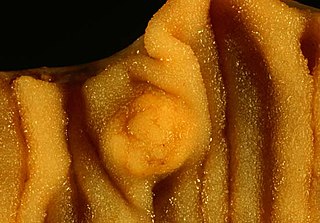
In anatomy, a polyp is an abnormal growth of tissue projecting from a mucous membrane. If it is attached to the surface by a narrow elongated stalk, it is said to be pedunculated; if it is attached without a stalk, it is said to be sessile. Polyps are commonly found in the colon, stomach, nose, ear, sinus(es), urinary bladder, and uterus. They may also occur elsewhere in the body where there are mucous membranes, including the cervix, vocal folds, and small intestine. Some polyps are tumors (neoplasms) and others are non-neoplastic, for example hyperplastic or dysplastic, which are benign. The neoplastic ones are usually benign, although some can be pre-malignant, or concurrent with a malignancy.

<span class="mw-page-title-main">Adenoma</span> Type of benign tumor

An adenoma is a benign tumor of epithelial tissue with glandular origin, glandular characteristics, or both. Adenomas can grow from many glandular organs, including the adrenal glands, pituitary gland, thyroid, prostate, and others. Some adenomas grow from epithelial tissue in nonglandular areas but express glandular tissue structure. Although adenomas are benign, they should be treated as pre-cancerous. Over time adenomas may transform to become malignant, at which point they are called adenocarcinomas. Most adenomas do not transform. However, even though benign, they have the potential to cause serious health complications by compressing other structures and by producing large amounts of hormones in an unregulated, non-feedback-dependent manner. Some adenomas are too small to be seen macroscopically but can still cause clinical symptoms.

<span class="mw-page-title-main">Gardner's syndrome</span> Medical condition

Gardner's syndrome is a subtype of familial adenomatous polyposis (FAP). Gardner syndrome is an autosomal dominant form of polyposis characterized by the presence of multiple polyps in the colon together with tumors outside the colon. The extracolonic tumors may include osteomas of the skull, thyroid cancer, epidermoid cysts, fibromas, as well as the occurrence of desmoid tumors in approximately 15% of affected individuals.

<span class="mw-page-title-main">Familial adenomatous polyposis</span> Medical condition

Familial adenomatous polyposis (FAP) is an autosomal dominant inherited condition in which numerous adenomatous polyps form mainly in the epithelium of the large intestine. While these polyps start out benign, malignant transformation into colon cancer occurs when they are left untreated. Three variants are known to exist, FAP and attenuated FAP are caused by APC gene defects on chromosome 5 while autosomal recessive FAP is caused by defects in the MUTYH gene on chromosome 1. Of the three, FAP itself is the most severe and most common; although for all three, the resulting colonic polyps and cancers are initially confined to the colon wall. Detection and removal before metastasis outside the colon can greatly reduce and in many cases eliminate the spread of cancer.

<span class="mw-page-title-main">Peutz–Jeghers syndrome</span> Medical condition

Peutz–Jeghers syndrome is an autosomal dominant genetic disorder characterized by the development of benign hamartomatous polyps in the gastrointestinal tract and hyperpigmented macules on the lips and oral mucosa (melanosis). This syndrome can be classed as one of various hereditary intestinal polyposis syndromes and one of various hamartomatous polyposis syndromes. It has an incidence of approximately 1 in 25,000 to 300,000 births.

<span class="mw-page-title-main">Benign tumor</span> Mass of cells which cannot spread throughout the body

A benign tumor is a mass of cells (tumor) that does not invade neighboring tissue or metastasize. Compared to malignant (cancerous) tumors, benign tumors generally have a slower growth rate. Benign tumors have relatively well differentiated cells. They are often surrounded by an outer surface or stay contained within the epithelium. Common examples of benign tumors include moles and uterine fibroids.

<span class="mw-page-title-main">Small intestine cancer</span> Medical condition

Small intestine cancer starts when cells in the small intestine start to grow out of control. The small intestine is the largest part of the gastrointestinal tract, which processes food for energy and rids the body of solid waste. The small intestine is not as common as other cancers of the GI tract such as colon, rectal, stomach, and esophageal cancers in the United States. They account for fewer than 1 in 10 cancers in the GI tract, and fewer than 1 in 100 cancers overall.

<span class="mw-page-title-main">Fundic gland polyposis</span> Medical condition

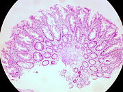
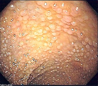
Fundic gland polyposis is a medical syndrome where the fundus and the body of the stomach develop many fundic gland polyps. The condition has been described both in patients with familial adenomatous polyposis (FAP) and attenuated variants (AFAP), and in patients in whom it occurs sporadically.

<span class="mw-page-title-main">Cronkhite–Canada syndrome</span> Medical condition

Cronkhite–Canada syndrome is a rare syndrome characterized by multiple polyps of the digestive tract. It is sporadic, and it is currently considered acquired and idiopathic.

<span class="mw-page-title-main">Juvenile polyposis syndrome</span> Medical condition

Juvenile polyposis syndrome is an autosomal dominant genetic condition characterized by the appearance of multiple juvenile polyps in the gastrointestinal tract. Polyps are abnormal growths arising from a mucous membrane. These usually begin appearing before age 20, but the term juvenile refers to the type of polyp, not to the age of the affected person. While the majority of the polyps found in juvenile polyposis syndrome are non-neoplastic, hamartomatous, self-limiting and benign, there is an increased risk of adenocarcinoma.

Attenuated familial adenomatous polyposis is a form of familial adenomatous polyposis, a cancer syndrome. It is a pre-malignant disease that can develop into colorectal cancer. A patient will have fewer than a hundred polyps located typically in right side of the colon. Cancer might develop as early as the age of five, though typically presents later than classical FAP.

<span class="mw-page-title-main">Sessile serrated lesion</span> Medical condition

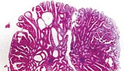
A sessile serrated lesion (SSL) is a premalignant flat lesion of the colon, predominantly seen in the cecum and ascending colon.

<span class="mw-page-title-main">Hyperplastic polyp</span> Type of colorectal polyp

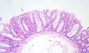
A hyperplastic polyp is a type of colorectal polyp.

<span class="mw-page-title-main">Colorectal adenoma</span> Medical condition

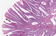
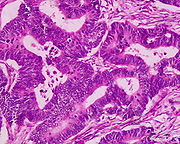
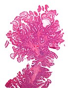
The colorectal adenoma is a benign glandular tumor of the colon and the rectum. It is a precursor lesion of the colorectal adenocarcinoma. They often manifest as colorectal polyps.

McKittrick-Wheelock syndrome is an uncommon syndrome caused by large, villous adenomas that secrete high quantities of electrolyte-rich mucin. This may lead to pre-renal acute kidney injury, secretory diarrhea, and dehydration. It is estimated that 2-3% of large villous adenomas, typically greater than 4 cm in diameter, will present with this hypersecretory pattern.

<span class="mw-page-title-main">Histopathology of colorectal adenocarcinoma</span> Analysis of tissue from a biopsy or surgery to identify colorectal cancer characteristics

The histopathology of colorectal cancer of the adenocarcinoma type involves analysis of tissue taken from a biopsy or surgery. A pathology report contains a description of the microscopical characteristics of the tumor tissue, including both tumor cells and how the tumor invades into healthy tissues and finally if the tumor appears to be completely removed. The most common form of colon cancer is adenocarcinoma, constituting between 95% and 98% of all cases of colorectal cancer. Other, rarer types include lymphoma, adenosquamous and squamous cell carcinoma. Some subtypes have been found to be more aggressive.

<span class="mw-page-title-main">Serrated polyposis syndrome</span> Medical condition

Serrated polyposis syndrome (SPS), previously known as hyperplastic polyposis syndrome, is a disorder characterized by the appearance of serrated polyps in the colon. While serrated polyposis syndrome does not cause symptoms, the condition is associated with a higher risk of colorectal cancer (CRC). The lifelong risk of CRC is between 25 and 40%. SPS is the most common polyposis syndrome affecting the colon, but is under recognized due to a lack of systemic long term monitoring. Diagnosis requires colonoscopy, and is defined by the presence of either of two criteria: ≥5 serrated lesions/polyps proximal to the rectum, or >20 serrated lesions/polyps of any size distributed throughout the colon with 5 proximal to the rectum.

Polymerase proofreading-associated polyposis (PPAP) is an autosomal dominant hereditary cancer syndrome, which is characterized by numerous polyps in the colon and an increased risk of colorectal cancer. It is caused by germline mutations in DNA polymerase ε (POLE) and δ (POLD1). Affected individuals develop numerous polyps called colorectal adenomas. Compared with other polyposis syndromes, Polymerase proofreading-associated polyposis is rare. Genetic testing can help exclude similar syndromes, such as Familial adenomatous polyposis and MUTYH-associated polyposis. Endometrial cancer, duodenal polyps and duodenal cancer may also occur.

<span class="mw-page-title-main">Traditional serrated adenoma</span> Medical condition

Traditional serrated adenoma is a premalignant type of polyp found in the colon, often in the distal colon. Traditional serrated adenomas are a type of serrated polyp, and may occur sporadically or as a part of serrated polyposis syndrome. Traditional serrated adenomas are relatively rare, accounting for less than 1% of all colon polyps. Usually, traditional serrated adenomas are found in the distal colon and are usually less than 10 mm in size.

Juvenile polyps are a type of polyp found in the colon. While juvenile polyps are typically found in children, they may be found in people of any age. Juvenile polyps are a type of hamartomatous polyps, which consist of a disorganized mass of tissue. They occur in about two percent of children. Juvenile polyps often do not cause symptoms (asymptomatic); when present, symptoms usually include gastrointestinal bleeding and prolapse through the rectum. Removal of the polyp (polypectomy) is warranted when symptoms are present, for treatment and definite histopathological diagnosis. In the absence of symptoms, removal is not necessary. Recurrence of polyps following removal is relatively common. Juvenile polyps are usually sporadic, occurring in isolation, although they may occur as a part of juvenile polyposis syndrome. Sporadic juvenile polyps may occur in any part of the colon, but are usually found in the distal colon. In contrast to other types of colon polyps, juvenile polyps are not premalignant and are not usually associated with a higher risk of cancer; however, individuals with juvenile polyposis syndrome are at increased risk of gastric and colorectal cancer., Unlike juvenile polyposis syndrome, solitary juvenile polyps do not require follow up with surveillance colonoscopy.

References

  1. Marks, Jay W.; Anand, Bhupinder. "Colon Polyps: Symptoms, Causes, Cancer Risk, Treatment, and Prevention". Colon polyps center. MedicineNet. Retrieved 18 Jan 2020.
  2. 1 2 3 Phillips, Michael M.; Zieve, David; Conaway, Brenda (September 25, 2019). "Colorectal polyps". Medical Encyclopedia. MedlinePlus . Retrieved 18 Jan 2020.
  3. 1 2 3 4 5 "Colon polyps". Mayo Clinic. Mayo Foundation for Medical Education and Research. Retrieved 18 Jan 2020.
  4. 1 2 3 Quick CR, Reed JB, Harper SJ, Saeb-Parsy K, Burkitt HG (2014). Essential Surgery: Problems, Diagnosis and Management. Edinburgh: Elsevier. ISBN   9780702054839. OCLC   842350865.[ page needed ]
  5. Classen, Meinhard; Tytgat, G.N.J.; Lightdale, Charles J. (2002). Gastroenterological Endoscopy. Thieme. p. 303. ISBN   1-58890-013-4.
  6. 1 2 Townsend CM, Beauchamp RD, Evers BM, Mattox KL, eds. (2015). Sabiston Textbook of Surgery E-Book (19th ed.). Philadelphia, Pennsylvania: Elsevier Saunders. ISBN   978-1-4377-1560-6 via Google Books (Preview).
  7. "Familial Adenomatous Polyposis". The Lecturio Medical Concept Library. Retrieved 22 July 2021.
  8. Valle L, de Voer RM, Goldberg Y, Sjursen W, Försti A, Ruiz-Ponte C, Caldés T, Garré P, Olsen MF, Nordling M, Castellvi-Bel S, Hemminmki K (October 2019). "Update on genetic predisposition to colorectal cancer and polyposis". Molecular Aspects of Medicine. Elsevier. 69: 10–26. doi: 10.1016/j.mam.2019.03.001 . hdl: 2445/171217 . PMID   30862463.
  9. 1 2 3 4 5 6 7 8 9 10 11 12 13 14 15 16 17 18 19 20 21 22 23 24 25 26 27 28 29 30 31 32 33 34 35 36 37 Mahmoud, Najjia N.; Bleier, Joshua I.S.; Aarons, Cary B.; Paulson, E. Carter; Shanmugan, Skandan; Fry, Robert D. (2017). Sabiston Textbook of Surgery (20th ed.). Elsevier. ISBN   9780323401630.[ page needed ]
  10. 1 2 3 Finlay A Macrae. "Overview of colon polyps". UpToDate. This topic last updated: Dec 10, 2018.
  11. 1 2 3 Robert V Rouse (2010-01-31). "Hyperplastic Polyp of the Colon and Rectum". Stanford University School of Medicine. Archived from the original on 2019-12-11. Retrieved 2019-10-31. Last updated 6/2/2015
  12. Minhhuyen Nguyen. "Polyps of the Colon and Rectum". MSD Manual. Last full review/revision June 2019
  13. Robert V Rouse. "Adenoma of the Colon and Rectum". Archived from the original on 2019-09-11. Retrieved 2019-10-31. Original posting/last update : 1/31/10, 1/19/14
  14. 1 2 3 Bosman, F. T. (2010). WHO classification of tumours of the digestive system. Lyon: International Agency for Research on Cancer. ISBN   978-92-832-2432-7. OCLC   688585784.
  15. 1 2 Amersi, Farin; Agustin, Michelle; Ko, Clifford Y (2005). "Colorectal Cancer: Epidemiology, Risk Factors, and Health Services". Clinics in Colon and Rectal Surgery. 18 (3): 133–140. doi:10.1055/s-2005-916274. ISSN   1531-0043. PMC   2780097 . PMID   20011296.
  16. Alnoor Ramji (22 December 2022). "Villous Adenoma Follow-up". Medscape. Updated: Oct 24, 2016
  17. Rosty, C; Hewett, D. G.; Brown, I. S.; Leggett, B. A.; Whitehall, V. L. (2013). "Serrated polyps of the large intestine: Current understanding of diagnosis, pathogenesis, and clinical management". Journal of Gastroenterology. 48 (3): 287–302. doi:10.1007/s00535-012-0720-y. PMC   3698429 . PMID   23208018.
  18. 1 2 3 4 5 6 Kumar, Vinay (2010). "17—Polyps". Robbins and Cotran pathologic basis of disease (8th ed.). Philadelphia, PA: Saunders/Elsevier. ISBN   978-1-4160-3121-5.
  19. 1 2 Stoler, Mark A.; Mills, Stacey E.; Carter, Darryl; Joel K. Greenson; Reuter, Victor E. (2009). Sternberg's Diagnostic Surgical Pathology. Hagerstwon, MD: Lippincott Williams & Wilkins. ISBN   978-0-7817-7942-5.[ page needed ]
  20. Calva, Daniel; Howe, James R (2008). "Hamartomatous Polyposis Syndromes". Surgical Clinics of North America. 88 (4): 779–817, vii. doi:10.1016/j.suc.2008.05.002. PMC   2659506 . PMID   18672141.
  21. Tantamango, Yessenia M; Knutsen, Synnove F; Beeson, W Lawrence; Fraser, Gary; Sabate, Joan (2011). "Foods and Food Groups Associated with the Incidence of Colorectal Polyps: The Adventist Health Study". Nutrition and Cancer. 63 (4): 565–72. doi:10.1080/01635581.2011.551988. PMC   3427008 . PMID   21547850.
  22. 1 2 3 Summers, Ronald M (2010). "Polyp Size Measurement at CT Colonography: What Do We Know and What Do We Need to Know?". Radiology. 255 (3): 707–20. doi:10.1148/radiol.10090877. PMC   2875919 . PMID   20501711.
  23. Luis Bujanda Fernández de Piérola, Joaquin Cubiella Fernández, Fernando Múgica Aguinaga, Lander Hijona Muruamendiaraz and Carol Julyssa Cobián Malaver (2013). "Malignant Colorectal Polyps: Diagnosis, Treatment and Prognosis". Colonoscopy and Colorectal Cancer Screening: Future Directions. doi:10.5772/52697. ISBN   9789535109495.{{cite book}}: CS1 maint: multiple names: authors list (link) Creative Commons Attribution 3.0 License
  24. Hattori, Santa (2014). "Narrow-band imaging observation of colorectal lesions using NICE classification to avoid discarding significant lesions". World Journal of Gastrointestinal Endoscopy. 6 (12): 600–605. doi: 10.4253/wjge.v6.i12.600 . PMC   4265957 . PMID   25512769.
  25. Hoff, RT; Lakha, A (1 February 2020). "Rectal Tubulovillous Adenoma". The Journal of the American Osteopathic Association. 120 (2): 121. doi: 10.7556/jaoa.2020.024 . PMID   31985762.
  26. Fisichella, M. (2016). "Laparoscopic Cecal Wedge Resection Appendectomy". J Med Ins: 207. doi:10.24296/jomi/207.
  27. Saunders B, Ginsberg GG, Bjorkman DJ (2008). "How I do it: Removing large or sessile colonic polyps" (PDF). Munich: OMED. Archived from the original (PDF) on 2008-04-11.
  28. Gupta, Samir; Lieberman, David; Anderson, Joseph C.; Burke, Carol A.; Dominitz, Jason A.; Kaltenbach, Tonya; Robertson, Douglas J.; Shaukat, Aasma; Syngal, Sapna; Rex, Douglas K. (2020). "Recommendations for Follow-Up After Colonoscopy and Polypectomy: A Consensus Update by the US Multi-Society Task Force on Colorectal Cancer". Gastrointestinal Endoscopy. 91 (3): 463–485.e5. doi:10.1016/j.gie.2020.01.014. ISSN   0016-5107. PMC   7389642 . PMID   32044106.