Benign tumor

Last updated
Benign tumor
Other namesnon-cancerous tumor
Normal Epidermis and Dermis with Intradermal Nevus 10x.JPG
Normal epidermis and dermis with intradermal nevus, 10x-cropped
Specialty Oncology, Pathology

A benign tumor is a mass of cells (tumor) that does not invade neighboring tissue or metastasize (spread throughout the body). Compared to malignant (cancerous) tumors, benign tumors generally have a slower growth rate. Benign tumors have relatively well differentiated cells. They are often surrounded by an outer surface (fibrous sheath of connective tissue) or stay contained within the epithelium. Common examples of benign tumors include moles and uterine fibroids.

Contents

Some forms of benign tumors may be harmful to health. Benign tumor growth causes a mass effect that can compress neighboring tissues. This can lead to nerve damage, blood flow reduction (ischemia), tissue death (necrosis), or organ damage. The health effects of benign tumor growth may be more prominent if the tumor is contained within an enclosed space such as the cranium, respiratory tract, sinus, or bones. For example, unlike most benign tumors elsewhere in the body, benign brain tumors can be life-threatening. Tumors may exhibit behaviors characteristic of their cell type of origin; as an example, endocrine tumors such as thyroid adenomas and adrenocortical adenomas may overproduce certain hormones.

The word "benign" means "favourable, kind, fortunate, salutary, propitious". [1] However, a benign tumour is not benign in the usual sense; the name merely specifies that it is not "malignant", i.e. cancerous. While benign tumours usually do not pose a serious health risk, they can be harmful or fatal. [2] Many types of benign tumors have the potential to become cancerous (malignant) through a process known as tumor progression. For this reason and other possible harms, some benign tumors are removed by surgery. When removed, benign tumors usually do not return. Exceptions to this rule may indicate malignant transformation.

Signs and symptoms

Benign tumors are very diverse; they may be asymptomatic or may cause specific symptoms, depending on their anatomic location and tissue type. They grow outward, producing large, rounded masses which can cause what is known as a "mass effect". This growth can cause compression of local tissues or organs, leading to many effects, such as blockage of ducts, reduced blood flow (ischaemia), tissue death (necrosis) and nerve pain or damage. [3] Some tumors also produce hormones that can lead to life-threatening situations. Insulinomas can produce large amounts of insulin, causing hypoglycemia. [4] [5] Pituitary adenomas can cause elevated levels of hormones such as growth hormone and insulin-like growth factor-1, which cause acromegaly; prolactin; ACTH and cortisol, which cause Cushing's disease; TSH, which causes hyperthyroidism; and FSH and LH. [6] Bowel intussusception can occur with various benign colonic tumors. [7] Cosmetic effects can be caused by tumors, especially those of the skin, possibly causing psychological or social discomfort for the person with the tumor. [8] Vascular tissue tumors can bleed, in some cases leading to anemia. [9]

Causes

PTEN hamartoma syndrome

PTEN hamartoma syndrome encompasses hamartomatous disorders characterized by genetic mutations in the PTEN tumor suppressor gene, [10] including Cowden syndrome, Bannayan–Riley–Ruvalcaba syndrome, Proteus syndrome and Proteus-like syndrome. Absent or dysfunctional PTEN protein allows cells to over-proliferate, causing hamartomas. [11] Cowden syndrome is an autosomal dominant genetic disorder characterized by multiple benign hamartomas (trichilemmomas and mucocutaneous papillomatous papules) as well as a predisposition for cancers of multiple organs including the breast and thyroid. [12] [13] Bannayan–Riley–Ruvalcaba syndrome is a congenital disorder characterized by hamartomatous intestinal polyposis, macrocephaly, lipomatosis, hemangiomatosis and glans penis macules. [11] [14] Proteus syndrome is characterized by nevi, asymmetric overgrowth of various body parts, adipose tissue dysregulation, cystadenomas, adenomas, vascular malformation. [15] [16]

Endoscopic image of sigmoid colon of a patient with familial adenomatous polyposis. Familial adenomatous polyposis as seen on sigmoidoscopy.jpg
Endoscopic image of sigmoid colon of a patient with familial adenomatous polyposis.

Familial adenomatous polyposis

Familial adenomatous polyposis (FAP) is a familial cancer syndrome caused by mutations in the APC gene. In FAP, adenomatous polyps are present in the colon. The polyps progress into colon cancer unless removed. [17] The APC gene is a tumor suppressor. Its protein product is involved in many cellular processes. Inactivation of the APC gene leads to the buildup of a protein called β-catenin. This protein activates two transcription factors: T-cell factor (TCF) and lymphoid enhancer factor (LEF). These factors cause the upregulation of many genes involved in cell proliferation, differentiation, migration and apoptosis (programmed cell death), causing the growth of benign tumors. [18]

Tuberous sclerosis complex

Tuberous sclerosis complex (TSC) is an autosomal dominant genetic disorder caused by mutations in the genes TSC1 and TSC2. TSC1 produces the protein hamartin. TSC2 produces the protein tuberin. This disorder presents with many benign hamartomatous tumors including angiofibromas, renal angiomyolipomas, and pulmonary lymphangiomyomatosis. Tuberin and hamartin inhibit the mTOR protein in normal cellular physiology. Inactivation of the TSC tumor suppressors causes an increase in mTOR activity. This leads to the activation of genes and the production of proteins that increase cell growth. [19] [20] [21]

Von Hippel–Lindau disease

Von Hippel–Lindau disease is a dominantly inherited cancer syndrome that significantly increases the risk of various tumors. This includes benign hemangioblastomas and malignant pheochromocytomas, renal cell carcinomas, pancreatic endocrine tumors, and endolymphatic sac tumors. It is caused by genetic mutations in the Von Hippel–Lindau tumor suppressor gene. The VHL protein (pVHL) is involved in cellular signaling in oxygen starved (hypoxic) cells. One role of pVHL is to cause the cellular degradation of another protein, HIF1α. Dysfunctional pVHL leads to accumulation of HIF1α. This activates several genes responsible for the production of substances involved in cell growth and blood vessel production: VEGF, PDGFβ, TGFα and erythropoietin. [22]

Bone tumors

Benign tumors of bone can be similar macroscopically and require a combination of a clinical history with cytogenetic, molecular, and radiologic tests for diagnosis. [23] Three common forms of benign bone tumors with are giant cell tumor of bone, osteochondroma, and enchondroma; other forms of benign bone tumors exist but may be less prevalent.

Giant cell tumors

Giant cell tumors of bone frequently occur in long bone epiphyses of the appendicular skeleton or the sacrum of the axial skeleton. Local growth can cause destruction of neighboring cortical bone and soft tissue, leading to pain and limiting range of motion. The characteristic radiologic finding of giant cell tumors of bone is a lytic lesion that does not have marginal sclerosis of bone. On histology, giant cells of fused osteoclasts are seen as a response to neoplastic mononucleated cells. Notably, giant cells are not unique among benign bone tumors to giant cell tumors of bone. Molecular characteristics of the neoplastic cells causing giant cell tumors of bone indicate an origin of pluripotent mesenchymal stem cells that adopt preosteoblastic markers. Cytogenetic causes of giant cell tumors of bone involve telomeres. Treatment involves surgical curettage with adjuvant bisphosphonates.

Osteochondroma

Osteochondromas form cartilage-capped projections of bone. Structures such as the marrow cavity and cortical bone of the osteochondroma are contiguous to those of the originating bone. Sites of origin often involve metaphyses of long bones. While many osteochondromas occur spontaneously, there are cases in which several osteochondromas can occur in the same individual; these may be linked to a genetic condition known as hereditary multiple osteochondromas. Osteochondroma appears on X-ray as a projecting mass that often points away from joints. [23] These tumors stop growing with the closure of the parental bone's growth plates. Failure to stop growth can be indicative of transformation to malignant chondrosarcoma. Treatment is not indicated unless symptomatic. In that case, surgical excision is often curative.

Enchondroma

Enchondromas are benign tumors of hyaline cartilage. Within a bone, enchondromas are often found in metaphyses. They can be found in many types of bone, including small bones, long bones, and the axial skeleton. X-ray of enchondromas shows well-defined borders and a stippled appearance. [23] Presentation of multiple enchondromas is consistent with multiple enchondromatosis (Ollier Disease). Treatment of enchondromas involves surgical curettage and grafting.

Benign soft tissue tumors

Lipomas

Lipomas are benign, subcutaneous tumors of fat cells (adipocytes). They are usually painless, slow-growing, and mobile masses that can occur anywhere in the body where there are fat cells, but are typically found on the trunk and upper extremities. [24]

[25] Although lipomas can develop at any age, they more commonly appear between the ages of 40 and 60. [24] Lipomas affect about 1% of the population, with no documented sex bias, and about 1 in every 1000 people will have a lipoma within their lifetime. [25] [26] The cause of lipomas is not well defined. Genetic or inherited causes of lipomas play a role in around 2-3% of patients. [25] In individuals with inherited familial syndromes such as Proteus syndrome or Familial multiple lipomatosis, it is common to see multiple lipomas across the body. [25] These syndromes are also associated with specific symptoms and sub-populations. Mutations in chromosome 12 have been identified in around 65% of lipoma cases. [25] Lipomas have also been shown to be increased in those with obesity, hyperlipidemia, and diabetes mellitus. [25]

Lipomas are usually diagnosed clinically, although imaging (ultrasound, computed tomography, or magnetic resonance imaging) may be utilized to assist with the diagnosis of lipomas in atypical locations. [24] The main treatment for lipomas is surgical excision, after which the tumor is examined with histopathology to confirm the diagnosis. [24] The prognosis for benign lipomas is excellent and recurrence after excision is rare, but may occur if the removal was incomplete. [25]

Mechanism

Benign (L) vs Malignant tumor (R). Types of tumor cells.jpg
Benign (L) vs Malignant tumor (R).

Benign vs malignant

Diagram showing two epithelial tumors. The upper tumor is a benign tumor that is non-invasive. Benign tumors are usually round in shape and encapsulated by fibrous connective tissue. The lower picture depicts a malignant tumor. It is irregularly shaped, vascular, and it is invasive, crossing the basement membrane. BENIGN AND MALIGNANT TUMOUR.png
Diagram showing two epithelial tumors. The upper tumor is a benign tumor that is non-invasive. Benign tumors are usually round in shape and encapsulated by fibrous connective tissue. The lower picture depicts a malignant tumor. It is irregularly shaped, vascular, and it is invasive, crossing the basement membrane.

One of the most important factors in classifying a tumor as benign or malignant is its invasive potential. If a tumor lacks the ability to invade adjacent tissues or spread to distant sites by metastasizing then it is benign, whereas invasive or metastatic tumors are malignant. [3] For this reason, benign tumors are not classed as cancer. [27] Benign tumors will grow in a contained area usually encapsulated in a fibrous connective tissue capsule. The growth rates of benign and malignant tumors also differ; benign tumors generally grow more slowly than malignant tumors. Although benign tumors pose a lower health risk than malignant tumors, they both can be life-threatening in certain situations. There are many general characteristics which apply to either benign or malignant tumors, but sometimes one type may show characteristics of the other. For example, benign tumors are mostly well differentiated and malignant tumors are often undifferentiated. However, undifferentiated benign tumors and differentiated malignant tumors can occur. [28] [29] Although benign tumors generally grow slowly, cases of fast-growing benign tumors have also been documented. [30] Some malignant tumors are mostly non-metastatic such as in the case of basal-cell carcinoma. [31] CT and chest radiography can be a useful diagnostic exam in visualizing a benign tumor and differentiating it from a malignant tumor. The smaller the tumor on a radiograph the more likely it is to be benign as 80% of lung nodules less than 2 cm in diameter are benign. Most benign nodules are smoothed radiopaque densities with clear margins but these are not exclusive signs of benign tumors. [32]

Multistage carcinogenesis

Tumors are formed by carcinogenesis, a process in which cellular alterations lead to the formation of cancer. Multistage carcinogenesis involves the sequential genetic or epigenetic changes to a cell's DNA, where each step produces a more advanced tumor. It is often broken down into three stages; initiation, promotion and progression, and several mutations may occur at each stage. Initiation is where the first genetic mutation occurs in a cell. Promotion is the clonal expansion (repeated division) of this transformed cell into a visible tumor that is usually benign. Following promotion, progression may take place where more genetic mutations are acquired in a sub-population of tumor cells. Progression changes the benign tumor into a malignant tumor. [33] [34] A prominent and well studied example of this phenomenon is the tubular adenoma, a common type of colon polyp which is an important precursor to colon cancer. The cells in tubular adenomas, like most tumors that frequently progress to cancer, show certain abnormalities of cell maturation and appearance collectively known as dysplasia. These cellular abnormalities are not seen in benign tumors that rarely or never turn cancerous, but are seen in other pre-cancerous tissue abnormalities which do not form discrete masses, such as pre-cancerous lesions of the uterine cervix.

Diagnosis

Classification

Tumors and cell origin
Cell originCell typeTumor
Endodermal Biliary tree Cholangioma
Colon Colonic polyp
Glandular Adenoma
Papilloma
Cystadenoma
Liver Liver cell adenoma
Placental Hydatiform mole
Renal Renal tubular adenoma
Squamous Squamous cell papilloma
Stomach Gastric polyp
Mesenchymal Blood vessel Hemangioma, Cardiac myxoma
Bone Osteoma
Cartilage Chondroma
Fat tissue Lipoma
Fibrous tissue Fibroma
Lymphatic vessel Lymphangioma
Smooth muscle Leiomyoma
Striated muscle Rhabdomyoma
Ectodermal Glia Astrocytoma, Schwannoma
Melanocytes Nevus
Meninges Meningioma
Nerve cells Ganglioneuroma
Reference [35]

Benign neoplasms are typically, but not always, composed of cells which bear a strong resemblance to a normal cell type in their organ of origin. These tumors are named for the cell or tissue type from which they originate. The suffix "-oma" (but not -carcinoma, -sarcoma, or -blastoma, which are generally cancers) is applied to indicate a benign tumor. For example, a lipoma is a common benign tumor of fat cells (lipocytes), and a chondroma is a benign tumor of cartilage-forming cells (chondrocytes). Adenomas are benign tumors of gland-forming cells, and are usually specified further by their cell or organ of origin, as in hepatic adenoma (a benign tumor of hepatocytes, or liver cells). Teratomas contain many cell types such as skin, nerve, brain and thyroid, among others, because they are derived from germ cells. [36] Hamartomas are a group of benign tumors that have relatively normal cellular differentiation but exhibit disorganized tissue organization. [19]

Exceptions to the nomenclature rules exist for historical reasons; malignant examples include melanoma (a cancer of pigmented skin cells, or melanocytes) and seminoma (a cancer of male reproductive cells). [37]

Benign tumors do not encompass all benign growths. Skin tags, vocal chord polyps, and hyperplastic polyps of the colon are often referred to as benign, but they are overgrowths of normal tissue rather than neoplasms. [36]

Treatment

Benign tumors typically need no treatment unless if they cause problems such as seizures, discomfort or cosmetic concerns. Surgery is usually the most effective approach and is used to treat most benign tumors. In some cases, other treatments may be used. Adenomas of the rectum may be treated with sclerotherapy, in which chemicals are used to shrink blood vessels in order to cut off the blood supply. [38] Most benign tumors do not respond to chemotherapy or radiation therapy, although there are exceptions; benign intercranial tumors are sometimes treated with radiation therapy and chemotherapy under certain circumstances. [39] [40] Radiation can also be used to treat hemangiomas in the rectum. [38] Benign skin tumors are usually surgically resected but other treatments such as cryotherapy, curettage, electrodesiccation, laser therapy, dermabrasion, chemical peels and topical medication are used. [41] [42]


Related Research Articles

<span class="mw-page-title-main">Lipoma</span> Benign tumor made of fat tissue

A lipoma is a benign tumor made of fat tissue. They are generally soft to the touch, movable, and painless. They usually occur just under the skin, but occasionally may be deeper. Most are less than 5 cm (2.0 in) in size. Common locations include upper back, shoulders, and abdomen. It is possible to have a number of lipomas.

<span class="mw-page-title-main">Proteus syndrome</span> Human genetic disorder

Proteus syndrome is a rare disorder with a genetic background that can cause tissue overgrowth involving all three embryonic lineages. Patients with Proteus syndrome tend to have an increased risk of embryonic tumor development. The clinical and radiographic symptoms of Proteus syndrome are highly variable, as are its orthopedic manifestations.

<span class="mw-page-title-main">Malignancy</span> Tendency of a medical condition to become progressively worse

Malignancy is the tendency of a medical condition to become progressively worse; the term is most familiar as a characterization of cancer.

<span class="mw-page-title-main">Pituitary adenoma</span> Human disease

Pituitary adenomas are tumors that occur in the pituitary gland. Most pituitary tumors are benign, approximately 35% are invasive and just 0.1% to 0.2% are carcinomas. Pituitary adenomas represent from 10% to 25% of all intracranial neoplasms and the estimated prevalence rate in the general population is approximately 17%.

<span class="mw-page-title-main">Neoplasm</span> Abnormal mass of tissue as a result of abnormal growth or division of cells

A neoplasm is a type of abnormal and excessive growth of tissue. The process that occurs to form or produce a neoplasm is called neoplasia. The growth of a neoplasm is uncoordinated with that of the normal surrounding tissue, and persists in growing abnormally, even if the original trigger is removed. This abnormal growth usually forms a mass, when it may be called a tumour or tumor.

<span class="mw-page-title-main">Enchondroma</span> Medical condition

Enchondroma is a type of benign bone tumor belonging to the group of cartilage tumors. There may be no symptoms, or it may present typically in the short tubular bones of the hands with a swelling, pain or pathological fracture.

<span class="mw-page-title-main">Hamartoma</span> Tumour-like overgrowth due to a systemic genetic condition

A hamartoma is a mostly benign, local malformation of cells that resembles a neoplasm of local tissue but is usually due to an overgrowth of multiple aberrant cells, with a basis in a systemic genetic condition, rather than a growth descended from a single mutated cell (monoclonality), as would typically define a benign neoplasm/tumor. Despite this, many hamartomas are found to have clonal chromosomal aberrations that are acquired through somatic mutations, and on this basis the term hamartoma is sometimes considered synonymous with neoplasm. Hamartomas are by definition benign, slow-growing or self-limiting, though the underlying condition may still predispose the individual towards malignancies.

<span class="mw-page-title-main">Osteochondroma</span> Medical condition

Osteochondromas are the most common benign tumors of the bones. The tumors take the form of cartilage-capped bony projections or outgrowth on the surface of bones exostoses. It is characterized as a type of overgrowth that can occur in any bone where cartilage forms bone. Tumors most commonly affect long bones about the knee and in the forearm. Additionally, flat bones such as the pelvis and scapula may be affected. Hereditary multiple exostoses usually present during childhood. Yet, the vast majority of affected individuals become clinically manifest by the time they reach adolescence. Osteochondromas occur in 3% of the general population and represent 35% of all benign tumors and 8% of all bone tumors. The majority of these tumors are solitary non-hereditary lesions and approximately 15% of osteochondromas occur as hereditary multiple exostoses preferably known as hereditary multiple osteochondromas (HMOs). Osteochondromas do not result from injury and the exact cause remains unknown. Recent research has indicated that multiple osteochondromas is an autosomal dominant inherited disease. Germ line mutations in EXT1 and EXT2 genes located on chromosomes 8 and 11 have been associated with the cause of the disease. The treatment choice for osteochondroma is surgical removal of solitary lesion or partial excision of the outgrowth, when symptoms cause motion limitations or nerve and blood vessel impingements. In hereditary multiple exostoses the indications of surgery are based upon multiple factors that are taken collectively, namely: patient's age, tumor location and number, accompanying symptomatology, esthetic concerns, family history and underlying gene mutation. A variety of surgical procedures have been employed to remedy hereditary multiple exostoses such as osteochondroma excision, bone lengthening, corrective osteotomy and hemiepiphysiodesis. Sometimes a combination of the previous procedures is used. The indicators of surgical success in regard to disease and patient characteristics are greatly disputable. Because most studies of hereditary multiple exostoses are retrospective and of limited sample size with missing data, the best evidence for each of the currently practiced surgical procedures is lacking.

<span class="mw-page-title-main">Multiple endocrine neoplasia type 1</span> Medical condition

Multiple endocrine neoplasia type 1 (MEN-1) is one of a group of disorders, the multiple endocrine neoplasias, that affect the endocrine system through development of neoplastic lesions in pituitary, parathyroid gland and pancreas. Individuals suffering from this disorder are prone to developing multiple endocrine and nonendocrine tumors. It was first described by Paul Wermer in 1954.

<span class="mw-page-title-main">Cowden syndrome</span> Medical condition

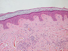
Cowden syndrome is an autosomal dominant inherited condition characterized by benign overgrowths called hamartomas as well as an increased lifetime risk of breast, thyroid, uterine, and other cancers. It is often underdiagnosed due to variability in disease presentation, but 99% of patients report mucocutaneous symptoms by age 20–29. Despite some considering it a primarily dermatologic condition, Cowden's syndrome is a multi-system disorder that also includes neurodevelopmental disorders such as macrocephaly.

<span class="mw-page-title-main">Metachondromatosis</span> Medical condition

Metachondromatosis is an autosomal dominant, incompletely penetrant genetic disease affecting the growth of bones, leading to exostoses primarily in the hands and feet as well as enchondromas of long bone metaphyses and iliac crests. This syndrome affects mainly tubular bones, though it can also involve the vertebrae, small joints, and flat bones. The disease is thought to affect exon 4 of the PTPN11 gene. Metachondromatosis is believed to be caused by an 11 base pair deletion resulting in a frameshift and nonsense mutation. The disease was discovered and named in 1971 by Pierre Maroteaux, a French physician, when he observed two families with skeletal radiologic features with exostoses and Ollier disease. The observation of one family with five affected people led to the identification of the disease as autosomal dominant. There have been less than 40 cases of the disease reported to date.

<i>PTEN</i> (gene) Tumor suppressor gene

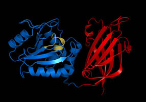
Phosphatase and tensin homolog (PTEN) is a phosphatase in humans and is encoded by the PTEN gene. Mutations of this gene are a step in the development of many cancers, specifically glioblastoma, lung cancer, breast cancer, and prostate cancer. Genes corresponding to PTEN (orthologs) have been identified in most mammals for which complete genome data are available.

<span class="mw-page-title-main">Bannayan–Riley–Ruvalcaba syndrome</span> Medical condition

Bannayan–Riley–Ruvalcaba syndrome (BRRS) is a rare overgrowth syndrome and hamartomatous disorder with occurrence of multiple subcutaneous lipomas, macrocephaly and hemangiomas. The disease is inherited in an autosomal dominant manner. The disease belongs to a family of hamartomatous polyposis syndromes, which also includes Peutz–Jeghers syndrome, juvenile polyposis and Cowden syndrome. Mutation of the PTEN gene underlies this syndrome, as well as Cowden syndrome, Proteus syndrome, and Proteus-like syndrome, these four syndromes are referred to as PTEN Hamartoma-Tumor Syndromes.

<span class="mw-page-title-main">Ollier disease</span> Medical condition

Ollier disease is a rare sporadic nonhereditary skeletal disorder in which typically benign cartilaginous tumors (enchondromas) develop near the growth plate cartilage. This is caused by cartilage rests that grow and reside within the metaphysis or diaphysis and eventually mineralize over time to form multiple enchondromas. Key signs of the disorder include asymmetry and shortening of the limb as well as an increased thickness of the bone margin. These symptoms are typically first visible during early childhood with the mean age of diagnosis being 13 years of age. Many patients with Ollier disease are prone to develop other malignancies including bone sarcomas that necessitate treatment and the removal of malignant bone neoplasm. Cases in patients with Ollier disease has shown a link to IDH1, IDH2, and PTH1R gene mutations. Currently, there are no forms of treatment for the underlying condition of Ollier disease but complications such as fractures, deformities, malignancies that arise from it can be treated through surgical procedures. The prevalence of this condition is estimated at around 1 in 100,000. It is unclear whether the men or women are more affected by this disorder due to conflicting case studies.

<span class="mw-page-title-main">Maffucci syndrome</span> Medical condition

Maffucci syndrome is a very rare disorder in which multiple benign tumors of cartilage develop within the bones. The tumors most commonly appear in the bones of the hands, feet, and limbs, causing bone deformities and short limbs.

<span class="mw-page-title-main">Endometrial intraepithelial neoplasia</span>

Endometrial intraepithelial neoplasia (EIN) is a premalignant lesion of the uterine lining that predisposes to endometrioid endometrial adenocarcinoma. It is composed of a collection of abnormal endometrial cells, arising from the glands that line the uterus, which have a tendency over time to progress to the most common form of uterine cancer—endometrial adenocarcinoma, endometrioid type.

<span class="mw-page-title-main">Juvenile polyposis syndrome</span> Medical condition

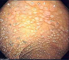
Juvenile polyposis syndrome is an autosomal dominant genetic condition characterized by the appearance of multiple juvenile polyps in the gastrointestinal tract. Polyps are abnormal growths arising from a mucous membrane. These usually begin appearing before age 20, but the term juvenile refers to the type of polyp, not to the age of the affected person. While the majority of the polyps found in juvenile polyposis syndrome are non-neoplastic, hamartomatous, self-limiting and benign, there is an increased risk of adenocarcinoma.

<span class="mw-page-title-main">Colorectal polyp</span> Growth found in bowel wall

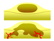
A colorectal polyp is a polyp occurring on the lining of the colon or rectum. Untreated colorectal polyps can develop into colorectal cancer.

Vulvar tumors are those neoplasms of the vulva. Vulvar and vaginal neoplasms make up a small percentage (3%) of female genital cancers. They can be benign or malignant. Vulvar neoplasms are divided into cystic or solid lesions and other mixed types. Vulvar cancers are those malignant neoplasms that originate from vulvar epithelium, while vulvar sarcomas develop from non-epithelial cells such as bone, cartilage, fat, muscle, blood vessels, or other connective or supportive tissue. Epithelial and mesenchymal tissue are the origin of vulvar tumors.

CYLD cutaneous syndrome (CCS) is the recently designated term for three rare inherited cutaneous adnexal tumor syndromes: multiple familial trichoepithelioma (MFT1), Brooke–Spiegler syndrome (BSS), and familial cylindromatosis (FC). Cutaneous adnexal tumors are a large group of skin tumors that consist of tissues that have differentiated towards one of the four primary adnexal structures found in normal skin: hair follicles, sebaceous sweat glands, apocrine sweat glands, and eccrine sweat glands. CCS tumors are hair follicle tumors.

References

  1. "Benign" . Oxford English Dictionary (Online ed.). Oxford University Press.(Subscription or participating institution membership required.)
  2. Rao AK (February 2021). "Overview of Heart Tumors - Treatment of noncancerous (benign) heart tumors". MSD Manual Consumer Version. Children with this type of [inoperable, benign] tumor usually die of an abnormal heart rhythm at an early age.
  3. 1 2 Wilson KA, Waugh A, Chambers G, Grant A, Ross J (2006). Ross and Wilson anatomy and physiology in health and illness. Edinburgh: Churchill Livingstone. pp. 53–54. ISBN   0-443-10101-9.
  4. Marks V, Teale JD (June 1991). "Tumours producing hypoglycaemia". Diabetes/Metabolism Reviews. 7 (2): 79–91. doi:10.1002/dmr.5610070202. PMID   1665409. S2CID   19272145.
  5. Grant CS (October 2005). "Insulinoma". Best Practice & Research. Clinical Gastroenterology. 19 (5): 783–798. doi:10.1016/j.bpg.2005.05.008. PMID   16253900.
  6. Eng C, DeLellis RA, Lloyd RV, Heitz PU (2004). Pathology and genetics of tumours of endocrine organs. Lyon: IARC Press. ISBN   92-832-2416-7.
  7. Gill SS, Heuman DM, Mihas AA (October 2001). "Small intestinal neoplasms". Journal of Clinical Gastroenterology. 33 (4): 267–282. doi:10.1097/00004836-200110000-00004. PMID   11588539.
  8. Tromberg J, Bauer B, Benvenuto-Andrade C, Marghoob AA (2005). "Congenital melanocytic nevi needing treatment". Dermatologic Therapy. 18 (2): 136–150. doi: 10.1111/j.1529-8019.2005.05012.x . PMID   15953143. S2CID   20915929.
  9. Zuber M, Harder F (2001). Benign tumors of the colon and rectum. Munich: Zuckschwerdt: Surgical Treatment: Evidence-Based and Problem-Oriented.
  10. Pilarski R (June 2019). "PTEN Hamartoma Tumor Syndrome: A Clinical Overview". Cancers. 11 (6): 844. doi: 10.3390/cancers11060844 . PMC   6627214 . PMID   31216739.
  11. 1 2 Hobert JA, Eng C (October 2009). "PTEN hamartoma tumor syndrome: an overview". Genetics in Medicine. 11 (10): 687–694. doi: 10.1097/GIM.0b013e3181ac9aea . PMID   19668082.
  12. Pilarski R, Eng C (May 2004). "Will the real Cowden syndrome please stand up (again)? Expanding mutational and clinical spectra of the PTEN hamartoma tumour syndrome". Journal of Medical Genetics. 41 (5): 323–326. doi:10.1136/jmg.2004.018036. PMC   1735782 . PMID   15121767.
  13. Eng C (November 2000). "Will the real Cowden syndrome please stand up: revised diagnostic criteria". Journal of Medical Genetics. 37 (11): 828–830. doi:10.1136/jmg.37.11.828. PMC   1734465 . PMID   11073535.
  14. Eng C (September 2003). "PTEN: one gene, many syndromes". Human Mutation. 22 (3): 183–198. doi: 10.1002/humu.10257 . PMID   12938083. S2CID   13417857.
  15. Blumenthal GM, Dennis PA (November 2008). "PTEN hamartoma tumor syndromes". European Journal of Human Genetics. 16 (11): 1289–1300. doi:10.1038/ejhg.2008.162. PMC   6939673 . PMID   18781191.
  16. Cohen MM (August 2005). "Proteus syndrome: an update". American Journal of Medical Genetics. Part C, Seminars in Medical Genetics. 137C (1): 38–52. doi: 10.1002/ajmg.c.30063 . PMID   16010681. S2CID   31873101.
  17. Galiatsatos P, Foulkes WD (February 2006). "Familial adenomatous polyposis". The American Journal of Gastroenterology. 101 (2): 385–398. doi:10.1111/j.1572-0241.2006.00375.x. PMID   16454848. S2CID   8516051.
  18. Aoki K, Taketo MM (October 2007). "Adenomatous polyposis coli (APC): a multi-functional tumor suppressor gene". Journal of Cell Science. 120 (Pt 19): 3327–3335. doi:10.1242/jcs.03485. PMID   17881494. S2CID   8743.
  19. 1 2 Inoki K, Corradetti MN, Guan KL (January 2005). "Dysregulation of the TSC-mTOR pathway in human disease". Nature Genetics. 37 (1): 19–24. doi:10.1038/ng1494. PMID   15624019. S2CID   205344131.
  20. Crino PB, Nathanson KL, Henske EP (September 2006). "The tuberous sclerosis complex". The New England Journal of Medicine. 355 (13): 1345–1356. doi:10.1056/NEJMra055323. PMID   17005952. S2CID   3579356.
  21. Kwiatkowski DJ (January 2003). "Tuberous sclerosis: from tubers to mTOR". Annals of Human Genetics. 67 (Pt 1): 87–96. doi:10.1046/j.1469-1809.2003.00012.x. PMID   12556239. S2CID   41992893.
  22. Maher ER (December 2004). "Von Hippel–Lindau disease". Current Molecular Medicine. 4 (8): 833–842. doi:10.2174/1566524043359827. PMID   15579030.
  23. 1 2 3 Garcia RA, Inwards CY, Unni KK (February 2011). "Benign bone tumors--recent developments". Seminars in Diagnostic Pathology. Orthopaedic Pathology. 28 (1): 73–85. doi:10.1053/j.semdp.2011.02.013. PMID   21675379.
  24. 1 2 3 4 Lichon, Sabrina; Khachemoune, Amor (September 2018). "Clinical presentation, diagnostic approach, and treatment of hand lipomas: a review". Acta Dermatovenerologica Alpina, Pannonica, et Adriatica. 27 (3): 137–139. ISSN   1581-2979. PMID   30244263.
  25. 1 2 3 4 5 6 7 Kolb, Logan; Yarrarapu, Siva Naga S.; Ameer, Muhammad Atif; Rosario-Collazo, Juan A. (2022), "Lipoma", StatPearls, Treasure Island (FL): StatPearls Publishing, PMID   29939683 , retrieved 2022-09-19
  26. Charifa, Ahmad; Azmat, Chaudhary Ehtsham; Badri, Talel (2022), "Lipoma Pathology", StatPearls, Treasure Island (FL): StatPearls Publishing, PMID   29493968 , retrieved 2022-09-19
  27. Nunn LS, Silverstein A, Silverstein VB (2006). Cancer . Brookfield, Conn: Twenty-First Century Books. pp.  11–12. ISBN   0-7613-2833-5.
  28. Skorić T, Korsić M, Zarković K, Plavsić V, Besenski N, Breskovac L, et al. (June 1999). "Clinical and morphological features of undifferentiated monomorphous GH/TSH-secreting pituitary adenoma". European Journal of Endocrinology. 140 (6): 528–537. doi: 10.1530/eje.0.1400528 . PMID   10366409.
  29. Song HJ, Xue YL, Qiu ZL, Luo QY (2012). "Uncommon metastases from differentiated thyroid carcinoma". Hellenic Journal of Nuclear Medicine. 15 (3): 233–240. PMID   23106056.
  30. Sagel SS, Ablow RC (November 1968). "Hamartoma: on occasion a rapidly growing tumor of the lung". Radiology. 91 (5): 971–972. doi:10.1148/91.5.971. PMID   5681331.
  31. Strayer DL, Rubin R, Rubin E (2008). Rubin's pathology: clinicopathologic foundations of medicine. Philadelphia: Wolters Kluwer/Lippincott Williams & Wilkins. pp. 138–139. ISBN   978-1605479682.
  32. Erasmus JJ, Connolly JE, McAdams HP, Roggli VL (2000). "Solitary pulmonary nodules: Part I. Morphologic evaluation for differentiation of benign and malignant lesions". Radiographics. 20 (1): 43–58. doi:10.1148/radiographics.20.1.g00ja0343. PMID   10682770.
  33. Clark WH (October 1991). "Tumour progression and the nature of cancer". British Journal of Cancer. 64 (4): 631–644. doi:10.1038/bjc.1991.375. PMC   1977704 . PMID   1911211.
  34. Barrett JC (April 1993). "Mechanisms of multistep carcinogenesis and carcinogen risk assessment". Environmental Health Perspectives. 100: 9–20. doi:10.1289/ehp.931009. PMC   1519586 . PMID   8354184.
  35. Wujcik D, Yarbro CH, Gobel BH (2011). Cancer nursing: principles and practice. Boston: Jones and Bartlett Publishers. ISBN   978-0-7637-6357-2.
  36. 1 2 Strayer DL, Rubin R, Rubin E (2008). Rubin's pathology: clinicopathologic foundations of medicine. Philadelphia: Wolters Kluwer/Lippincott Williams & Wilkins. pp. 138–139. ISBN   978-0-7817-9516-6.
  37. Cotran R, Kumar V, Collins T (1999). Robbins Pathologic Basis of Disease (6th ed.). W.B. Saunders. ISBN   0-7216-7335-X.
  38. 1 2 Zuber M, Harder F (2001). Benign tumors of the colon and rectum. Munich: Zuckschwerdt: Surgical Treatment: Evidence-Based and Problem-Oriented.
  39. Brada M (February 2013). "Radiotherapy for benign brain tumours coming of age; example of vestibular schwannoma". Radiotherapy and Oncology. 106 (2): 157–160. doi:10.1016/j.radonc.2013.01.009. PMID   23462704.
  40. Sioka C, Kyritsis AP (March 2009). "Chemotherapy, hormonal therapy, and immunotherapy for recurrent meningiomas". Journal of Neuro-Oncology. 92 (1): 1–6. doi:10.1007/s11060-008-9734-y. PMID   19023520. S2CID   28106960.
  41. Luba MC, Bangs SA, Mohler AM, Stulberg DL (February 2003). "Common benign skin tumors". American Family Physician. 67 (4): 729–738. PMID   12613727.
  42. Marghoob AA, Borrego JP, Halpern AC (December 2007). "Congenital melanocytic nevi: treatment modalities and management options". Seminars in Cutaneous Medicine and Surgery. 26 (4): 231–240. doi:10.1016/j.sder.2008.03.007. PMID   18395671.