Related Research Articles

Metastasis is a pathogenic agent's spread from an initial or primary site to a different or secondary site within the host's body; the term is typically used when referring to metastasis by a cancerous tumor. The newly pathological sites, then, are metastases (mets). It is generally distinguished from cancer invasion, which is the direct extension and penetration by cancer cells into neighboring tissues.

A soft-tissue sarcoma (STS) is a malignant tumor, a type of cancer, that develops in soft tissue. A soft-tissue sarcoma is often a painless mass that grows slowly over months or years. They may be superficial or deep-seated. Any such unexplained mass must be diagnosed by biopsy. Treatment may include surgery, radiotherapy, chemotherapy, and targeted drug therapy. Bone sarcomas are the other class of sarcomas.

A sarcoma is a malignant tumor, a type of cancer that arises from cells of mesenchymal origin. Connective tissue is a broad term that includes bone, cartilage, fat, vascular, or other structural tissues, and sarcomas can arise in any of these types of tissues. As a result, there are many subtypes of sarcoma, which are classified based on the specific tissue and type of cell from which the tumor originates. Sarcomas are primary connective tissue tumors, meaning that they arise in connective tissues. This is in contrast to secondary connective tissue tumors, which occur when a cancer from elsewhere in the body spreads to the connective tissue. Sarcomas are one of five different types of cancer, classified by the cell type from which they originate. The word sarcoma is derived from the Greek σάρκωμα sarkōma 'fleshy excrescence or substance', itself from σάρξsarx meaning 'flesh'.
Kaposi's sarcoma-associated herpesvirus (KSHV) is the ninth known human herpesvirus; its formal name according to the International Committee on Taxonomy of Viruses (ICTV) is Human gammaherpesvirus 8, or HHV-8 in short. Like other herpesviruses, its informal names are used interchangeably with its formal ICTV name. This virus causes Kaposi's sarcoma, a cancer commonly occurring in AIDS patients, as well as primary effusion lymphoma, HHV-8-associated multicentric Castleman's disease and KSHV inflammatory cytokine syndrome. It is one of seven currently known human cancer viruses, or oncoviruses. Even after many years since the discovery of KSHV/HHV8, there is no known cure for KSHV associated tumorigenesis.
An oncovirus or oncogenic virus is a virus that can cause cancer. This term originated from studies of acutely transforming retroviruses in the 1950–60s, when the term "oncornaviruses" was used to denote their RNA virus origin. With the letters "RNA" removed, it now refers to any virus with a DNA or RNA genome causing cancer and is synonymous with "tumor virus" or "cancer virus". The vast majority of human and animal viruses do not cause cancer, probably because of longstanding co-evolution between the virus and its host. Oncoviruses have been important not only in epidemiology, but also in investigations of cell cycle control mechanisms such as the retinoblastoma protein.

Devil facial tumour disease (DFTD) is an aggressive non-viral clonally transmissible cancer which affects Tasmanian devils, a marsupial native to the Australian island of Tasmania. The cancer manifests itself as lumps of soft and ulcerating tissue around the mouth, which may invade surrounding organs and metastasise to other parts of the body. Severe genetic abnormalities exist in cancer cells—for example, DFT2 cells are tetraploid, containing twice as much genetic material as normal cells. DFTD is most often spread by bites, when teeth come into contact with cancer cells; less important pathways of transmission are ingesting of infected carcasses and sharing of food. Adult Tasmanian devils who are otherwise the fittest are most susceptible to the disease.

A canine transmissible venereal tumor (CTVT), also known as a transmissible venereal tumor (TVT), canine transmissible venereal sarcoma (CTVS), sticker tumor and infectious sarcoma, is a histiocytic tumor of the external genitalia of the dog and other canines, and is transmitted from animal to animal during mating. It is one of only three known transmissible cancers in mammals; the others are devil facial tumor disease, a cancer which occurs in Tasmanian devils, and contagious reticulum cell sarcoma of the Syrian hamster.
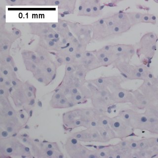
Primary effusion lymphoma (PEL) is classified as a diffuse large B cell lymphoma. It is a rare malignancy of plasmablastic cells that occurs in individuals that are infected with the Kaposi's sarcoma-associated herpesvirus. Plasmablasts are immature plasma cells, i.e. lymphocytes of the B-cell type that have differentiated into plasmablasts but because of their malignant nature do not differentiate into mature plasma cells but rather proliferate excessively and thereby cause life-threatening disease. In PEL, the proliferating plasmablastoid cells commonly accumulate within body cavities to produce effusions, primarily in the pleural, pericardial, or peritoneal cavities, without forming a contiguous tumor mass. In rare cases of these cavitary forms of PEL, the effusions develop in joints, the epidural space surrounding the brain and spinal cord, and underneath the capsule which forms around breast implants. Less frequently, individuals present with extracavitary primary effusion lymphomas, i.e., solid tumor masses not accompanied by effusions. The extracavitary tumors may develop in lymph nodes, bone, bone marrow, the gastrointestinal tract, skin, spleen, liver, lungs, central nervous system, testes, paranasal sinuses, muscle, and, rarely, inside the vasculature and sinuses of lymph nodes. As their disease progresses, however, individuals with the classical effusion-form of PEL may develop extracavitary tumors and individuals with extracavitary PEL may develop cavitary effusions.

Patrick S. Moore is an American virologist and epidemiologist who co-discovered together with his wife, Yuan Chang, two different human viruses causing the AIDS-related cancer Kaposi's sarcoma and the skin cancer Merkel cell carcinoma. Moore and Chang have discovered two of the seven known human viruses causing cancer. The couple met while in medical school together and were married in 1989 while they pursued fellowships at different universities.

Yuan Chang is a Taiwanese-born American virologist and pathologist who co-discovered together with her husband, Patrick S. Moore, the Kaposi's sarcoma-associated herpesvirus (KSHV) and Merkel cell polyomavirus, two of the seven known human oncoviruses.
Rous sarcoma virus (RSV) is a retrovirus and is the first oncovirus to have been described. It causes sarcoma in chickens.

Francis Peyton Rous was an American pathologist at the Rockefeller University known for his works in oncoviruses, blood transfusion and physiology of digestion. A medical graduate from the Johns Hopkins University, he was discouraged to become a practicing physician due to severe tuberculosis. After three years of working as an instructor of pathology at the University of Michigan, he became dedicated researcher at the Rockefeller Institute for Medical Research for the rest of his career.
Merkel cell polyomavirus was first described in January 2008 in Pittsburgh, Pennsylvania. It was the first example of a human viral pathogen discovered using unbiased metagenomic next-generation sequencing with a technique called digital transcriptome subtraction. MCV is one of seven currently known human oncoviruses. It is suspected to cause the majority of cases of Merkel cell carcinoma, a rare but aggressive form of skin cancer. Approximately 80% of Merkel cell carcinoma (MCC) tumors have been found to be infected with MCV. MCV appears to be a common—if not universal—infection of older children and adults. It is found in respiratory secretions, suggesting that it might be transmitted via a respiratory route. However, it has also been found elsewhere, such as in shedded healthy skin and gastrointestinal tract tissues, thus its precise mode of transmission remains unknown. In addition, recent studies suggest that this virus may latently infect the human sera and peripheral blood mononuclear cells.

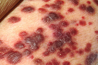
Kaposi's sarcoma (KS) is a type of cancer that can form masses in the skin, in lymph nodes, in the mouth, or in other organs. The skin lesions are usually painless, purple and may be flat or raised. Lesions can occur singly, multiply in a limited area, or may be widespread. Depending on the sub-type of disease and level of immune suppression, KS may worsen either gradually or quickly. Except for Classical KS where there is generally no immune suppression, KS is caused by a combination of immune suppression and infection by Human herpesvirus 8.

miR-138 is a family of microRNA precursors found in animals, including humans. MicroRNAs are typically transcribed as ~70 nucleotide precursors and subsequently processed by the Dicer enzyme to give a ~22 nucleotide product. The excised region or, mature product, of the miR-138 precursor is the microRNA mir-138.

Chickens and their eggs have been used extensively as research models throughout the history of biology. Today they continue to serve as an important model for normal human biology as well as pathological disease processes.
Tumour heterogeneity describes the observation that different tumour cells can show distinct morphological and phenotypic profiles, including cellular morphology, gene expression, metabolism, motility, proliferation, and metastatic potential. This phenomenon occurs both between tumours and within tumours. A minimal level of intra-tumour heterogeneity is a simple consequence of the imperfection of DNA replication: whenever a cell divides, a few mutations are acquired—leading to a diverse population of cancer cells. The heterogeneity of cancer cells introduces significant challenges in designing effective treatment strategies. However, research into understanding and characterizing heterogeneity can allow for a better understanding of the causes and progression of disease. In turn, this has the potential to guide the creation of more refined treatment strategies that incorporate knowledge of heterogeneity to yield higher efficacy.
Elizabeth Murchison is a British-Australian geneticist, Professor of Comparative Oncology and Genetics at the University of Cambridge, UK. The ongoing research of her group focuses on the known existing clonally transmissible cancers arising in mammals. These are cancers that can be passed on between individuals by the transfer of living cancer cells that somehow manage to evade the immune system of their hosts.
Anne-Maree Pearse is an Australian cytogeneticist who is credited with the theory that some cancer cells can be transmissible between individuals. This is known as the allograft theory. Her work has focussed on devil facial tumour disease (DFTD), a contagious cancer that affects Tasmanian devils. For this she has won multiple awards, including the 2012 Prince Hitachi Prize for Comparative Oncology.
Contagious reticulum cell sarcoma is a reticulum-cell sarcoma found in Syrian hamsters that can be transmitted from one hamster to another. It was first described in 1945.
References
- 1 2 3 Ostrander EA, Davis BW, Ostrander GK (January 2016). "Transmissible Tumors: Breaking the Cancer Paradigm". Trends in Genetics. 32 (1): 1–15. doi:10.1016/j.tig.2015.10.001. PMC 4698198 . PMID 26686413.
- 1 2 3 4 Welsh JS (2011). "Contagious cancer". The Oncologist. 16 (1): 1–4. doi:10.1634/theoncologist.2010-0301. PMC 3228048 . PMID 21212437.
- 1 2 Dujon AM, Gatenby RA, Bramwell G, MacDonald N, Dohrmann E, Raven N, et al. (July 2020). "Transmissible Cancers in an Evolutionary Perspective". iScience. 23 (7): 101269. Bibcode:2020iSci...23j1269D. doi:10.1016/j.isci.2020.101269. PMC 7327844 . PMID 32592998.
- 1 2 3 Ujvari B, Gatenby RA, Thomas F (2016-04-01). "The evolutionary ecology of transmissible cancers". Infection, Genetics and Evolution. 39: 293–303. doi:10.1016/j.meegid.2016.02.005. ISSN 1567-1348. PMID 26861618.
- ↑ Nguyen DX, Bos PD, Massagué J (April 2009). "Metastasis: from dissemination to organ-specific colonization". Nature Reviews Cancer. 9 (4): 274–284. doi:10.1038/nrc2622. ISSN 1474-175X. PMID 19308067.
- ↑ Gatenby RA, Gillies RJ (January 2008). "A microenvironmental model of carcinogenesis". Nature Reviews Cancer. 8 (1): 56–61. doi:10.1038/nrc2255. ISSN 1474-175X. PMID 18059462.
- ↑ Barozzi P, Luppi M, Facchetti F, Mecucci C, Alù M, Sarid R, et al. (May 2003). "Post-transplant Kaposi sarcoma originates from the seeding of donor-derived progenitors". Nature Medicine. 9 (5): 554–61. doi:10.1038/nm862. PMID 12692543. S2CID 2527251.
- ↑ Matser YA, Terpstra ML, Nadalin S, Nossent GD, de Boer J, van Bemmel BC, et al. (July 2018). "Transmission of breast cancer by a single multiorgan donor to 4 transplant recipients". American Journal of Transplantation. 18 (7): 1810–1814. doi: 10.1111/ajt.14766 . PMID 29633548.
- ↑ Muehlenbachs A, Bhatnagar J, Agudelo CA, Hidron A, Eberhard ML, Mathison BA, et al. (November 2015). "Malignant Transformation of Hymenolepis nana in a Human Host". The New England Journal of Medicine. 373 (19): 1845–52. doi: 10.1056/NEJMoa1505892 . PMID 26535513.
- ↑ Gärtner HV, Seidl C, Luckenbach C, Schumm G, Seifried E, Ritter H, et al. (November 1996). "Genetic analysis of a sarcoma accidentally transplanted from a patient to a surgeon". The New England Journal of Medicine. 335 (20): 1494–6. doi: 10.1056/NEJM199611143352004 . PMID 8890100.
- ↑ Gugel EA, Sanders ME (December 1986). "Needle-stick transmission of human colonic adenocarcinoma". The New England Journal of Medicine. 315 (23): 1487. doi:10.1056/NEJM198612043152314. PMID 3785302.
- ↑ Weiss RA, Fassati A, Murgia C (2006). "A sexually transmitted parasitic cancer". Retrovirology . 3 (Supplement 1): S92. doi: 10.1186/1742-4690-3-S1-S92 . PMC 1717007 .
- ↑ Ní Leathlobhair M, Gulland FM, Murchison EP (2017-06-22). "No evidence for clonal transmission of urogenital carcinoma in California sea lions ( Zalophus californianus)". Wellcome Open Research. 2: 46. doi: 10.12688/wellcomeopenres.11483.1 . PMC 5527528 . PMID 28948233.
- ↑ Rebbeck CA, Thomas R, Breen M, Leroi AM, Burt A (September 2009). "Origins and evolution of a transmissible cancer". Evolution; International Journal of Organic Evolution. 63 (9): 2340–9. doi: 10.1111/j.1558-5646.2009.00724.x . PMID 19453727.
- ↑ Strakova A, Murchison EP (February 2015). "The cancer which survived: insights from the genome of an 11000 year-old cancer" (PDF). Current Opinion in Genetics & Development. 30: 49–55. doi:10.1016/j.gde.2015.03.005. PMID 25867244. S2CID 21195930.
- ↑ Baez-Ortega A, Gori K, Strakova A, Allen JL, Allum KM, Bansse-Issa L, et al. (August 2019). "Somatic evolution and global expansion of an ancient transmissible cancer lineage". Science. 365 (6452): eaau9923. doi:10.1126/science.aau9923. PMC 7116271 . PMID 31371581.
- ↑ Belov K (February 2011). "The role of the Major Histocompatibility Complex in the spread of contagious cancers". Mammalian Genome. 22 (1–2): 83–90. doi:10.1007/s00335-010-9294-2. PMID 20963591. S2CID 8303843.
- ↑ Siddle HV, Kreiss A, Eldridge MD, Noonan E, Clarke CJ, Pyecroft S, et al. (October 2007). "Transmission of a fatal clonal tumor by biting occurs due to depleted MHC diversity in a threatened carnivorous marsupial". Proceedings of the National Academy of Sciences of the United States of America. 104 (41): 16221–6. doi: 10.1073/pnas.0704580104 . PMC 1999395 . PMID 17911263.
- 1 2 3 4 Harrison C (May 2018). "Clonally transmissible cancers in nature". Cancer Therapy Advisor. Retrieved 2019-10-03.
- ↑ Murgia C, Pritchard JK, Kim SY, Fassati A, Weiss RA (August 2006). "Clonal origin and evolution of a transmissible cancer". Cell. 126 (3): 477–87. doi:10.1016/j.cell.2006.05.051. PMC 2593932 . PMID 16901782.
- ↑ Copper HL, Mackay CM, Banfield WG (October 1964). "Chromosome studies of a contagious reticulum cell sarcoma of the Syrian hamster". Journal of the National Cancer Institute. 33 (4): 691–706. doi:10.1093/jnci/33.4.691. PMID 14220251.
- ↑ Banfield WG, Woke PA, Mackay CM, Cooper HL (May 1965). "Mosquito transmission of a reticulum cell sarcoma of hamsters". Science. 148 (3674): 1239–40. Bibcode:1965Sci...148.1239B. doi:10.1126/science.148.3674.1239. PMID 14280009. S2CID 12611674.
- ↑ Pearse AM, Swift K (February 2006). "Allograft theory: transmission of devil facial-tumour disease". Nature. 439 (7076): 549. Bibcode:2006Natur.439..549P. doi: 10.1038/439549a . PMID 16452970. S2CID 4409863.
- ↑ Epstein B, Jones M, Hamede R, Hendricks S, McCallum H, Murchison EP, et al. (August 2016). "Rapid evolutionary response to a transmissible cancer in Tasmanian devils". Nature Communications. 7 (1): 12684. Bibcode:2016NatCo...712684E. doi: 10.1038/ncomms12684 . PMC 5013612 . PMID 27575253.
- ↑ Yong E (2015-04-09). "Selfish shellfish cells cause contagious clam cancer". National Geographic . Archived from the original on 2015-09-05. Retrieved 2015-04-10.
- ↑ Metzger MJ, Reinisch C, Sherry J, Goff SP (April 2015). "Horizontal transmission of clonal cancer cells causes leukemia in soft-shell clams". Cell. 161 (2): 255–63. doi:10.1016/j.cell.2015.02.042. PMC 4393529 . PMID 25860608.
- ↑ Metzger MJ, Villalba A, Carballal MJ, Iglesias D, Sherry J, Reinisch C, et al. (June 2016). "Widespread transmission of independent cancer lineages within multiple bivalve species". Nature. 534 (7609): 705–9. Bibcode:2016Natur.534..705M. doi:10.1038/nature18599. PMC 4939143 . PMID 27338791.
- ↑ Frierman EM, Andrews JD (February 1976). "Occurrence of hematopoietic neoplasms in Virginia oysters (Crassostrea virginica)". Journal of the National Cancer Institute. 56 (2): 319–24. doi:10.1093/jnci/56.2.319. PMID 1255763.