Sarcoma

Last updated

Sarcoma
Other namesSarcomas, sarcomata
Nibib 030207 105309 sarcoma.jpg
Optical coherence tomography (OCT) image of a sarcoma
Specialty Oncology

A sarcoma is a malignant tumor, a type of cancer that arises from cells of mesenchymal (connective tissue) origin. [1] [2] Connective tissue is a broad term that includes bone, cartilage, fat, vascular, or other structural tissues, and sarcomas can arise in any of these types of tissues. [2] [3] As a result, there are many subtypes of sarcoma, which are classified based on the specific tissue and type of cell from which the tumor originates. [4]

Contents

Sarcomas are primary connective tissue tumors, meaning that they arise in connective tissues. [2] This is in contrast to secondary (or "metastatic") connective tissue tumors, which occur when a cancer from elsewhere in the body (such as the lungs, breast tissue or prostate) spreads to the connective tissue. [5] Sarcomas are one of five different types of cancer, classified by the cell type from which they originate. [6] The word sarcoma is derived from the Greek σάρκωμαsarkōma 'fleshy excrescence or substance', itself from σάρξ sarx meaning 'flesh'. [7] [8] [9]

Classification

Sarcomas are typically divided into two major groups: bone sarcomas and soft-tissue sarcomas, [2] each of which has multiple subtypes. In the United States, the American Joint Committee on Cancer (AJCC) publishes guidelines that classify the subtypes of sarcoma. [4] These subtypes are as follows:

Subtypes of bone sarcoma

Subtypes of soft-tissue sarcoma

Signs and symptoms

Symptoms of bone sarcomas typically include bone pain, especially at night, and swelling around the site of the tumor. [2]

Symptoms of soft-tissue sarcomas vary, but they often present as firm, often times painless lumps or nodules. [2] Gastrointestinal stromal tumors (GIST, a subtype of soft-tissue sarcoma) often are asymptomatic, but can be associated with vague complaints of abdominal pain, bleeding into the intestines, a feeling of fullness, or other signs of intestinal obstruction. [2]

Cause

Causes and risk factors

The cause of most bone sarcomas is not known, [3] but several factors are associated with an increased risk of developing bone sarcoma. Previous exposure to ionizing radiation (such as prior radiation therapy) is one such risk factor. [2] Therapeutic radiation is associated with sarcoma after 10 to 20 years. [10] Exposure to alkylating agents, such as those found in certain cancer chemotherapeutic medicines, also increases the risk of bone sarcoma. [3] Certain inherited genetic syndromes, including Li-Fraumeni syndrome, inherited RB1 gene mutations, and Paget's disease of bone are associated with an increased risk of developing bone sarcomas. [2]

Most soft-tissue sarcomas arise from what doctors call "sporadic" (or random) genetic mutations within an affected person's cells. [3] Nevertheless, there are certain risk factors associated with an increased risk of developing soft-tissue sarcoma. Previous exposure to ionizing radiation is one such risk factor. [2] Exposure to vinyl chloride (e.g., such as the fumes encountered in the production of polyvinyl chloride (PVC)), arsenic and Thorotrast all are associated with an increased risk of angiosarcoma. [2] [3] Lymphedema, such as that resulting from certain types of breast cancer treatment, also is a risk factor for development of angiosarcoma. [3] As with bone sarcomas, certain inherited genetic syndromes also are associated with an increased risk of developing soft-tissue sarcoma, including Li-Fraumeni syndrome, familial adenomatous polyposis, neurofibromatosis type 1, and heritable RB1 gene mutations. [3] Kaposi sarcoma is caused by Kaposi sarcoma-associated herpesvirus (HHV-8).

Mechanisms

The mechanisms by which healthy cells transform into cancer cells are described in detail elsewhere (see Cancer main page; Carcinogenesis main page). The precise molecular changes that result in sarcoma are not always known, but certain types of sarcomas are associated with particular genetic mutations. [2] [3] Examples include:

Diagnosis

Bone sarcomas

Diagnosis of bone sarcomas begins with a thorough history and physical examination which may reveal characteristic signs and symptoms (see Signs and Symptoms above). [3] Laboratory studies are not particularly useful in diagnosis, although some bone sarcomas (such as osteosarcoma) may be associated with elevated alkaline phosphatase levels, while others (such as Ewing sarcoma) can be associated with elevated erythrocyte sedimentation rate. [11] Importantly, however, none of these laboratory findings are specific to bone sarcomas, meaning that elevations in these lab values are associated with many other conditions as well as sarcoma, and thus cannot be relied upon to conclusively diagnose sarcoma. [3]

Imaging studies are critically important in diagnosis, and most clinicians will order a plain radiograph (X-ray) initially. [3] Other imaging studies commonly used in diagnosis include magnetic resonance imaging (MRI) studies and radioisotope bone scans. [11] [3] A CT scan is typically not used in diagnosis of most types of bone sarcoma, although it is an important tool for staging (see below). [3] Definitive diagnosis requires biopsy of the tumor and careful review of the biopsy specimen by an experienced pathologist. [3]

Soft-tissue sarcomas

Diagnosis of soft-tissue sarcomas also begins with a thorough history and physical examination. [3] Imaging studies can include either CT or MRI, although CT tends to be preferred for soft-tissue sarcomas located in the thorax, abdomen, or retroperitoneum. [3] Positron emission tomography (PET) also may be useful in diagnosis, although its most common use is for staging (see below). [3] As with bone sarcomas, definitive diagnosis requires biopsy of the tumor with evaluation of histology by a trained pathologist. [3] [12]

Staging

In general, cancer staging refers to how advanced a cancer is, and usually it is based upon factors such as tumor size and whether it has spread to other parts of the body. [3] [13] Staging is important because the stage affects the prognosis (likely outcome), as well as the types of treatments that are likely to be effective against the cancer. [2] [4] With sarcomas, staging requires a determination of whether the tumor has grown into surrounding tissues ("local invasion"), as well as imaging to determine whether it has spread (a process known as "metastasis") to lymph nodes (forming "nodal metastases") or to other tissues or organs in the body (forming "distant metastases"). [4]

The most common imaging tools used for staging bone sarcomas are MRI or CT to evaluate the primary tumor, contrast-enhanced CT of the chest to evaluate whether the cancer has spread (i.e., metastasized) to the lungs, and radioisotope bone scan to evaluate whether the cancer has spread to other bones. [4] Staging for soft-tissue sarcomas typically includes imaging of the primary tumor by MRI or CT to determine tumor size, as well as contrast-enhanced CT of the chest to evaluate for metastatic tumors in the lungs. [4]

Grade

Like some other cancers, sarcomas are assigned a grade (low, intermediate, or high) based on the appearance of the tumor cells under a microscope. [14] In general, grade refers to how aggressive the cancer is and how likely it is to spread to other parts of the body ("metastasize"). [14] Low-grade sarcomas have a better prognosis than higher-grade sarcomas, and are usually treated surgically, although sometimes radiation therapy or chemotherapy are used. [3] [4] Intermediate- and high-grade sarcomas are more frequently treated with a combination of surgery, chemotherapy, or radiation therapy. [15] Since high-grade tumors are more likely to undergo metastasis (invasion and spread to locoregional and distant sites), they are treated more aggressively. The recognition that many sarcomas are sensitive to chemotherapy has dramatically improved the survival of patients. For example, in the era before chemotherapy, long-term survival for pediatric patients with localized osteosarcoma was only about 20%, but now has risen to 60–70%. [16]

Screening

In the US, the US Preventive Services Task Force (USPSTF) publishes guidelines recommending preventive screening for certain types of common cancers and other diseases. [17] As of March 2019, the USPSTF does not recommend screening for sarcoma, [17] possibly because it is a very rare type of cancer (see Epidemiology below).

The American Cancer Society (ACS) also publishes guidelines recommending preventive screening for certain types of common cancers. [18] Like the USPSTF, as of March 2019 ACS does not recommend preventive screening for sarcoma. [18]

However, patients with some inherited conditions, such as neurofibromatosis, may benefit from screening for development of cancers from pre-existing benign tumors called neurofibromas.

Treatment

Surgery is the most common form of the treatment for most sarcomas that have not spread to other parts of the body, and for most sarcomas, surgery is the only curative treatment. [3] [19] Limb-sparing surgery, as opposed to amputation, can now be used to save the limbs of patients in at least 90% of extremity (arm or leg) sarcoma cases. [19] Additional treatments, including chemotherapy, radiation therapy (also called "radiotherapy"), which includes proton therapy, may be administered before surgery (called "neoadjuvant" chemotherapy or radiotherapy) or after surgery (called "adjuvant" chemotherapy or radiotherapy). [3] [15] The use of neoadjuvant or adjuvant chemotherapy and radiotherapy significantly improves the prognosis for many sarcoma patients. [3] [20] Treatment can be a long and arduous process, lasting about a year for many patients. [15]

It was believed that higher doses of chemotherapy might improve survival. However, high doses of chemotherapy stop the production of blood cells in the bone marrow and can be harmful. Stem cells collected from people before high‐dose chemotherapy can be transplanted back to the person if the blood cell count gets too low; this is called autologous hematopoietic stem cell transplantation, or high dose therapy with stem cell rescue. Research to investigate if using high‐dose chemotherapy followed by autologous hematopoietic stem cell transplantation was more favourable than standard‐dose chemotherapy [25] found only one RCT and this did not favour either of the two treatment arms with respect to overall survival. As a result, high dose chemotherapy with stem cell rescue is generally considered appropriate only in the research setting.

Prognosis

Factors that affect prognosis

The AJCC has identified several factors that affect prognosis of bone sarcomas: [4]

For soft-tissue sarcomas other than GISTs, factors that affect prognosis include: [4]

For GISTs, the key factor that affects prognosis is: [4]

Outcome data

According to data published by the US National Cancer Institute (NCI), the overall 5-year survival for bone sarcomas is 66.9%. [26] The American Cancer Society (ACS) estimates that 2,140 people in the US will die in 2023 from bone sarcomas, accounting for 0.3% of all cancer deaths. [27] The median age at death is 61 years old, although death can occur in any age group. [26] Thus, 12.3% of bone sarcoma deaths occur in people under 20 years old, 13.8% occur in people 20–34 years old, 5.5% occur in people 35–44 years old, 9.3% occur in people 45–54 years old, 13.5% occur in people 55–64 years old, 16.2% occur in people 65–74 years old, 16.4% occur in people 75–84 years old, and 13.1% occur in people 85 years or older. [26]

For soft-tissue sarcomas, the overall 5-year survival (irrespective of stage) is 64.5%, but survival is affected by many factors, including stage. [28] Thus, the 5-year survival is 80.8% for soft-tissue sarcomas that have not spread beyond the primary tumor ("localized" tumors), 58.0% for soft-tissue sarcomas that have spread only to nearby lymph nodes, and 16.4% for soft-tissue sarcomas that have spread to distant organs. [28] The ACS estimates that 5,140 people will die from soft-tissue sarcoma in 2023, accounting for 0.9% of all cancer deaths. [27]

Epidemiology

Sarcomas are rare cancers. [2] The risk of a previously healthy person receiving a new diagnosis of bone cancer is less than 0.001%, while the risk of receiving a new diagnosis of soft-tissue sarcoma is between 0.0014 and 0.005%. [3] The American Cancer Society estimates that in the United States there will be 3,970 new cases of bone sarcoma in 2023, and 13,400 new cases of soft-tissue sarcoma. [27] Considering that the total estimated number of new cancer diagnoses (all types of cancer) is 1,958,310, this means bone sarcomas represent only 0.2% of all new cancer diagnoses (making them the 30th most common type of cancer [26] ) and soft-tissue sarcomas represent only 0.7% (making them the 22nd most common type of cancer [28] ) of all new cancer diagnoses in the US in 2023. [27] These estimates are similar to previously reported data. [3]

Sarcomas affect people of all ages. Around 50% of bone sarcomas and 20% of soft-tissue sarcomas are diagnosed in people under the age of 35. [29] Some sarcomas, such as leiomyosarcoma, chondrosarcoma, and gastrointestinal stromal tumor (GIST), are more common in adults than in children. [2] Most high-grade bone sarcomas, including Ewing sarcoma and osteosarcoma, are much more common in children and young adults. [2]

In fossils

In 2016, scientists reported the discovery of an osteosarcoma tumor in a 1.6–1.8 million-year-old fossil from the skeleton of the now-extinct hominin species Australopithecus sediba , making it the earliest-known case of human cancer. [30] [31]

Research

Treatment of sarcoma, especially when the sarcoma has spread, or "metastasized", often requires chemotherapy but existing chemotherapeutic medicines are associated with significant toxicities and are not highly effective in killing cancer cells. [3] Therefore, research to identify new medications to treat sarcoma is being conducted as of 2019. [3] One new type of therapy still under investigation is the use of cancer immunotherapy (e.g., immune checkpoint inhibitors like anti-PD1, anti-PDL1, and anti-CTLA4 agents) to treat sarcomas. [32] These drugs are not yet FDA- or other regulator-approved treatment, except PDL1 inhibitor atezolizumab for the ultra-rate diagnosis of alveolar soft part sarcoma. [32] [33] Other strategies, such as small-molecule targeted therapy, biologic agents (e.g., small interfering RNA molecules), and nanoparticle-directed therapy, also are under active investigated. [3]

Research to understand the specific genetic and molecular factors that cause sarcoma to develop is underway. [3] This could allow for the design of new targeted therapies and allow physicians to more accurately predict a patient's prognosis. [3]

Awareness

In the US, July is widely recognized as Sarcoma Awareness Month. [34] The UK has a Sarcoma Awareness Week in July led by Sarcoma UK, the bone and soft-tissue cancer charity. [35]

American YouTuber Technoblade was diagnosed with sarcoma in August 2021, and died from his illness in June 2022 after the sarcoma metastasized. He had raised over $500,000 in a charity stream. Many YouTubers have raised awareness and donated to charities such as the Sarcoma Foundation of America after Technoblade's diagnosis and passing. To date, Technoblade's fans have raised over $1,000,000 for sarcoma research. [36]

TikTok has provided a voice for many creators to chronicle their experiences with sarcoma. "Dance You Outta My Head", by American singer Cat Janice went viral on TikTok in early 2024 before the singer died of sarcoma, prompting awareness of this rare disease. [37]

Kimberley Nix, a Canadian physician, chronicled her journey with undifferentiated pleomorphic sarcoma, from her diagnosis to eventual death, on TikTok under the username @cancerpatientmd. [38] Nix died on May 8, 2024 at the age of 31, and her death was announced in a video uploaded posthumously to her TikTok page. [39] In many of her videos, she links viewers to her Own.Cancer fundraiser, which has raised almost $118,000 CAD as of May 17, 2024. [40]

Related Research Articles

<span class="mw-page-title-main">Brain tumor</span> Neoplasm in the brain

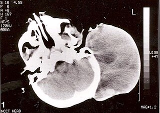
A brain tumor occurs when abnormal cells form within the brain. There are two main types of tumors: malignant (cancerous) tumors and benign (non-cancerous) tumors. These can be further classified as primary tumors, which start within the brain, and secondary tumors, which most commonly have spread from tumors located outside the brain, known as brain metastasis tumors. All types of brain tumors may produce symptoms that vary depending on the size of the tumor and the part of the brain that is involved. Where symptoms exist, they may include headaches, seizures, problems with vision, vomiting and mental changes. Other symptoms may include difficulty walking, speaking, with sensations, or unconsciousness.

<span class="mw-page-title-main">Soft-tissue sarcoma</span> Medical condition

A soft-tissue sarcoma (STS) is a malignant tumor, a type of cancer, that develops in soft tissue. A soft-tissue sarcoma is often a painless mass that grows slowly over months or years. They may be superficial or deep-seated. Any such unexplained mass must be diagnosed by biopsy. Treatment may include surgery, radiotherapy, chemotherapy, and targeted drug therapy. Bone sarcomas are the other class of sarcomas.

<span class="mw-page-title-main">Bone tumor</span> Medical condition

A bone tumor is an abnormal growth of tissue in bone, traditionally classified as noncancerous (benign) or cancerous (malignant). Cancerous bone tumors usually originate from a cancer in another part of the body such as from lung, breast, thyroid, kidney and prostate. There may be a lump, pain, or neurological signs from pressure. A bone tumor might present with a pathologic fracture. Other symptoms may include fatigue, fever, weight loss, anemia and nausea. Sometimes there are no symptoms and the tumour is found when investigating another problem.

<span class="mw-page-title-main">Osteosarcoma</span> Cancerous tumour in a bone

An osteosarcoma (OS) or osteogenic sarcoma (OGS) is a cancerous tumor in a bone. Specifically, it is an aggressive malignant neoplasm that arises from primitive transformed cells of mesenchymal origin and that exhibits osteoblastic differentiation and produces malignant osteoid.

<span class="mw-page-title-main">Rhabdomyosarcoma</span> Medical condition

Rhabdomyosarcoma (RMS) is a highly aggressive form of cancer that develops from mesenchymal cells that have failed to fully differentiate into myocytes of skeletal muscle. Cells of the tumor are identified as rhabdomyoblasts.

<span class="mw-page-title-main">Liposarcoma</span> Medical condition

Liposarcomas are the most common subtype of soft tissue sarcomas, accounting for at least 20% of all sarcomas in adults. Soft tissue sarcomas are rare neoplasms with over 150 different histological subtypes or forms. Liposarcomas arise from the precursor lipoblasts of the adipocytes in adipose tissues. Adipose tissues are distributed throughout the body, including such sites as the deep and more superficial layers of subcutaneous tissues as well as in less surgically accessible sites like the retroperitoneum and visceral fat inside the abdominal cavity.

<span class="mw-page-title-main">Malignant peripheral nerve sheath tumor</span> Medical condition

A malignant peripheral nerve sheath tumor (MPNST) is a form of cancer of the connective tissue surrounding nerves. Given its origin and behavior it is classified as a sarcoma. About half the cases are diagnosed in people with neurofibromatosis; the lifetime risk for an MPNST in patients with neurofibromatosis type 1 is 8–13%. MPNST with rhabdomyoblastomatous component are called malignant triton tumors.

The International Classification of Diseases for Oncology (ICD-O) is a domain-specific extension of the International Statistical Classification of Diseases and Related Health Problems for tumor diseases. This classification is widely used by cancer registries.

<span class="mw-page-title-main">Undifferentiated pleomorphic sarcoma</span> Medical condition

Undifferentiated pleomorphic sarcoma (UPS), also termed pleomorphic myofibrosarcoma, high-grade myofibroblastic sarcoma, and high-grade myofibrosarcoma, is characterized by the World Health Organization (WHO), 2020, as a rare, poorly differentiated neoplasm, i.e. an abnormal growth of cells that have an unclear identity and/or cell of origin. WHO classified it as one of the undifferentiated/unclassified sarcomas in the category of tumors of uncertain differentiation. Sarcomas are cancers known or thought to derive from mesenchymal stem cells that typically develop in bone, muscle, fat, blood vessels, lymphatic vessels, tendons, and ligaments. More than 70 sarcoma subtypes have been described. The UPS subtype of these sarcomas consists of tumor cells that are poorly differentiated and may appear as spindle-shaped cells, histiocytes, and giant cells. UPS is considered a diagnosis that defies formal sub-classification after thorough histologic, immunohistochemical, and ultrastructural examinations fail to identify the type of cells involved.

<span class="mw-page-title-main">Ewing sarcoma</span> Type of cancer

Ewing sarcoma is a type of pediatric cancer that forms in bone or soft tissue. Symptoms may include swelling and pain at the site of the tumor, fever, and a bone fracture. The most common areas where it begins are the legs, pelvis, and chest wall. In about 25% of cases, the cancer has already spread to other parts of the body at the time of diagnosis. Complications may include a pleural effusion or paraplegia.

Soft tissue sarcoma refers to a broad group of tumors that originate from connective tissues. They tend to have similar histologic appearance and biological behavior, and can be either benign or malignant. Soft tissue sarcomas can arise in any part of the pet's body but skin and subcutaneous tumors are the most commonly observed. Soft-tissue sarcomas comprise approximately 15% of all skin and subcutaneous tumors in dogs and approximately 7% of all skin and subcutaneous tumors in cats. The variety of different tumors that fall under the category of soft tissue sarcomas includes fibrosarcoma, hemangiopericytoma, liposarcoma, rhabdomyosarcoma, leiomyosarcoma, malignant fibrous histiocytoma, malignant nerve sheath tumors, myxosarcoma, myxofibrosarcoma, mesenchymoma, and spindle cell tumor.

<span class="mw-page-title-main">Epithelioid sarcoma</span> Medical condition

Epithelioid sarcoma is a rare soft tissue sarcoma arising from mesenchymal tissue and characterized by epithelioid-like features. It accounts for less than 1% of all soft tissue sarcomas. It was first definitively characterized by F.M. Enzinger in 1970. It commonly presents itself in the distal limbs of young adults as a small, soft mass or a cluster of bumps. A proximal version has also been described, frequently occurring in the upper extremities. Less commonly, cases are reported in the pelvis, vulva, penis, and spine.

<span class="mw-page-title-main">Clear cell sarcoma</span> Rare form of cancer

Clear cell sarcoma is a rare form of cancer called a sarcoma. It is known to occur mainly in the soft tissues and dermis. Rare forms were thought to occur in the gastrointestinal tract before they were discovered to be different and redesignated as gastrointestinal neuroectodermal tumors.

Embryonal rhabdomyosarcoma (EMRS) is a rare histological form of cancer in the connective tissue wherein the mesenchymally-derived malignant cells resemble the primitive developing skeletal muscle of the embryo. It is the most common soft tissue sarcoma occurring in children. Embryonal rhabdomyosarcoma is also known as PAX-fusion negative or fusion-negative rhabdomyosarcoma, as tumors of this subtype are unified by their lack of a PAX3-FOXO1 fusion oncogene. Fusion status refers to the presence or absence of a fusion gene, which is a gene formed from joining two different genes together through DNA rearrangements. These types of tumors are classified as embryonal rhabdomyosarcoma "because of their remarkable resemblance to developing embryonic and fetal skeletal muscle."

<span class="mw-page-title-main">Low-grade fibromyxoid sarcoma</span> Medical condition

Low-grade fibromyxoid sarcoma (LGFMS) is a rare type of low-grade sarcoma first described by H. L. Evans in 1987. LGFMS are soft tissue tumors of the mesenchyme-derived connective tissues; on microscopic examination, they are found to be composed of spindle-shaped cells that resemble fibroblasts. These fibroblastic, spindle-shaped cells are neoplastic cells that in most cases of LGFMS express fusion genes, i.e. genes composed of parts of two different genes that form as a result of mutations. The World Health Organization (2020) classified LGFMS as a specific type of tumor in the category of malignant fibroblastic and myofibroblastic tumors.

A rhabdomyoblast is a cell type which is found in some rhabdomyosarcomas. When found histologically, a rhabdomyoblast aids the diagnosis of embryonal, alveolar, spindle cell/sclerosing, and pleomorphic rhabdomyosarcomas; however, in a tumor, expression of the rhabdomyoblast phenotype is not the only factor in diagnosing a rhabdomyosarcoma. Mesenchymal malignancies can exhibit this phenotype as well. Immunohistochemistry techniques allow for the sensitive detection of desmin, vimentin, muscle specific actin, and MyoD1. Similarly the rhabdomyoblast phenotype can be detected morphologically. Rhabdomyoblasts are early stage mesenchymal cells, having the potential to differentiate into a wide range of skeletal cells. Each stage of differentiation exhibits unique and distinguishable histological characteristics. In its initial from, stellate cells with amphiphilic cytoplasm and ovular central nuclei are observed. Commonly referred to as rhabdoid features, the maturing rhabdomyoblast will likely exhibit low levels of eosinophilic cytoplasm in proximal distances to the nucleus. As maturation and differentiation progress, the cell's cytoplasmic levels of white blood cells increase; additionally, elongated shapes, commonly depicted as “tadpole”, “strap” and "spider cells", are observed. In the concluding phase of differentiation, the white blood cell rich cytoplasm appears bright and exhibits cross-striation. The highly regulated organization of actin and myosin microfilaments in contractile proteins results in this appearance.

Myxofibrosarcoma (MFS), although a rare type of tumor, is one of the most common soft tissue sarcomas, i.e. cancerous tumors, that develop in the soft tissues of elderly individuals. Initially considered to be a type of histiocytoma termed fibrous histiocytoma or myxoid variant of malignant fibrous histiocytoma, Angervall et al. termed this tumor myxofibrosarcoma in 1977. In 2020, the World Health Organization reclassified MFS as a separate and distinct tumor in the category of malignant fibroblastic and myofibroblastic tumors.

Sclerosing epithelioid fibrosarcoma (SEF) is a very rare malignant tumor of soft tissues that on microscopic examination consists of small round or ovoid neoplastic epithelioid fibroblast-like cells, i.e. cells that have features resembling both epithelioid cells and fibroblasts. In 2020, the World Health Organization classified SEF as a distinct tumor type in the category of malignant fibroblastic and myofibroblastic tumors. However, current studies have reported that low-grade fibromyxoid sarcoma (LGFMS) has many clinically and pathologically important features characteristic of SEF; these studies suggest that LGSFMS may be an early form of, and over time progress to become, a SEF. Since the World Health Organization has classified LGFMS as one of the malignant fibroblastic and myofibroblastic tumors that is distinctly different than SEF, SEF and LGFMS are here regarded as different tumor forms.

Low-grade myofibroblastic sarcoma (LGMS) is a subtype of the malignant sarcomas. As it is currently recognized, LGMS was first described as a rare, atypical myofibroblastic tumor by Mentzel et al. in 1998. Myofibroblastic sarcomas had been divided into low-grade myofibroblastic sarcomas, intermediate‐grade myofibroblasic sarcomas, i.e. IGMS, and high‐grade myofibroblasic sarcomas, i.e. HGMS based on their microscopic morphological, immunophenotypic, and malignancy features. LGMS and IGMS are now classified together by the World Health Organization (WHO), 2020, in the category of intermediate fibroblastic and myofibroblastic tumors. WHO, 2020, classifies HGMS as a soft tissue tumor in the category of tumors of uncertain differentiation. This article follows the WHO classification: here, LGMS includes IGMS but not HGMS which is a more aggressive and metastasizing tumor than LGMS and consists of cells of uncertain origin.

References

  1. Yang J, Ren Z, Du X, Hao M, Zhou W (27 October 2014). "The role of mesenchymal stem/progenitor cells in sarcoma: update and dispute". Stem Cell Investigation. 1: 18. doi:10.3978/j.issn.2306-9759.2014.10.01. PMC   4923508 . PMID   27358864.
  2. 1 2 3 4 5 6 7 8 9 10 11 12 13 14 15 16 17 18 19 Tobias J (2015). Cancer and its Management (Seventh ed.). Chichester, West Sussex, PO198SQ, UK: John Wiley & Sons, Ltd. p. 446. ISBN   978-1-118-46875-3.{{cite book}}: CS1 maint: location (link)
  3. 1 2 3 4 5 6 7 8 9 10 11 12 13 14 15 16 17 18 19 20 21 22 23 24 25 26 27 28 29 30 31 32 33 34 35 36 DeVita Jr V (2015). DeVita, Hellman and Rosenberg's Cancer: Principles & Practice of Oncology (10th ed.). Philadelphia, PA: Wolters Kluwer Health. pp. 1241–1313. ISBN   978-1-4511-9294-0.
  4. 1 2 3 4 5 6 7 8 9 10 11 12 Amin MB (2017). AJCC Cancer Staging Manual, Eight Edition. Chicago, IL: Springer International Publishing AG Switzerland. pp. 471–548. ISBN   978-3-319-40617-6.
  5. "Metastatic Cancer". National Cancer Institute. 12 May 2015. Retrieved 22 March 2019.
  6. "Defining Cancer". National Cancer Institute. 17 September 2007. Retrieved 10 June 2014.
  7. σάρκωμα, σάρξ . Liddell, Henry George ; Scott, Robert ; A Greek–English Lexicon at the Perseus Project.
  8. "Definition of SARCOMA". www.merriam-webster.com. Retrieved 22 March 2019.
  9. Harper D. "sarcoma". Online Etymology Dictionary .
  10. "UpToDate". www.uptodate.com. Retrieved 19 March 2023.
  11. 1 2 Unni K (2010). Dahlin's Bone Tumors. Philadelphia, PA: Lippincott Williams & Wilkins. pp. 1–8. ISBN   978-0-7817-6242-7.
  12. Rastogi S, Aggarwal A, Shishak S, Barwad A, Dhamija E, Pandey R, et al. (9 August 2019). "Discordance of Histo-pathological Diagnosis of Patients with Soft Tissue Sarcoma Referred to Tertiary Care Center". Asian Pacific Journal of Cancer Care. 4 (4): 119–123. doi: 10.31557/apjcc.2019.4.4.119-123 .
  13. "Staging". National Cancer Institute. 9 March 2015. Retrieved 21 March 2019.
  14. 1 2 "Tumor Grade". National Cancer Institute. 9 May 2013. Retrieved 21 March 2019.
  15. 1 2 3 Buecker P (2005). "Sarcoma: A Diagnosis of Patience". ESUN. 2 (5). Archived from the original on 4 December 2020. Retrieved 15 April 2009.
  16. Longhi A, Errani C, De Paolis M, Mercuri M, Bacci G (October 2006). "Primary bone osteosarcoma in the pediatric age: state of the art". Cancer Treatment Reviews. 32 (6): 423–436. doi:10.1016/j.ctrv.2006.05.005. PMID   16860938.
  17. 1 2 "Published Recommendations - US Preventive Services Task Force". www.uspreventiveservicestaskforce.org. Retrieved 20 March 2019.
  18. 1 2 "Cancer Screening Guidelines | Detecting Cancer Early". www.cancer.org. Retrieved 20 March 2019.
  19. 1 2 Morris C (2005). "Malignant Fibrous Histiocytoma (MFH)". ESUN. 2 (2). Archived from the original on 11 November 2012. Retrieved 19 October 2011.
  20. Baker L (2006). "A Rose is a Rose or a Thorn is a Thorn". ESUN. 3 (5). Archived from the original on 19 January 2013. Retrieved 19 October 2011.
  21. Liposarcoma Treatment & Management~treatment at eMedicine
  22. "Rhabdomyosarcoma". Boston Children's Hospital. Archived from the original on 23 May 2011. Retrieved 2 March 2010.
  23. Wexler L (2004). "Rhabdomyosarcoma". ESUN. 1 (4). Archived from the original on 27 October 2012. Retrieved 19 October 2011.
  24. Osteosarcoma Treatment & Management~treatment at eMedicine
  25. Peinemann F, Enk H, Smith LA (April 2017). "Autologous hematopoietic stem cell transplantation following high-dose chemotherapy for nonrhabdomyosarcoma soft tissue sarcomas". The Cochrane Database of Systematic Reviews. 4 (7): CD008216. doi:10.1002/14651858.cd008216.pub5. PMC   6478255 . PMID   28407197.
  26. 1 2 3 4 "Bone and Joint Cancer - Cancer Stat Facts". SEER. Retrieved 27 March 2019.
  27. 1 2 3 4 Siegel RL, Miller KD, Wagle NS, Jemal A (2023). "Cancer statistics, 2023". CA: A Cancer Journal for Clinicians. 73 (1): 17–48. doi: 10.3322/caac.21763 . ISSN   1542-4863. PMID   36633525.
  28. 1 2 3 "Soft Tissue Cancer - Cancer Stat Facts". SEER. Retrieved 27 March 2019.
  29. Darling J (2007). "A Different View of Sarcoma Statistics". ESUN. 4 (6). Archived from the original on 25 July 2013. Retrieved 6 October 2012.
  30. Willingham AJ (28 July 2016). "Scientists find cancer in million-year-old fossil". CNN. Retrieved 27 March 2019.
  31. Randolph-Quinney PS, Williams SA, Steyn M, Meyer MR, Smilg JS, Churchill SE, et al. (2016). "Osteogenic tumour in Australopithecus sediba: Earliest hominin evidence for neoplastic disease". South African Journal of Science. 112 (7/8): 7. doi: 10.17159/sajs.2016/20150470 .
  32. 1 2 Thanindratarn P, Dean DC, Nelson SD, Hornicek FJ, Duan Z (April 2019). "Advances in immune checkpoint inhibitors for bone sarcoma therapy". Journal of Bone Oncology. 15: 100221. doi: 10.1016/j.jbo.2019.100221 . PMC   6365405 . PMID   30775238.
  33. Chen AP, Sharon E, O'Sullivan-Coyne G, Moore N, Foster JC, Hu JS, et al. (7 September 2023). "Atezolizumab for Advanced Alveolar Soft Part Sarcoma". The New England Journal of Medicine. 389 (10): 911–921. doi:10.1056/NEJMoa2303383. ISSN   1533-4406. PMC   10729808 . PMID   37672694.
  34. "Cancer Awareness Dates". American Society of Clinical Oncology. 19 December 2013.
  35. "Sarcoma Awareness Week 2018". Sarcoma UK. 25 January 2016. Archived from the original on 22 June 2018. Retrieved 13 April 2018.
  36. Saunders C (1 July 2022). "Technoblade Tribute". Sarcoma Foundation of America. Retrieved 10 November 2022.
  37. "Singer Cat Janice, who went viral after dedicating her last song to her young son, dies". NBC News. 29 February 2024. Retrieved 17 May 2024.
  38. Archive VA, feed Ga (10 May 2024). "TikTok star Kimberley Nix announces own death in farewell video" . Retrieved 17 May 2024.{{cite web}}: |first= has generic name (help)
  39. "Obituary of Kimberley Marie Nix | McInnis & Holloway, Park Memorial". mhfh.com. Retrieved 17 May 2024.
  40. "OWNP2P". donate.owncancer.ca. Retrieved 17 May 2024.