Crucibulum

Last updated
Crucibulum
Crucibulum-laeve-DSCF0307.JPG
Crucibulum laeve
Scientific classification OOjs UI icon edit-ltr.svg
Domain: Eukaryota
Kingdom: Fungi
Division: Basidiomycota
Class: Agaricomycetes
Order: Agaricales
Family: Nidulariaceae
Genus: Crucibulum (fungus)
Tul. & C.Tul. (1844)
Type species
Crucibulum laeve
(Huds.) Kambly (1936) [1]
Species

Crucibulum cyathiforme
Crucibulum laeve
Crucibulum parvulum

Contents

Crucibulum
Information icon.svg
Gleba icon.png Glebal hymenium
Infundibuliform cap icon.svg Cap is infundibuliform
NA cap icon.svg Hymenium attachment is not applicable
NA cap icon.svgLacks a stipe
Saprotrophic fungus.svgEcology is saprotrophic
Mycomorphbox Inedible.pngEdibility is inedible

Crucibulum is a genus in the Nidulariaceae, a family of fungi whose fruiting bodies resemble tiny egg-filled bird's nests. Often called "splash cups", the fruiting bodies are adapted for spore dispersal by using the kinetic energy of falling drops of rain. [2] The "eggs" inside the bird's nests (technically known as peridioles) are hard waxy shells containing spores, and tend to stick to whatever nearby herbage they land on, thus increasing the odds of being consumed and dispersed by herbivorous animals. [3] Members of this genus are saprobic, obtaining nutrients from dead organic matter, and are typically found growing on decayed wood and wood debris. The three known Crucibulum species (C. laeve, C. parvulum, and C. cyathiforme) are distinguished from other genera of the Nidulariaceae by their relatively simple funiculus – a cord of hyphae that connects the peridiole (the "eggs") to the exterior of the bird's nest.

History

Bird's nest fungi were first mentioned by Flemish botanist Carolus Clusius in Rariorum plantarum historia (1601). Over the next couple of centuries, these fungi were the subject of some controversy regarding whether the peridioles were seeds, and the mechanism by which they were dispersed in nature. For example, the French botanist Jean-Jacques Paulet, in his work Traité des champignons (1790–3), erroneously suggested that peridioles were ejected from the fruiting bodies by some sort of spring mechanism. [4]

The structure and biology of the genus Crucibulum was better known by the mid-19th century, when the brothers Louis René and Charles Tulasne published a monograph on the bird's nest fungi. [5] Subsequently, monographs were written in 1902 by Violet S. White (American species), [6] Curtis Gates Lloyd in 1906, [7] Gordon Herriot Cunningham in 1924 (New Zealand species), [8] and Harold J. Brodie in 1975. [9]

The type species for the genus Crucibulum described by the Tulasne brothers was Crucibulum vulgare, an older synonym of the species known today as C. laeve. However, this naming choice was later deemed invalid by rules of fungal nomenclature; the first name validly applied to the species was C. laeve, use by De Candolle, who had based his species upon Nidularia laevis as it appeared in Bulliard's Histoire des Champignons de la France (Paris, 1791). [10] Kambly and Lee published the first taxonomically valid description of the genus in 1936. [1] In their 1844 monograph on the Nidulariaceae, [5] the brothers Louis René and Charles Tulasne used the name Crucibulum vulgare, and the species was known by this name until the International Commission on the Taxonomy of Fungi (ICTF) changed the starting-point date for the naming of fungi, and C. vulgare was deemed invalid. [10] The etymology of the specific epithet is derived from the Latin laeve, meaning "smooth". [10]

Description

Crucibulum species have light tan to cinnamon-colored fruiting bodies, known as a peridium, that are cup- or crucible-shaped. Depending on the species, the size of the peridium may range from 2–4 tall by 1.5–3 mm wide at the mouth (for C. parvulum) [11] to 5–10 mm tall by 5–8 mm wide (for C. laeve). [12] Viewed microscopically, the wall of the peridium is made of a single layer of tissue, in contrast to the three-layered peridium wall in Cyathus species. The outer surface of the peridium has hyphae that agglutinate so as to form a texture with visible filaments, a condition known as fibrillose; this outer layers of hairs typically wears off with age to leave a relatively smooth surface. [13]

Cross section of C. laeve fruiting bodies in various stages of development Crucibulum laeve drawing.jpg
Cross section of C. laeve fruiting bodies in various stages of development

Young specimens have a thin layer of tissue called an epiphragm that covers the top of the peridium; it wears off at maturity to expose the peridioles within. There are usually 4–6 peridioles (up to 15 have been noted for C. laeve) [12] that are disc-shaped, whitish in color, and attached to the endoperidium by a strand called a funicular cord. Made of mycelia, the funicular cord tends to wither away and disappear as the fruiting body ages. [14] Spores from Crucibulum species typically have an elliptical or roughly spherical shape, and are thick-walled, translucent (hyaline) or light yellow-brown in color, with dimensions of 5–15 by 5–8  µm. [15] the spores of C. cyathiforme are notably slightly or strongly curved. [11]

Because the basic fruiting body structure in all genera of the family Nidulariaceae is essentially similar, Crucibulum may be readily confused with species of Nidula or Cyathus , especially older, weathered specimens of Cyathus that may have the hairy ectoperidium worn off. [13] It distinguished from Nidula by the presence of a funiculus, a cord of hyphae attaching the peridiole to the endoperidium. Cyathus differs from genus Crucibulum by having a distinct three-layered wall and a more intricate funiculus. [16]

Peridiole structure

Derived from the Greek word peridion, meaning "small leather pouch", [17] the peridiole is the "egg" of the bird's nest. It is a mass of basidiospores and glebal tissue enclosed by a hard and waxy outer shell. In Crucibulum, the disc-shaped peridioles are light buff or white; the white colouring is due to a persistent layer of tissue surrounding the peridioles, called a tunica. Inside the peridiole is a spore-bearing tissue (the hymenium) that is made of spore-bearing cells (basidia), sterile (non-reproductive) structures, and spores.

Peridioles are attached to the fruiting body by a funiculus, a structure of hyphae that may be differentiated into three regions: the basal piece, which attaches it to the inner wall of the peridium, the middle piece, and an upper sheath, called the purse, connected to the lower surface of the peridiole. In the purse and middle piece is a coiled thread of interwoven hyphae called the funicular cord, attached at one end to the peridiole and at the other end to an entangled mass of hyphae called the hapteron. In Crucibulum species the peridioles is covered by a whitish tunica. [18] The funiculus of Crucibulum species is markedly different from those of Cyathus species: in Crucibulum, the purse is a rounded knob 0.3–0.5 mm wide, attached to the underside of the peridiole. Attaching the purse directly to the wall of the peridium is a stout yellow-grey cord 0.1 mm wide and about 2.5 mm long. [19]

Spore dispersal

C. laeve growing on a Douglas-fir cone Crucibulum laeve PG.jpg
C. laeve growing on a Douglas-fir cone

Spores are dispersed when a peridiole is dislodged by raindrops or water dripping off an over-hanging leaf. The smooth inner walls of the fruiting body consistently form an angle of 70–75° with the horizontal; it has been demonstrated experimentally that the combined effect of the crucible shape and internal wall angle produce a good splash action. [20] The force of the falling water splashes out the peridiole, uncoiling and snapping the funiculus, the cord that connects it to the fruiting body. As the peridiole continues its flight, the funiculus extends to its full length. The sticky end of the funiculus may adhere to a leaf or a twig some distance away, and the peridiole may end up being wrapped around or hanging down the object to which the funiculus is stuck. The spores can germinate when the thick outer wall of the peridiole wears away, or the peridiole may be eaten by a herbivorous animal, and ultimately passed through its digestive system. This method of spore dispersal, first suggested by John Ray in the late 17th century, was tested experimentally by Martin (1927), [14] and more thoroughly by Buller and Brodie in the 1940s. [21]

Life cycle

The life cycle of Crucibulum, which contains both haploid and diploid stages, is typical of the species of Basidiomycota that can reproduce both asexually (via vegetative spores), or sexually (with meiosis). Like other wood-decay fungi, this life cycle may be considered as two functionally different phases: the vegetative stage for the spread of mycelia, and the reproductive stage for the establishment of spore-producing structures, the fruiting bodies. [22]

Cross section of C. laeve fruiting bodies in various stages of development Crucibulum laeve drawing.jpg
Cross section of C. laeve fruiting bodies in various stages of development

The vegetative stage encompasses those phases of the life cycle involved with the germination, spread, and survival of the mycelium. Spores germinate under suitable conditions of moisture and temperature, and grow into branching filaments called hyphae, pushing out like roots into the rotting wood. These hyphae are homokaryotic, containing a single nucleus in each compartment; they increase in length by adding cell-wall material to a growing tip. As these tips expand and spread to produce new growing points, a network called the mycelium develops. Mycelial growth occurs by mitosis and the synthesis of hyphal biomass. When two homokaryotic hyphae of different mating compatibility groups fuse with one another, they form a dikaryotic mycelia in a process called plasmogamy. Prerequisites for mycelial survival and colonization a substrate (like rotting wood) include suitable humidity and nutrient availability. Crucibulum laeve is saprobic, so mycelial growth in rotting wood is made possible by the secretion of enzymes that break down complex polysaccharides (such as cellulose and lignin) into simple sugars that can be used as nutrients. [23]

After a period of time and under the appropriate environmental conditions, the dikaryotic mycelia may enter the reproductive stage of the life cycle. Fruiting body formation is influenced by external factors such as season (which affects temperature and air humidity), nutrients and light. As fruiting bodies develop they produce peridioles containing the basidia upon which new basidiospores are made. Young basidia contain a pair of haploid sexually compatible nuclei which fuse, and the resulting diploid fusion nucleus undergoes meiosis to produce basidiospores, each containing a single haploid nucleus. The dikaryotic mycelia from which the fruiting bodies are produced is long lasting, and will continue to produce successive generations of fruiting bodies as long as the environmental conditions are favorable. [24]

Development

The initial studies on the development of the fruiting bodies in Crucibulum were performed by the brothers Tulasne (1844), [5] Sachs (1855), [25] DeBary (1866), [26] Eidam (1877), [27] and Walker (1920). [28] Collectively, these early researchers determined that basidiospores are produced on club-shaped basidia which line the internal cavity of the peridiole. Basidia typically have 4 spores, attached by a short projection (a sterigma ); after being detached from the basidia the spores migrate towards the center of the peridiole concurrently with the collapse and gelatinization of the underlying tissues.

Bioactive compounds

Skeletal formula of salfredin B11. A total synthesis of the compound was achieved in 1998. Salfredin B11.svg
Skeletal formula of salfredin B11. A total synthesis of the compound was achieved in 1998.

Grown in liquid culture, Crucibulum laeve produces bioactive chemicals called salfredins that are structurally related to benzofuran and chromene, molecules that contain cyclic amide or lactone five-membered ring structures; these compounds are unique to this species. [30] Salfredin B11 was first identified in 1995, [31] while later research confirmed the presence of additional salfredin-type metabolites. These compounds are inhibitors of aldose reductase, an enzyme that has been implicated in the formation of cataracts in advanced stages of diabetes mellitus. [32] [33] The salfredin compounds may have therapeutic use in the treatment of this disorder. [30]

Habitat and distribution

Like other bird's nest fungi, Crucibulum species are saprobic and derive their nutrients from decomposing organic matter. They are typically found growing on wood and woody debris such as stems, twigs, wood chips, old nut shells, and old matting; [34] they are sometimes found on "dried manure cakes". [7] Brodie notes (of C. laeve) they are "never" found on soil or large logs. [11] C. parvulum has been found on the roots and stems of old or dead dry land plants such as Juniperus horizontalis and Artemisia species.

C. laeve, the most well-known species of Crucibulum, is a temperate-zone species with a circumpolar distribution. It has been collected in most European countries and the Canary Islands; in North America it has been found from Alaska to Mexico, while South American locations include Chile and Tierra del Fuego. It has also been found in Australia, [35] Iceland, [36] Japan, and New Zealand. [10] C. parvulum has a primarily North American distribution, having been found in Alaska, southern Alberta Badlands, the Canadian Rocky Mountains and in semi-deserts of Idaho; [37] in 2004 it was collected in China. [38] C. cyathiforme is only known from Colombia—where it was discovered growing at an elevation of nearly 7000 feet (2146 metres)— [39] and Armenia. [40]

Edibility

Species in the family Nidulariaceae, including Crucibulum, are considered inedible, as they are "not sufficiently large, fleshy, or odorous to be of interest to humans as food". [41] However, there have not been reports of poisonous alkaloids or other substances considered toxic to humans.

Species

Until the 1970s Crucibulum was thought to be monotypic, containing the single species C. laeve (formerly C. vulgaris). This was in part due to the stance that Curtis Gates Lloyd and other mycologists had taken in the early 20th century, believing that the designation of new species was not justifiable due to the existence of intermediate forms in similar habitats and close proximity. [13] In 1970–71, Brodie discovered and reported two variants that differed from C. laeve sufficiently to justify naming them as new species.

C. cyathiforme

The specific epithet of this species refers to the vase-like or Cyathus -like (obconic) form of the peridia. It differs from Crucibulum laeve in the shape and pink-color of its peridia, as well as its slightly or strongly curved spores (typically 6.5–8 x 11–17 µm). It was found growing on rotten wood and soil in Colombia by mycologist Gastón Guzmán. [39]

C. laeve

Close-up of the peridioles of C. laeve Crucibulum laeve.jpg
Close-up of the peridioles of C. laeve

Crucibulum laeve or the white-egg bird's nest [42] have peridia that are 3–7 mm in diameter x 3–8 mm tall, cup-shaped, short and cylindrical with roughly parallel side walls. The tomentose exterior surface is tan to yellow when young and whiter in age. Young specimens have a coarsely tomentose epiphragm (membranous cover) that soon disappears. The peridioles are 1–2 mm broad, tan to white in color, disc-shaped, and wrinkled when dry. This species grows on material like twigs, lignin-rich vegetable debris, wood chips, old matting, or manure. [43]

The immature fruiting body of Crucibulum laeve (technically, the peridium), is roughly spherical in shape, but in maturity the base is narrowed slightly relative to the top, so that it appears like a cup, or crucible. The fruiting bodies are usually 5–8 mm tall and almost as wide at the mouth. [10] When young, the mouth is enclosed by a thin membrane called an epiphragm, which is covered with surface hairs. As the fruiting body matures and the fruiting body expands, the epiphragm ruptures, exposing the internal contents. The wall of the fruiting body is made of a single uniform layer of closely interwoven hyphae (the threadlike filaments that form the mycelium) roughly 0.25–0.5 mm thick; this wall structure is in contrast to species from the bird's nest fungus genus Cyathus , which have a distinctly three-layered wall. Young species have a yellowish velvety cover of fine hairs, but this external surface becomes sloughed off and becomes smooth as the fruiting body matures; the color changes to brown, although some old weathered specimens may be bleached grey or dirty white. [7] The inner surface of the fruiting body is smooth and shiny. The cups contain tiny pale ochraceous or white "eggs," technically termed peridioles, usually 1–2 mm in diameter. In each peridiole is a spore-producing layer of tissue, the hymenium. This layer is largely composed of basidia (spore-producing cells) mixed with paraphyses (non-spore producing elements interspersed between basidia). Peridioles are covered by a thin membrane of loosely woven hyphae known as a tunica; separated from the light-colored tunica, the peridioles are black. The peridioles are attached to the inner wall of the peridium by a thin, elastic cord of mycelium, a funiculus, which can be extended at length when moist.

Crucibulum laeve has spores that are elliptical, hyaline (translucent), and smooth, with dimensions of 7–10 by 4–6  µm. [44]

C. parvulum

This species, which is also known as the scanty bird's nest, [42] is characterized by its very small peridia (dimensions 1.5–3 mm wide at the mouth x 2–4 mm tall) with a color that may range from white to grey to pale buff, but never yellow – helping distinguish it from C. laeve. The peridia are obconic, thin-walled (150–180 µm at the lip, approximately 300 µm thick at the edge of the lip), tomentose on the outer side and smooth on the inner side, and taper to a narrow base. The peridioles range in width between 0.5 and 1.25 mm broad. Basidiospores have dimensions of 4–5 by 7–8 µm. [37]

Related Research Articles

<span class="mw-page-title-main">Basidiomycota</span> Division of fungi

Basidiomycota is one of two large divisions that, together with the Ascomycota, constitute the subkingdom Dikarya within the kingdom Fungi. Members are known as basidiomycetes. More specifically, Basidiomycota includes these groups: agarics, puffballs, stinkhorns, bracket fungi, other polypores, jelly fungi, boletes, chanterelles, earth stars, smuts, bunts, rusts, mirror yeasts, and Cryptococcus, the human pathogenic yeast.

<span class="mw-page-title-main">Basidium</span> Fungal structure

A basidium is a microscopic spore-producing structure found on the hymenophore of reproductive bodies of basidiomycete fungi. These bodies also called tertiary mycelia, which are highly coiled versions of secondary mycelia. The presence of basidia is one of the main characteristic features of the genus. A basidium usually bears four sexual spores called basidiospores. Occasionally the number may be two or even eight. Each reproductive spore is produced at the tip of a narrow prong or horn called a sterigma (pl. sterigmata), and is forcefully expelled at full growth.

<span class="mw-page-title-main">Gleba</span> Spore-bearing part of certain fungi

Gleba is the fleshy spore-bearing inner mass of certain fungi such as the puffball or stinkhorn.

<i>Scleroderma</i> (fungus) Genus of fungi

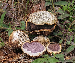
Scleroderma is a genus of fungi, commonly known as earth balls, now known to belong to the Boletales order, in suborder Sclerodermatineae. The best known species are S. citrinum and S. verrucosum. They are found worldwide. Various members of this genus are used as inoculation symbionts to colonize and promote the growth of tree seedlings in nurseries. They are not edible.

Funiculus is any cord-like structure in anatomy or biology, and may refer to:

<i>Cyathus striatus</i> Species of fungus

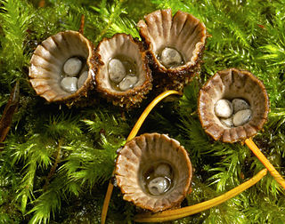
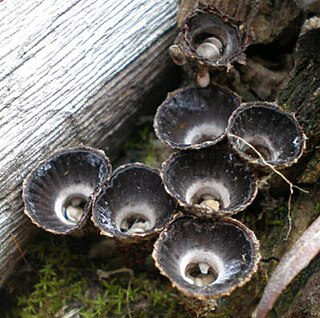
Cyathus striatus, commonly known as the fluted bird's nest, is a common saprobic bird's nest fungus with a widespread distribution throughout temperate regions of the world. This fungus resembles a miniature bird's nest with numerous tiny "eggs"; the eggs, or peridioles, are actually lens-shaped bodies that contain spores. C. striatus can be distinguished from most other bird's nest fungi by its hairy exterior and grooved inner walls. Although most frequently found growing on dead wood in open forests, it also grows on wood chip mulch in urban areas. The fruiting bodies are encountered from summer until early winter. The color and size of this species can vary somewhat, but they are typically less than a centimeter wide and tall, and grey or brown in color. Another common name given to C. striatus, splash cups, alludes to the method of spore dispersal: the sides of the cup are angled such that falling drops of water can dislodge the peridioles and eject them from the cup. The specific epithet is derived from the Latin stria, meaning "with fine ridges or grooves".

<i>Nidularia</i> Genus of fungi

Nidularia is a genus of nine species of fungi in the family Agaricaceae. Their fruit bodies resemble tiny egg-filled bird nests. The name comes from the Latin nidus meaning nest. The related genus Mycocalia was segregated from Nidularia in 1961 based on differences in the microscopic structure of the peridium.

<i>Nidula</i> Genus of fungi

Nidula is a genus of fungi in the family Agaricaceae. Their fruit bodies resemble tiny egg-filled birds' nests, from which they derive their common name "bird's nest fungi". Originally described in 1902, the genus differs from the related genera Cyathus and Crucibulum by the absence of a cord that attaches the eggs to the inside of the fruit body. The life cycle of this genus allows it to reproduce both sexually, with meiosis, and asexually via spores. Species in this genus produce a number of bioactive compounds, including 4-(p-hydroxyphenyl)-2-butanone, a major component of raspberry flavor and insect attractor used in pesticides.

Mycocalia is a genus of fungi in the family Nidulariaceae. Basidiocarps are minute and irregularly spherical. Each produces one or more peridioles which contain the spores and are released from the disintegrating fruit bodies at maturity. Species are usually found growing on herbaceous stems and other plant debris. The genus was originally described in 1961 by British mycologist J.T. Palmer and has a north temperate distribution.

<i>Cyathus</i> Genus of fungi in the Nidulariaceae

Cyathus is a genus of fungi in the Nidulariaceae, which is a family collectively known as the bird's nest fungi. They are given this name as they resemble tiny bird's nests filled with "eggs" – structures large enough to have been mistaken in the past for seeds. However, these are now known to be reproductive structures containing spores. The "eggs", or peridioles, are firmly attached to the inner surface of this fruit body by an elastic cord of mycelia known as a funiculus. The 45 species are widely distributed throughout the world and some are found in most countries, although a few exist in only one or two locales. Cyathus stercoreus is considered endangered in a number of European countries. Some species of Cyathus are also known as splash cups, which refers to the fact that falling raindrops can knock the peridioles out of the open-cup fruit body. The internal and external surfaces of this cup may be ridged longitudinally ; this is one example of a taxonomic characteristic that has traditionally served to distinguish between species.

<i>Cyathus olla</i> Species of fungus

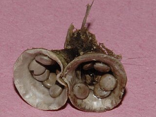
Cyathus olla also known as the field bird's nest is a species of saprobic fungus in the genus Cyathus of the family Nidulariaceae. The fruit bodies resemble tiny bird's nests filled with "eggs" – spore-containing structures called peridioles. Like other bird's nest fungi, C. olla relies on the force of falling water to dislodge peridioles from fruiting bodies to eject and disperse their spores. The life cycle of this fungus allows it to reproduce both sexually, with meiosis, and asexually via spores. C. olla is a relatively common fungus, with a worldwide distribution. It is the subject of agricultural research to determine its potential as a means to accelerate the breakdown of crop residue, and reduce the population of plant pathogens. The specific epithet is derived from the Latin word olla, meaning "pot".

<i>Cyathus stercoreus</i> Species of fungus

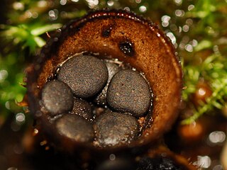
Cyathus stercoreus, commonly known as the dung-loving bird's nest or the dung bird's nest, is a species of fungus in the genus Cyathus, family Nidulariaceae. Like other species in the Nidulariaceae, the fruiting bodies of C. stercoreus resemble tiny bird's nests filled with eggs. The fruiting bodies are referred to as splash cups, because they are developed to use the force of falling drops of water to dislodge and disperse their spores. The species has a worldwide distribution, and prefers growing on dung, or soil containing dung; the specific epithet is derived from the Latin word stercorarius, meaning "of dung".

<i>Cyathus helenae</i> Species of fungus

Cyathus helenae or Helena's bird's nest is a species of fungus in the genus Cyathus, family Nidulariaceae. Like other members of the Nidulariaceae, C. helenae resembles a tiny bird's nest filled with 'eggs'—spore-containing structures known as peridioles. It was initially described by mycologist Harold Brodie in 1965, who found it growing on mountain scree in Alberta, Canada. C. helenae's life cycle allows it to reproduce both sexually and asexually. One of the smaller species of Cyathus, C. helenae produces a number of chemically unique diterpenoid molecules known as cyathins. The specific epithet of this species was given by Brodie in tribute to his late wife Helen.

<span class="mw-page-title-main">Coprophilous fungi</span> Fungi that grow on animal dung

Coprophilous fungi are a type of saprobic fungi that grow on animal dung. The hardy spores of coprophilous species are unwittingly consumed by herbivores from vegetation, and are excreted along with the plant matter. The fungi then flourish in the feces, before releasing their spores to the surrounding area.

Limnoperdon is a fungal genus in the monotypic family Limnoperdaceae. The genus is also monotypic, as it contains a single species, the aquatic fungus Limnoperdon incarnatum. The species, described as new to science in 1976, produces fruit bodies that lack specialized structures such as a stem, cap and gills common in mushrooms. Rather, the fruit bodies—described as aquatic or floating puffballs—are small balls of loosely interwoven hyphae. The balls float on the surface of the water above submerged twigs. Experimental observations on the development of the fruit body, based on the growth on the fungus in pure culture, suggest that a thin strand of mycelium tethers the ball above water while it matures. Fruit bodies start out as a tuft of hyphae, then become cup-shaped, and eventually enclose around a single chamber that contains reddish spores. Initially discovered in a marsh in the state of Washington, the fungus has since been collected in Japan, South Africa, and Canada.

<i>Sebacina</i> Genus of fungi

Sebacina is a genus of fungi in the family Sebacinaceae. Its species are mycorrhizal, forming a range of associations with trees and other plants. Basidiocarps are produced on soil and litter, sometimes partly encrusting stems of living plants. The fruit bodies are cartilaginous to rubbery-gelatinous and variously effused (corticioid) to coral-shaped (clavarioid). The genus has a cosmopolitan distribution.

<i>Geastrum quadrifidum</i> Species of fungus in the family Geastraceaea

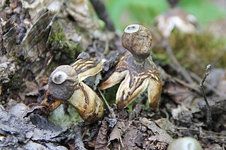
Geastrum quadrifidum, commonly known as the rayed earthstar or four-footed earthstar, is an inedible species of mushroom belonging to the genus Geastrum, or earthstar fungi. First described scientifically by Christian Hendrik Persoon in 1794, G. quadrifidum is a cosmopolitan—but not common—species of Europe, the Americas, Africa, Asia, and Australasia. The fungus is a saprobe, feeding off decomposing organic matter present in the soil and litter of coniferous forests.

<span class="mw-page-title-main">Nidulariaceae</span> Family of fungi

The Nidulariaceae are a family of fungi in the order Agaricales. Commonly known as the bird's nest fungi, their fruiting bodies resemble tiny egg-filled birds' nests. As they are saprobic, feeding on decomposing organic matter, they are often seen growing on decaying wood and in soils enriched with wood chips or bark mulch; they have a widespread distribution in most ecological regions. The five genera within the family, namely, Crucibulum, Cyathus, Mycocalia, Nidula, and Nidularia, are distinguished from each other by differences in morphology and peridiole structure; more recently, phylogenetic analysis and comparison of DNA sequences is guiding new decisions in the taxonomic organization of this family.

<i>Spongiforma</i> Genus of fungi

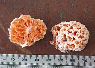
Spongiforma is a genus of sponge-like fungi in the family Boletaceae. Newly described in 2009, the genus contains two species: S. thailandica and S. squarepantsii. The type species S. thailandica is known only from Khao Yai National Park in central Thailand, where it grows in soil in old-growth forests dominated by dipterocarp trees. The rubbery fruit bodies, which has a strong odour of coal-tar similar to Tricholoma sulphureum, consists of numerous internal cavities lined with spore-producing tissue. S. squarepantsii, described as new to science in 2011, is found in Malaysia. It produces sponge-like, rubbery orange fruit bodies with a fruity or musky odour. These fruit bodies will—like a sponge—resume their original shape if water is squeezed out. The origin of the specific name derives from its perceived resemblance to the cartoon character SpongeBob SquarePants. Apart from differences in distribution, S. squarepantsii differs from S. thailandica in its colour, odour, and spore structure.

References

  1. 1 2 Kambly PE, Lee RE (1936). "The Gasteromycetes of Iowa". University of Iowa Studies in Natural History. 17 (4): 121–185.
  2. "The Nidulariaceae (MushroomExpert.Com)" . Retrieved 2009-01-04.
  3. Buller AH (1942). "The splash-cups of the birds-nest fungi, liverworts and mosses". Transactions of the Royal Society of Canada. 36 (5): 1–159.
  4. Brodie, p. 15.
  5. 1 2 3 Tulasne LR, Tulasne C (1844). "Recherches sur l'organisation et le mode de fructification des champignons de la tribu des Nidulariées, suivies d'un essai monographique". Annales des Sciences Naturelles. 3rd series (in French). 1: 41–107.
  6. White VS (1902). "The Nidulariaceae of North America". Bulletin of the Torrey Botanical Club. 29 (5): 251–280. doi:10.2307/2478721. JSTOR   2478721.
  7. 1 2 3 Lloyd CG (1906). "The Nidulariaceae". Mycological Writings. 2: 1–30.
  8. Cunningham GH (1924). "A revision of the New Zealand Nidulariales, or 'bird's-nest fungi'". Transactions of the New Zealand Institute. 55: 55–66.
  9. Brodie, The Bird's Nest Fungi.
  10. 1 2 3 4 5 Brodie, The Bird's Nest Fungi, p. 148.
  11. 1 2 3 Brodie, p. 149.
  12. 1 2 Ellis JB, Ellis MB (1990). Fungi without Gills (Hymenomycetes and Gasteromycetes): An Identification Handbook. London, UK: Chapman and Hall. p. 224. ISBN   0-412-36970-2.
  13. 1 2 3 Brodie, p. 147.
  14. 1 2 Martin GW (1927). "Basidia and spores of the Nidulariaceae". Mycologia. 19 (5): 239–247. doi:10.2307/3753710. JSTOR   3753710.
  15. Miller HR, Miller OK (1988). Gasteromycetes: Morphological and Developmental Features, with Keys to the Orders, Families, and Genera. Eureka, California: Mad River Press. p. 71. ISBN   0-916422-74-7.
  16. Brodie, p. 150.
  17. Alexopoulos CJ, Mims CW, Blackwell M (1996). Introductory Mycology. John Wiley and Sons. p. 545. ISBN   0-471-52229-5.
  18. Brodie, p. 129.
  19. Brodie, p. 86.
  20. Brodie, The Bird's Nest Fungi, pp. 88–89, 96–97.
  21. Brodie, The Bird's Nest Fungi, pp. 93–100.
  22. Schmidt O. (2006). Wood and Tree Fungi: Biology, Damage, Protection, and Use. Berlin: Springer. pp. 10–11. ISBN   3-540-32138-1.
  23. Deacon, pp. 231–234.
  24. Deacon, pp. 31–32.
  25. Sachs J. (1855). "Morphologie d. Crucibulum vulgare Tul". Botanische Zeitung. 13: 833–845, 849–861.
  26. DeBary A. (1866). Beiträge zur Morphologie und Physiologi der Pilze. Leipzig.
  27. Eidam E. (1876–7). "Keimung der Sporen und die Entdehung der Fruchtkörper bie den Nidularien". Cohn's Beiträge Biologie2: 221–45.
  28. Walker LB (1920). "Development of Cyathus fascicularis, C. striatus, and Crucibulum vulgare". Botanical Gazette. 70 (1): 1–24. doi: 10.1086/332706 .
  29. Mali RS, Babu KN (1998). "Naturally occurring prenylated phthalides: First total synthesis of salfredin B11". Journal of Chemical Research. 6 (6): 292–93. doi:10.1039/a707767j.
  30. 1 2 Neumann T, Schlegel B, Hoffmann P, Heinze S, Grafe U (1999). "Isolation and structure elucidation of new salfredin-type metabolites from Crucibulum laeve DSM 1653 and DSM 8519". Journal of Basic Microbiology. 39 (5–6): 357–363. doi:10.1002/(SICI)1521-4028(199912)39:5/6<357::AID-JOBM357>3.0.CO;2-8. S2CID   84363223.
  31. Matsumoto K, Nagashima K, Kamigauchi T, Kawamura Y, Yasuda Y, Ishii K, Uotani N, Sato T, Nakai H, Terui Y (1995). "Salfredins, new aldose reductase inhibitors produced by Crucibulum sp. RF-3817. I. Fermentation, isolation and structures of salfredins". Journal of Antibiotics. 48 (6): 439–446. doi: 10.7164/antibiotics.48.439 . PMID   7622427.
  32. Srivastava SK, Ramana KV, Bhatnagar A (2005). "Role of aldose reductase and oxidative damage in diabetes and the consequent potential for therapeutic options". Endocrine Reviews. 26 (3): 380–392. doi: 10.1210/er.2004-0028 . PMID   15814847.
  33. Kyselova Z, Stefek M, Bauer V (2004). "Pharmacological prevention of diabetic cataract". Journal of Diabetes Complications. 18 (2): 129–140. doi:10.1016/S1056-8727(03)00009-6. PMID   15120709.
  34. Healy RA, Huffman DR, Tiffany LH, Knaphaus G (2008). Mushrooms and Other Fungi of the Midcontinental United States . Bur Oak Guide. Iowa City: University of Iowa Press. p.  239. ISBN   978-1-58729-627-7.
  35. "Fungi of Australia - Crucibulum laeve". Archived from the original on 2008-08-21. Retrieved 2009-01-04.
  36. Hallgrimsson H, Jensson E, Kristinsson H (1992). "Three new gasteromycetes discovered in Iceland". Natturufraedingurinn. 61 (3–4): 219–227.
  37. 1 2 Brodie HJ (1970). "Crucibulum parvulum, a very small new bird's nest fungus from northwestern North America". Canadian Journal of Botany. 48 (5): 847–849. doi:10.1139/b70-116.
  38. Zhou TX, Zhao LZ, Zhao RL, Chen YH (2004). "Bird's nest fungi from China" (PDF). Fungal Diversity. 17 (17): 243–251.
  39. 1 2 Brodie HJ (1971). "Crucibulum cyathiforme a new species of bird's nest fungus from Colombia". Canadian Journal of Botany. 49 (11): 2009–2010. doi:10.1139/b71-281.
  40. Taslakhch'yan MG, Nanagyulyan SG (1989). "New Ascomycete and Basidiomycete species recorded for the Armenian SSR USSR". Biologicheskii Zhurnal Armenii (in Russian). 42 (12): 1081–1090.
  41. Brodie, p. 119.
  42. 1 2 "Standardized Common Names for Wild Species in Canada". National General Status Working Group. 2020.
  43. Rosanne AH, Huffman DR, Tiffany LH, Knaphaus G (2008). Mushrooms and Other Fungi of the Midcontinental United States (Bur Oak Guide). Iowa City: University of Iowa Press. ISBN   978-1-58729-627-7.
  44. Orr DB, Orr RT (1979). Mushrooms of Western North America. Berkeley: University of California Press. p. 118. ISBN   0-520-03656-5.

Cited texts