Cytoplasmic streaming

Last updated
Chloroplasts can be seen travelling around the central vacuole of a cell in Rhizomnium punctatum Rhizomnium punctatum lamina.jpeg
Chloroplasts can be seen travelling around the central vacuole of a cell in Rhizomnium punctatum
Cytoplasmic streaming in onion bulb scale epidermis cells

Cytoplasmic streaming, also called protoplasmic streaming and cyclosis, is the flow of the cytoplasm inside the cell, driven by forces from the cytoskeleton. [1] It is likely that its function is, at least in part, to speed up the transport of molecules and organelles around the cell. It is usually observed in large plant and animal cells, greater than approximately 0.1 mm[ vague ]. In smaller cells, the diffusion of molecules is more rapid, but diffusion slows as the size of the cell increases, so larger cells may need cytoplasmic streaming for efficient function. [1]

Contents

The green alga genus Chara possesses some very large cells, up to 10 cm in length, [2] and cytoplasmic streaming has been studied in these large cells. [3]

Cytoplasmic streaming is strongly dependent upon intracellular pH and temperature. It has been observed that the effect of temperature on cytoplasmic streaming created linear variance and dependence at different high temperatures in comparison to low temperatures. [4] This process is complicated, with temperature alterations in the system increasing its efficiency, with other factors such as the transport of ions across the membrane being simultaneously affected. This is due to cells homeostasis depending upon active transport which may be affected at some critical temperatures.

In plant cells, chloroplasts are transported within the cytoplasmic stream to optimize their exposure to light for photosynthesis. [5] This rate of motion is influenced by several factors including light intensity, temperature, and pH levels. [6] Cytoplasmic streaming is most efficient at a neutral pH and tends to decrease in efficiency under conditions of both low and high pH. [6]

Several methods exist to halt the flow of cytoplasm within cells. One approach involves the introduction of Lugol's iodine solution, which effectively immobilizes the cytoplasmic streaming.[ citation needed ] Alternatively, the compound Cytochalasin D, dissolved in dimethyl sulfoxide, can be employed to achieve a similar effect by disrupting the actin microfilaments responsible for facilitating cytoplasmic movement. [7]

Mechanism

What is clearly visible in plants cells which exhibit cytoplasmic streaming is the motion of the chloroplasts moving with the cytoplasmic flow. This motion results from fluid being entrained by moving motor molecules of the plant cell. [8] Myosin filaments connect cell organelles to actin filaments. These actin filaments are generally attached to the chloroplasts and/or membranes of plant cells. [8] As the myosin molecules "walk" along the actin filaments dragging the organelles with them, the cytoplasmic fluid becomes entrained and is pushed/pulled along. [8] Cytoplasmic flow rates can range between 1 and 100 micron/sec. [8] [9]

In Chara corallina

Chara corallina exhibits cyclic cytoplasmic flow around a large central vacuole. [8] The large central vacuole is one of the largest organelles in a plant cell and is generally used for storage. [10] In Chara coralina, cells can grow up to 10 cm long and 1 mm in diameter. [8] The diameter of the vacuole can occupy around 80% of the cell's diameter. [11] Thus for a 1 mm diameter cell, the vacuole can have a diameter of 0.8 mm, leaving only a path width of about 0.1 mm around the vacuole for cytoplasm to flow. The cytoplasm flows at a rate of 100 microns/sec, the fastest of all known cytoplasmic streaming phenomena. [8]

Characteristics

The flow of the cytoplasm in the cell of Chara corallina is belied by the "barber pole" movement of the chloroplasts. [8] Two sections of chloroplast flow are observed with the aid of a microscope. These sections are arranged helically along the longitudinal axis of the cell. [8] In one section, the chloroplasts move upward along one band of the helix, while in the other, the chloroplasts move downwardly. [8] The area between these sections are known as indifferent zones. Chloroplasts are never seen to cross these zones, [8] and as a result it was thought that cytoplasmic and vacuolar fluid flow are similarly restricted, but this is not true.

First, Kamiya and Kuroda, experimentally determined that cytoplasmic flow rate varies radially within the cell, a phenomenon not clearly depicted by the chloroplast movement. [12] Second, Raymond Goldstein and others developed a mathematical fluid model for the cytoplasmic flow which not only predicts the behavior noted by Kamiya and Kuroda, [8] but predicts the trajectories of cytoplasmic flow through indifferent zones. The Goldstein model ignores the vacuolar membrane, and simply assumes that shear forces are directly translated to the vacuolar fluid from the cytoplasm.

The Goldstein model predicts there is net flow toward one of the indifferent zones from the other. [8] This actually is suggested by the flow of the chloroplasts. At one indifferent zone, the section with the chloroplasts moving at a downward angle will be above the chloroplasts moving at an upward angle. This section is known as the minus different zone (IZ-). Here, if each direction is broken into components in the theta (horizontal) and z (vertical) directions, the sum of these components oppose each other in the z direction, and similarly diverges in theta direction. [8] The other indifferent zone has the upwardly angled chloroplast movement on top and is known as the positive indifferent zone (IZ+). Thus, while the z directional components oppose each other again, the theta components now converge. [8] The net effect of the forces is cytoplasmic/vacuolar flow moves from the minus indifferent zone to the positive indifferent zone. [8]

As stated, these directional components are suggested by chloroplast movement, but are not obvious. Further, the effect of this cytoplasmic/vacuolar flow from one indifferent zone to the other demonstrates that cytoplasmic particles do cross the indifferent zones even if the chloroplasts at the surface do not. Particles, as they rise in the cell, spiral around in a semicircular manner near the minus indifferent zone, cross one indifferent zone, and end up near a positive indifferent zone. [8]

Further experiments on the Characean cells support of the Goldstein model for vacuolar fluid flow. [11] However, due to the vacuolar membrane (which was ignored in the Goldstein model), the cytoplasmic flow follows a different flow pattern. Further, recent experiments have shown that the data collected by Kamiya and Kuroda which suggested a flat velocity profile in the cytoplasm are not fully accurate. [11] Kikuchi worked with Nitella flexillis cells, and found an exponential relationship between fluid flow velocity and distance from cell membrane. [11] Although this work is not on Characean cells, the flows between Nitella flexillis and Chara coralina are visually and structurally similar. [11]

Advantages

Enhanced nutrient transport

The Goldstein model predicts enhanced transport (over transport characterized by strictly longitudinal cytoplasmic flow) into the vacuolar cavity due to the complicated flow trajectories arising from the cytoplasmic streaming. [8] Although, a nutrient concentration gradient would result from longitudinally uniform concentrations and flows, the complicated flow trajectories predicted produce a larger concentration gradient across the vacuolar membrane. [8]

By Fick's laws of diffusion, it is known that larger concentration gradients lead to larger diffusive flows. [13] Thus, the unique flow trajectories of the cytoplasmic flow in Chara coralina lead to enhanced nutrient transport by diffusion into the storage vacuole. This allows for higher concentrations of nutrients inside the vacuole than would be allowed by strictly longitudinal cytoplasmic flows. Goldstein also demonstrated the faster the cytoplasmic flow along these trajectories, the larger the concentration gradient that arises, and the larger diffusive nutrient transport into the storage vacuole that occurs. The enhanced nutrient transport into the vacuole leads to striking differences in growth rate and overall growth size. [9]

Experiments have been performed in Arabidopsis thaliana . Wild type versions of this plant exhibit cytoplasmic streaming due to the entrainment of fluid similar to Chara coralina, only at slower flow rates. [9] One experiment removes the wild type myosin motor molecule from the plant and replaces it with a faster myosin molecule which moves along the actin filaments at 16 microns/sec. In another set of plants, the myosin molecule is replaced with the slower homo sapiens Vb myosin motor molecule. Human myosin Vb only moves at a rate of .19 microns/sec. Resulting cytoplasmic flows rates are 4.3 microns/sec for the wild type and 7.5 microns/sec for the plants implanted with the rapidly moving myosin protein.

The plants implanted with human myosin Vb do not exhibit continuous cytoplasmic streaming. The plants are then allowed to grow under similar conditions. Faster cytoplasmic rates produced larger plants with larger and more abundant leaves. [9] This suggests that the enhanced nutrient storage demonstrated by the Goldstein model allows for plants to grow larger and faster. [8] [9]

Increased photosynthetic activity

Photosynthesis converts light energy into chemical energy in the form of adenosine triphosphate (ATP). [14] This occurs in the chloroplasts of plants cells. Light photons interact with various intermembrane proteins of the cholorplast to accomplish this. However, these proteins can become saturated with photons, making them unable to function until the saturation is alleviated. This is known as the Kautsky effect and is a cause of inefficiency on the ATP production mechanism. Cytoplasmic streaming in Chara corallina, however, enables chloroplasts to move around the stem of the plant. Thus, the chloroplasts move into lighted regions and shaded regions. [14] This intermittent exposure to photons due to cytoplasmic streaming actually increases the photosynthetic efficiency of chloroplasts. [14] Photosynthetic activity is generally assessed using chlorophyll fluorescence analysis.

Gravisensing

Gravisensing is the ability to sense the gravitational force and react to it. Many plants use gravisensing to direct growth. For example, depending on root orientation, amyloplasts will settle within a plant cell differently. These different settling patterns cause the protein auxin to be distributed differently within the plant. This differences in the distribution pattern direct roots to grow downward or outward.

In most plants, gravisensing requires a coordinated multi-cellular effort, but in Chara corallina, one cell detects gravity and responds to it. [15] The barber pole chloroplast motion resulting from cytoplasmic streaming has one flow upward and another downward. [8] The downward motion of the chloroplasts moves a bit faster than the upward flow producing a ratio of speeds of 1.1. [8] [15] This ratio is known as the polar ratio and depends on the force of gravity. [15] This increase in speed is not a direct result of the force of gravity, but an indirect result. Gravity causes the plant protoplast to settle within the cell wall. Thus, the cell membrane is put into tension at the top, and into compression at the bottom. The resulting pressures on the membrane allow for gravisensing which result in the differing speeds of cytoplasmic flow observed in Chara coralina. This gravitational theory of gravisensing is directly opposed to the statolith theory exhibited by the settling of amyloplasts. [15]

Natural emergence

Cytoplasmic streaming occurs due to the motion of organelles attached to actin filaments via myosin motor proteins. [8] However, in Chara corallina, the organization of actin filaments is highly ordered. Actin is a polar molecule, which means that myosin only moves in one direction along the actin filament. [3] Thus, in Chara corallina, where motion of the chloroplasts and the myosin molecule follow a barber pole pattern, the actin filaments must all be similarly oriented within each section. [3] In other words, the section where the chloroplasts move upward will have all of the actin filaments oriented in the same upward direction, and the section where the chloroplasts move downward will have all the actin filaments oriented in the downward direction.

This organization emerges naturally from basic principles. With basic, realistic assumptions about the actin filament, Woodhouse demonstrated that the formation of two sets of actin filament orientations in a cylindrical cell is likely. His assumptions included a force keeping the actin filament in place once set down, an attractive force between filaments leading them to be more likely align as a filament already in place, and a repulsive force preventing alignment perpendicular to the length of the cylindrical cell. [3]

The first two assumptions derive from the molecular forces within the actin filament, while the last assumption was made due to the actin molecule's dislike of curvature. [3] Computer simulations run with these assumptions with varying parameters for the assumptive forces almost always leads to highly ordered actin organizations. [3] However, no order was as organized and consistent as the barber pole pattern found in nature, which suggests this mechanism plays role, but is not wholly responsible for the organization of actin filaments in Chara corallina.

Created by pressure gradients

Cytoplasmic streaming in some species is caused by pressure gradients along the length of the cell.

In Physarum polycephalum

Physarum polycephalum is a single-celled protist, belonging to a group of organisms informally referred to as 'slime molds'. Biological investigations into the myosin and actin molecules in this amoeboid have demonstrated striking physical and mechanistic similarities to human muscle myosin and actin molecules. Contraction and relaxation of these molecules leads to pressure gradients along the length of the cell. These contractions force cytoplasmic fluid in one direction and contributes to growth. [16] It has been demonstrated that while the molecules are similar to those in humans, the molecule blocking the binding site of myosin to actin is different. While, in humans, tropomyosin covers the site, only allowing contraction when calcium ions are present, in this amoeboid, a different molecule known as calmodulin blocks the site, allowing relaxation in the presence of high calcium ion levels. [16]

In Neurospora crassa

Neurospora crassa is a multicellular fungus with many off shooting hyphae. Cells can be up to 10 cm long, and are separated by a small septum. [17] Small holes in the septum allow cytoplasm and cytoplasmic contents to flow from cell to cell. Osmotic pressure gradients occur through the length of the cell to drive this cytoplasmic flow. Flows contribute to growth and the formation of cellular subcompartments. [17] [18]

Contribution to growth

Cytoplasmic flows created through osmotic pressure gradients flow longitudinally along the fungal hyphae and crash into the end causing growth. It has been demonstrated that the greater pressure at the hyphal tip corresponds to faster growth rates. Longer hyphae have greater pressure differences along their length allowing for faster cytoplasmic flow rates and larger pressures at the hyphal tip. [17] This is why longer hyphae grow faster than shorter ones. Tip growth increases as cytoplasmic flow rate increases over a 24-hour period until a max rate of 1 micron/second growth rate is observed. [17] Offshoots from the main hyphae are shorter and have slower cytoplasmic flow rates and correspondingly slower growth rates. [17]

Top: Idealized cytoplasmic flow in hyphae of Neurospora crassa. Bottom: Actual cytoplasmic flow in hyphae of Neurospora crassa. Microtubules (red) orient themselves perpendicular to flow as they exit the septal hole, deaccelerating the flow faster than the idealized case preventing the formation of eddies on the downstream side of the septum. Nuclei and other proteins aggregate on the upstream side which maintain the integrity of the septum. Microtubules Alters Idealized Pipeflow Preventing Eddy Formation on Downstream Side of Neurospora Crassa hypahe.jpg
Top: Idealized cytoplasmic flow in hyphae of Neurospora crassa. Bottom: Actual cytoplasmic flow in hyphae of Neurospora crassa. Microtubules (red) orient themselves perpendicular to flow as they exit the septal hole, deaccelerating the flow faster than the idealized case preventing the formation of eddies on the downstream side of the septum. Nuclei and other proteins aggregate on the upstream side which maintain the integrity of the septum.

Formation of cellular subcompartments

Cytoplasmic flow in Neurospora crassa carry microtubules. The presence of microtubules create interesting aspects to the flow. Modelling the fungal cells as a pipe separated at regular points with a septum with a hole in the center should produce very symmetrical flow. Basic fluid mechanics suggest that eddies should form both before and after each septum. [19] However, eddies only form before the septum in Neurospora crassa. This is because when microtubules enter the septal hole, they are arranged parallel to flow and contribute very little to flow characteristics, however, as the exit the septal hole, the orient themselves perpendicular to flow, slowing acceleration, and preventing eddy formation. [17] The eddies formed just before the septum allow for the formation of subcompartments where nuclei spotted with special proteins aggregate. [17] These proteins, one of which is called SPA-19, contribute to septum maintenance. Without it, the septum would degrade and the cell would leak large amounts of cytoplasm into the neighboring cell leading to cell death. [17]

In mouse oocytes

In many animal cells, centrioles and spindles keep nuclei centered within a cell for mitotic, meiotic, and other processes. Without such a centering mechanism, disease and death can result. While mouse oocytes do have centrioles, they play no role in nucleus positioning, yet, the nucleus of the oocyte maintains a central position. This is a result of cytoplasmic streaming. [20]

Microfilaments, independent of microtubules and myosin 2, form a mesh network throughout the cell. Nuclei, positioned in non-centered cell locations, have been demonstrated to migrate distances greater than 25 microns to the cell center. They will do this without going off course by more than 6 microns when the network is present. [20] This network of microfilaments has organelles bound to it by the myosin Vb molecule. [20] Cytoplasmic fluid is entrained by the motion of these organelles, however, no pattern of directionality is associated with the movement of the cytoplasm. In fact, the motion has been demonstrated to fulfill Brownian motion characteristics. For this reason, there is some debate as to whether this should be called cytoplasmic streaming.

Nonetheless, directional movement of organelles does result from this situation. Since the cytoplasm fills the cell, it is geometrically arranged into the shape of a sphere. As the radius of a sphere increases, surface area increases. Further, the motion in any given direction is proportional to the surface area. So thinking of the cell as a series of concentric spheres, it is clear that spheres with larger radii produce a greater amount of movement than spheres with smaller radii. Thus, the movement toward the center is greater than the movement away from the center, and net movement pushing the nucleus towards a central cellular location exists. In other words, the random motion of the cytoplasmic particles create a net force toward the center of the cell. [20]

Additionally, the increased motion with the cytoplasm reduces cytoplasmic viscosity allowing the nucleus to move more easily within the cell. These two factors of the cytoplasmic streaming center the nucleus in the oocyte cell. [20]

See also

Related Research Articles

<span class="mw-page-title-main">Cell (biology)</span> Basic unit of many life forms

The cell is the basic structural and functional unit of all forms of life. Every cell consists of cytoplasm enclosed within a membrane; many cells contain organelles, each with a specific function. The term comes from the Latin word cellula meaning 'small room'. Most cells are only visible under a microscope. Cells emerged on Earth about 4 billion years ago. All cells are capable of replication, protein synthesis, and motility.

<span class="mw-page-title-main">Cytoplasm</span> All of the contents of a eukaryotic cell except the nucleus

In cell biology, the cytoplasm describes all material within a eukaryotic cell, enclosed by the cell membrane, except for the cell nucleus. The material inside the nucleus and contained within the nuclear membrane is termed the nucleoplasm. The main components of the cytoplasm are the cytosol, the organelles, and various cytoplasmic inclusions. The cytoplasm is about 80% water and is usually colorless.

<span class="mw-page-title-main">Cytosol</span> Liquid found in cells

The cytosol, also known as cytoplasmic matrix or groundplasm, is one of the liquids found inside cells. It is separated into compartments by membranes. For example, the mitochondrial matrix separates the mitochondrion into many compartments.

<span class="mw-page-title-main">Endomembrane system</span> Membranes in the cytoplasm of a eukaryotic cell

The endomembrane system is composed of the different membranes (endomembranes) that are suspended in the cytoplasm within a eukaryotic cell. These membranes divide the cell into functional and structural compartments, or organelles. In eukaryotes the organelles of the endomembrane system include: the nuclear membrane, the endoplasmic reticulum, the Golgi apparatus, lysosomes, vesicles, endosomes, and plasma (cell) membrane among others. The system is defined more accurately as the set of membranes that forms a single functional and developmental unit, either being connected directly, or exchanging material through vesicle transport. Importantly, the endomembrane system does not include the membranes of plastids or mitochondria, but might have evolved partially from the actions of the latter.

<span class="mw-page-title-main">Vacuole</span> Membrane-bound organelle in cells containing fluid

A vacuole is a membrane-bound organelle which is present in plant and fungal cells and some protist, animal, and bacterial cells. Vacuoles are essentially enclosed compartments which are filled with water containing inorganic and organic molecules including enzymes in solution, though in certain cases they may contain solids which have been engulfed. Vacuoles are formed by the fusion of multiple membrane vesicles and are effectively just larger forms of these. The organelle has no basic shape or size; its structure varies according to the requirements of the cell.

<span class="mw-page-title-main">Cytoskeleton</span> Network of filamentous proteins that forms the internal framework of cells

The cytoskeleton is a complex, dynamic network of interlinking protein filaments present in the cytoplasm of all cells, including those of bacteria and archaea. In eukaryotes, it extends from the cell nucleus to the cell membrane and is composed of similar proteins in the various organisms. It is composed of three main components: microfilaments, intermediate filaments, and microtubules, and these are all capable of rapid growth or disassembly depending on the cell's requirements.

<span class="mw-page-title-main">Cleavage furrow</span> Plasma membrane invagination at the cell division site

In cell biology, the cleavage furrow is the indentation of the cell's surface that begins the progression of cleavage, by which animal and some algal cells undergo cytokinesis, the final splitting of the membrane, in the process of cell division. The same proteins responsible for muscle contraction, actin and myosin, begin the process of forming the cleavage furrow, creating an actomyosin ring. Other cytoskeletal proteins and actin binding proteins are involved in the procedure.

<span class="mw-page-title-main">Myofibril</span> Contractile element of muscle

A myofibril is a basic rod-like organelle of a muscle cell. Skeletal muscles are composed of long, tubular cells known as muscle fibers, and these cells contain many chains of myofibrils. Each myofibril has a diameter of 1–2 micrometres. They are created during embryonic development in a process known as myogenesis.

<span class="mw-page-title-main">Actin</span> Family of proteins

Actin is a family of globular multi-functional proteins that form microfilaments in the cytoskeleton, and the thin filaments in muscle fibrils. It is found in essentially all eukaryotic cells, where it may be present at a concentration of over 100 μM; its mass is roughly 42 kDa, with a diameter of 4 to 7 nm.

<span class="mw-page-title-main">Myosin</span> Superfamily of motor proteins

Myosins are a superfamily of motor proteins best known for their roles in muscle contraction and in a wide range of other motility processes in eukaryotes. They are ATP-dependent and responsible for actin-based motility.

<span class="mw-page-title-main">Plasmodesma</span> A pore connecting between adjacent plant cells

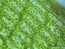
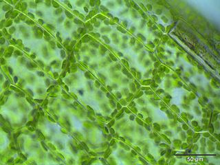
Plasmodesmata are microscopic channels which traverse the cell walls of plant cells and some algal cells, enabling transport and communication between them. Plasmodesmata evolved independently in several lineages, and species that have these structures include members of the Charophyceae, Charales, Coleochaetales and Phaeophyceae, as well as all embryophytes, better known as land plants. Unlike animal cells, almost every plant cell is surrounded by a polysaccharide cell wall. Neighbouring plant cells are therefore separated by a pair of cell walls and the intervening middle lamella, forming an extracellular domain known as the apoplast. Although cell walls are permeable to small soluble proteins and other solutes, plasmodesmata enable direct, regulated, symplastic transport of substances between cells. There are two forms of plasmodesmata: primary plasmodesmata, which are formed during cell division, and secondary plasmodesmata, which can form between mature cells.

<span class="mw-page-title-main">Contractile vacuole</span> Organelle used in regulating osmosis

A contractile vacuole (CV) is a sub-cellular structure (organelle) involved in osmoregulation. It is found predominantly in protists and in unicellular algae. It was previously known as pulsatile or pulsating vacuole.

A stromule is a microscopic structure found in plant cells. Stromules are highly dynamic structures extending from the surface of all plastid types, including proplastids, chloroplasts, etioplasts, leucoplasts, amyloplasts, and chromoplasts. Protrusions from and interconnections between plastids were observed in 1888 and 1908 and have been described sporadically in the literature since then. Stromules were recently rediscovered in 1997 and have since been reported to exist in a number of angiosperm species including Arabidopsis thaliana, wheat, rice and tomato, but their role is not yet fully understood.

<span class="mw-page-title-main">Motor protein</span> Class of molecular proteins

Motor proteins are a class of molecular motors that can move along the cytoskeleton of cells. They convert chemical energy into mechanical work by the hydrolysis of ATP. Flagellar rotation, however, is powered by a proton pump.

<span class="mw-page-title-main">Protein filament</span> Long chain of protein monomers

In biology, a protein filament is a long chain of protein monomers, such as those found in hair, muscle, or in flagella. Protein filaments form together to make the cytoskeleton of the cell. They are often bundled together to provide support, strength, and rigidity to the cell. When the filaments are packed up together, they are able to form three different cellular parts. The three major classes of protein filaments that make up the cytoskeleton include: actin filaments, microtubules and intermediate filaments.

<span class="mw-page-title-main">Outline of cell biology</span> Overview of and topical guide to cell biology

The following outline is provided as an overview of and topical guide to cell biology:

<span class="mw-page-title-main">Amoeboid movement</span> Mode of locomotion in eukaryotic cells

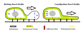
Amoeboid movement is the most typical mode of locomotion in adherent eukaryotic cells. It is a crawling-like type of movement accomplished by protrusion of cytoplasm of the cell involving the formation of pseudopodia ("false-feet") and posterior uropods. One or more pseudopodia may be produced at a time depending on the organism, but all amoeboid movement is characterized by the movement of organisms with an amorphous form that possess no set motility structures.

Force Spectrum Microscopy (FSM) is an application of active microrheology developed to measure aggregate random forces in the cytoplasm. Large, inert flow tracers are injected into live cells and become lodged inside the cytoskeletal mesh, wherein it is oscillated by repercussions from active motor proteins. The magnitude of these random forces can be inferred from the frequency of oscillation of tracer particles. Tracking the fluctuations of tracer particles using optical microscopy can isolate the contribution of active random forces to intracellular molecular transport from that of Brownian motion.

<span class="mw-page-title-main">Intracellular transport</span> Directed movement of vesicles and substances within a cell

Intracellular transport is the movement of vesicles and substances within a cell. Intracellular transport is required for maintaining homeostasis within the cell by responding to physiological signals. Proteins synthesized in the cytosol are distributed to their respective organelles, according to their specific amino acid’s sorting sequence. Eukaryotic cells transport packets of components to particular intracellular locations by attaching them to molecular motors that haul them along microtubules and actin filaments. Since intracellular transport heavily relies on microtubules for movement, the components of the cytoskeleton play a vital role in trafficking vesicles between organelles and the plasma membrane by providing mechanical support. Through this pathway, it is possible to facilitate the movement of essential molecules such as membrane‐bounded vesicles and organelles, mRNA, and chromosomes.

Cell mechanics is a sub-field of biophysics that focuses on the mechanical properties and behavior of living cells and how it relates to cell function. It encompasses aspects of cell biophysics, biomechanics, soft matter physics and rheology, mechanobiology and cell biology.

References

  1. 1 2 Goldstein RE, van de Meent JW (August 2015). "A physical perspective on cytoplasmic streaming". Interface Focus. 5 (4): 20150030. doi:10.1098/rsfs.2015.0030. PMC   4590424 . PMID   26464789.
  2. Beilby MJ, Casanova MT (2013-11-19). The Physiology of Characean Cells. Springer Science & Business Media. ISBN   978-3-642-40288-3.
  3. 1 2 3 4 5 6 Woodhouse FG, Goldstein RE (August 2013). "Cytoplasmic streaming in plant cells emerges naturally by microfilament self-organization". Proceedings of the National Academy of Sciences of the United States of America. 110 (35): 14132–7. arXiv: 1308.6422 . Bibcode:2013PNAS..11014132W. doi: 10.1073/pnas.1302736110 . PMC   3761564 . PMID   23940314.
  4. Shimmen T, Yokota E (February 2004). "Cytoplasmic streaming in plants". Current Opinion in Cell Biology. 16 (1): 68–72. doi:10.1016/j.ceb.2003.11.009. PMID   15037307.
  5. Bulychev AA, Dodonova SO (September 2011). "Effects of cyclosis on chloroplast-cytoplasm interactions revealed with localized lighting in Characean cells at rest and after electrical excitation". Biochimica et Biophysica Acta. 1807 (9): 1221–1230. doi:10.1016/j.bbabio.2011.06.009. PMID   21708122.
  6. 1 2 Goldstein RE, van de Meent JW (August 2015). "A physical perspective on cytoplasmic streaming". Interface Focus. 5 (4): 20150030. doi:10.1098/rsfs.2015.0030. PMC   4590424 . PMID   26464789.
  7. Foissner I, Wasteneys GO (March 1997). "A cytochalasin-sensitive actin filament meshwork is a prerequisite for local wound wall deposition in Nitella internodal cells". Protoplasma. 200 (1–2): 17–30. doi:10.1007/BF01280731. ISSN   0033-183X.
  8. 1 2 3 4 5 6 7 8 9 10 11 12 13 14 15 16 17 18 19 20 21 22 23 Goldstein RE, Tuval I, van de Meent JW (March 2008). "Microfluidics of cytoplasmic streaming and its implications for intracellular transport". Proceedings of the National Academy of Sciences of the United States of America. 105 (10): 3663–7. doi: 10.1073/pnas.0707223105 . PMC   2268784 . PMID   18310326.
  9. 1 2 3 4 5 Tominaga M, Kimura A, Yokota E, Haraguchi T, Shimmen T, Yamamoto K, et al. (November 2013). "Cytoplasmic streaming velocity as a plant size determinant". Developmental Cell. 27 (3): 345–52. doi: 10.1016/j.devcel.2013.10.005 . PMID   24229646.
  10. Cole L, Orlovich DA, Ashford AE (June 1998). "Structure, function, and motility of vacuoles in filamentous fungi". Fungal Genetics and Biology. 24 (1–2): 86–100. doi:10.1006/fgbi.1998.1051. PMID   9742195.
  11. 1 2 3 4 5 Kikuchi K, Mochizuki O (2015). "Diffusive Promotion by Velocity Gradient of Cytoplasmic Streaming (CPS) in Nitella Internodal Cells". PLOS ONE. 10 (12): e0144938. Bibcode:2015PLoSO..1044938K. doi: 10.1371/journal.pone.0144938 . PMC   4690613 . PMID   26694322.
  12. Kamiya N, Kuroda K (1956). "Velocity Distribution of the Protoplasmic Streaming in Nitella Cells". Shokubutsugaku Zasshi. 109 (822): 544–54. doi: 10.15281/jplantres1887.69.544 .
  13. Barrett C, Tetelman AS, Nix WD (1973). The Principles of Engineering Materials. Englewood Cliffs, NJ: Prentice-Hall. ISBN   978-0-137-09394-6.
  14. 1 2 3 Dodonova SO, Bulychev AA (2011). "Effect of Cytoplasmic Streaming on Photosynthetic Activity of Chloroplasts in Internodes of Chara Corallina". Russian Journal of Plant Physiology. 59: 35–41. doi:10.1134/S1021443711050050. S2CID   16387286.
  15. 1 2 3 4 Staves MP, Wayne R, Leopold AC (November 1997). "The effect of the external medium on the gravity-induced polarity of cytoplasmic streaming in Chara corallina (Characeae)". American Journal of Botany. 84 (11): 1516–1521. doi:10.2307/2446612. JSTOR   2446612. PMID   11541058. S2CID   15171541.
  16. 1 2 Yoshiyama S, Ishigami M, Nakamura A, Kohama K (December 2009). "Calcium wave for cytoplasmic streaming of Physarum polycephalum". Cell Biology International. 34 (1): 35–40. doi:10.1042/CBI20090158. PMID   19947949. S2CID   24802118.
  17. 1 2 3 4 5 6 7 8 Pieuchot L, Lai J, Loh RA, Leong FY, Chiam KH, Stajich J, et al. (August 2015). "Cellular Subcompartments through Cytoplasmic Streaming". Developmental Cell. 34 (4): 410–20. doi: 10.1016/j.devcel.2015.07.017 . PMID   26305593.
  18. Lew RR (June 2011). "How does a hypha grow? The biophysics of pressurized growth in fungi". Nature Reviews. Microbiology. 9 (7): 509–18. doi:10.1038/nrmicro2591. PMID   21643041. S2CID   6622913.
  19. White F (1986). Fluid Mechanics. New York, New York: McGraw Hill. ISBN   978-0-070-69673-0.
  20. 1 2 3 4 5 Almonacid M, Ahmed WW, Bussonnier M, Mailly P, Betz T, Voituriez R, et al. (April 2015). "Active diffusion positions the nucleus in mouse oocytes". Nature Cell Biology. 17 (4): 470–9. doi:10.1038/ncb3131. PMID   25774831. S2CID   10255963.

Sources