Dilated fundus examination

Last updated
Dilated fundus examination
Human eye in dim light.jpg
Dilated pupil of an eye, measuring 9 mm in diameter
Synonyms Dilated-pupil fundus examination
PurposeObtain a better view of the fundus of eye.

Dilated fundus examination (DFE) is a diagnostic procedure that uses mydriatic eye drops to dilate or enlarge the pupil in order to obtain a better view of the fundus of the eye. [1] Once the pupil is dilated, examiners use ophthalmoscopy to view the eye's interior, which makes it easier to assess the retina, optic nerve head, blood vessels, and other important features. DFE has been found to be a more effective method for evaluating eye health when compared to non-dilated examination, [2] [3] and is the best method of evaluating structures behind the iris. [4] It is frequently performed by ophthalmologists and optometrists as part of an eye examination. [5]

Contents

Examination

The most common agents used to dilate the pupil are phenylephrine (2.5% in pediatrics or 10% in adults) and tropicamide (0.5% or 1%). While phenylephrine stimulates receptors that contract the dilator muscle of the pupil, [6] tropicamide blocks stimulation of the pupillary sphincter muscle to allow for relaxation. [7] As the insertion of these drops may cause some stinging or burning, some practitioners choose to first insert a drop of topical anesthetic to numb the eye. [8] Pupil dilation typically begins within 15 minutes and may last for 3–8 hours. [6] [7]

After the pupil has been dilated, an ophthalmoscope can be used to examine the fundus. This allows for 15x magnification of the optic disc, also known as the optic nerve head, and retina to better evaluate for any ophthalmic pathology. [9] Evaluation of the optic disc may include noting the color of the disc, the sharpness of the disc outline and if any swelling is present, and signs of an enlarged or bulging optic cup, the central depression in the optic disc. Evaluation of the retina includes examining the arteries and veins for narrowing or arteriovenous nicking, as well as detecting any lesions, exudates, retinal hemorrhages, microaneurysms, or neovascularization. [9]

Following a dilated fundus examination, patients may experience blurred vision and light sensitivity until the effects of the mydriatic eye drops wear off; for this reason, the use of sunglasses is recommended to prevent damage to the eyes while dilated. [1]

Common conditions diagnosed during examination

Screening guidelines

Adults

The American Academy of Ophthalmology recommends comprehensive eye examinations, including dilated fundus examination, for asymptomatic patients without risk factors for eye disease at varying frequencies based on age: every 5–10 years in adults under 40 years, every 2–4 years in adults aged 40 to 54 years, every 1–3 years in adults aged 55 to 64 years, and every 1 to 2 years in adults aged 65 years and older. [4] However, routine DFE is relatively low yield for asymptomatic patients. [14]

In contrast, individuals with diabetes mellitus are at risk of diabetic retinopathy and therefore require more frequent screening. Those with type 1 diabetes should have their first comprehensive eye examination 5 years after diagnosis, followed by yearly exams. Those with type 2 diabetes should have their first comprehensive eye examination at the time of diagnosis, followed by yearly exams. Women with type 1 or type 2 diabetes who are planning a pregnancy should have a comprehensive eye examination prior to conception as well as early in the first trimester; women with gestational diabetes do not require screening for diabetic retinopathy during pregnancy. [4]

For individuals at risk of developing glaucoma, the American Academy of Ophthalmology recommends screening every 2–5 years in adults under 40 years, every 1–3 years in adults aged 40 to 54 years, every 1–2 years in adults aged 55 to 64 years, and every 1 to 2 years in adults aged 65 years and older. Screenings may be recommended even more frequently for individuals of African or Hispanic/Latino descent, as these ethnic groups have a further increased risk of developing glaucoma. [4]

Pediatrics

A child's first vision screening is typically done shortly after birth during the newborn exam, during which a pediatrician will perform simple vision testing such as assessing pupillary response and a red reflex. [15] However, if an infant is premature (gestational age under 32 weeks) or has a low birth weight (less than 1500 g) a dilated fundus examination is indicated due to risk of retinopathy of prematurity. [16]

A child will continue to be screened for vision issues, as well as normal eye alignment and movement, at regular intervals during their well child exams; if they fail a vision test or findings are inconclusive, the child can be referred for a more comprehensive eye examination including dilation. [15] A dilated fundus examination will allow for an ophthalmologist to calculate the refractive error more accurately than a non-dilated exam, as children tend to accommodate well; this allows for a more accurate prescription. [17]

Finally, children with certain medical conditions that place them at a higher risk for eye pathology (Down syndrome, juvenile idiopathic arthritis, neurofibromatosis) require comprehensive eye examinations with dilation. Similarly, children with a family history of amblyopia, strabismus, retinoblastoma, congenital cataracts or glaucoma may also require more frequent or comprehensive examinations. [15]

Contraindications

A dilated fundus examination is typically contraindicated in situations where mydriatic eye drops are contraindicated.

Absolute contraindications include: [8]

Relative contraindications include: [8] [9]

Related Research Articles

<span class="mw-page-title-main">Retinopathy</span> Medical condition

Retinopathy is any damage to the retina of the eyes, which may cause vision impairment. Retinopathy often refers to retinal vascular disease, or damage to the retina caused by abnormal blood flow. Age-related macular degeneration is technically included under the umbrella term retinopathy but is often discussed as a separate entity. Retinopathy, or retinal vascular disease, can be broadly categorized into proliferative and non-proliferative types. Frequently, retinopathy is an ocular manifestation of systemic disease as seen in diabetes or hypertension. Diabetes is the most common cause of retinopathy in the U.S. as of 2008. Diabetic retinopathy is the leading cause of blindness in working-aged people. It accounts for about 5% of blindness worldwide and is designated a priority eye disease by the World Health Organization.

<span class="mw-page-title-main">Diabetic retinopathy</span> Medical condition

Diabetic retinopathy, is a medical condition in which damage occurs to the retina due to diabetes mellitus. It is a leading cause of blindness in developed countries.

<span class="mw-page-title-main">Glaucoma</span> Group of eye diseases

Glaucoma is a group of eye diseases that lead to damage of the optic nerve, which transmits visual information from the eye to the brain. Glaucoma may cause vision loss if left untreated. It has been called the "silent thief of sight" because the loss of vision usually occurs slowly over a long period of time. A major risk factor for glaucoma is increased pressure within the eye, known as intraocular pressure (IOP). It is associated with old age, a family history of glaucoma, and certain medical conditions or medications. The word glaucoma comes from the Ancient Greek word γλαυκóς, meaning 'gleaming, blue-green, gray'.

<span class="mw-page-title-main">Mydriasis</span> Excessive dilation of the pupil

Mydriasis is the dilation of the pupil, usually having a non-physiological cause, or sometimes a physiological pupillary response. Non-physiological causes of mydriasis include disease, trauma, or the use of certain types of drug. It may also be of unknown cause.

This is a partial list of human eye diseases and disorders.

<span class="mw-page-title-main">Ophthalmoscopy</span> Part of an eye examination

Ophthalmoscopy, also called funduscopy, is a test that allows a health professional to see inside the fundus of the eye and other structures using an ophthalmoscope. It is done as part of an eye examination and may be done as part of a routine physical examination. It is crucial in determining the health of the retina, optic disc, and vitreous humor.

Optic neuropathy is damage to the optic nerve from any cause. The optic nerve is a bundle of millions of fibers in the retina that sends visual signals to the brain. [1].

The red reflex refers to the reddish-orange reflection of light from the back of the eye, or fundus, observed when using an ophthalmoscope or retinoscope. The reflex relies on the transparency of optical media and reflects off the fundus back through media into the aperture of the ophthalmoscope. The red reflex is considered abnormal if there is any asymmetry between the eyes, dark spots, or white reflex (Leukocoria).

Ocular ischemic syndrome is the constellation of ocular signs and symptoms secondary to severe, chronic arterial hypoperfusion to the eye. Amaurosis fugax is a form of acute vision loss caused by reduced blood flow to the eye; it may be a warning sign of an impending stroke, as both stroke and retinal artery occlusion can be caused by thromboembolism due to atherosclerosis elsewhere in the body. Consequently, those with transient blurring of vision are advised to urgently seek medical attention for a thorough evaluation of the carotid artery. Anterior segment ischemic syndrome is a similar ischemic condition of anterior segment usually seen in post-surgical cases. Retinal artery occlusion leads to rapid death of retinal cells, thereby resulting in severe loss of vision.

The swinging-flashlight test, also known as the swinging light test, is used in medical examinations to identify a relative afferent pupillary defect.

<span class="mw-page-title-main">Fundus photography</span> Medical imaging of the eyes

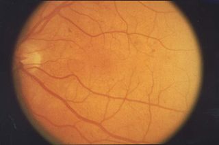
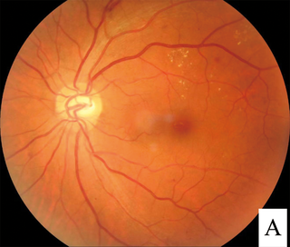
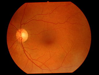
Fundus photography involves photographing the rear of an eye, also known as the fundus. Specialized fundus cameras consisting of an intricate microscope attached to a flash enabled camera are used in fundus photography. The main structures that can be visualized on a fundus photo are the central and peripheral retina, optic disc and macula. Fundus photography can be performed with colored filters, or with specialized dyes including fluorescein and indocyanine green.

<span class="mw-page-title-main">Intraocular hemorrhage</span> Medical condition

Intraocular hemorrhage is bleeding inside the eye. Bleeding can occur from any structure of the eye where there is vasculature or blood flow, including the anterior chamber, vitreous cavity, retina, choroid, suprachoroidal space, or optic disc.

<span class="mw-page-title-main">Intraretinal microvascular abnormalities</span> Medical condition

Intraretinal microvascular abnormalities (IRMA) are abnormalities of the blood vessels that supply the retina of the eye, a sign of diabetic retinopathy. IRMA can be difficult to distinguish from and is likely a precursor to retinal neovascularization. One way to distinguish IRMA from retinal neovascularization is to perform fluorescein angiography. Since IRMA blood vessels are patent, unlike neovascular vessels, they do not leak, and therefore exhibit hyperfluorescence on fluorescein angiography.

<span class="mw-page-title-main">Vitreous hemorrhage</span> Medical condition

Vitreous hemorrhage is the extravasation, or leakage, of blood into the areas in and around the vitreous humor of the eye. The vitreous humor is the clear gel that fills the space between the lens and the retina of the eye. A variety of conditions can result in blood leaking into the vitreous humor, which can cause impaired vision, floaters, and photopsia.

The eye is made up of the sclera, the iris, and the pupil, a black hole located at the center of the eye with the main function of allowing light to pass to the retina. Due to certain muscle spasms in the eye, the pupil can resemble a tadpole, which consists of a circular body, no arms or legs, and a tail.

<span class="mw-page-title-main">Teleophthalmology</span>

Teleophthalmology is a branch of telemedicine that delivers eye care through digital medical equipment and telecommunications technology. Today, applications of teleophthalmology encompass access to eye specialists for patients in remote areas, ophthalmic disease screening, diagnosis and monitoring; as well as distant learning.

Photostress recovery time (PSRT) is the time taken for visual acuity to return to normal levels after the retina has been bleached by a bright light source. Photostress recovery time measurement procedure is known as photostress test. Normal recovery time is about 15–30 seconds.

Sickle cell retinopathy can be defined as retinal changes due to blood vessel damage in the eye of a person with a background of sickle cell disease. It can likely progress to loss of vision in late stages due to vitreous hemorrhage or retinal detachment. Sickle cell disease is a structural red blood cell disorder leading to consequences in multiple systems. It is characterized by chronic red blood cell destruction, vascular injury, and tissue ischemia causing damage to the brain, eyes, heart, lungs, kidneys, spleen, and musculoskeletal system.

Diabetic papillopathy is an ocular complication of diabetes mellitus characterized by optic disc swelling and edema of optic nerve head. The condition may affect both type 1 and type 2 diabetic patients.

<span class="mw-page-title-main">Heidelberg Retinal Tomography</span>

The Heidelberg Retinal Tomography is a diagnostic procedure used in ophthalmology. The Heidelberg Retina Tomograph (HRT) is an ophthalmological confocal point scanning laser ophthalmoscope for examining the cornea and certain areas of the retina using different diagnostic modules. However, the most widely used area of application for HRT is the inspection of the optic nerve head (papilla) for early detection and follow-up of glaucoma. The procedure has established itself as an integral part of routine glaucoma diagnostics alongside the visual field examination (perimetry), the chamber angle examination (gonioscopy) and the measurement of intraocular pressure (tonometry). The HRT is the most widely used application of confocal scanning laser ophthalmoscopy.

References

  1. 1 2 "Get a Dilated Eye Exam". National Institute of Health: National Eye Institute. May 2021.
  2. Siegel, B. S.; Thompson, A. K.; Yolton, D. P.; Reinke, A. R.; Yolton, R. L. (January 1990). "A comparison of diagnostic outcomes with and without pupillary dilatation". Journal of the American Optometric Association. 61 (1): 25–34. ISSN   0003-0244. PMID   2319090.
  3. Parisi, M. L.; Scheiman, M.; Coulter, R. S. (May 1996). "Comparison of the effectiveness of a nondilated versus dilated fundus examination in the pediatric population". Journal of the American Optometric Association. 67 (5): 266–272. ISSN   0003-0244. PMID   8888844.
  4. 1 2 3 4 "Comprehensive Adult Medical Eye Evaluation PPP 2020". American Academy of Ophthalmology. 2020-11-13. Retrieved 2023-11-02.
  5. "Eye Health: Optometrist vs. Ophthalmologist" (PDF). American Diabetes Association. March 2022.
  6. 1 2 "Highlights of Prescribing Information" (PDF). FDA. January 2015.
  7. 1 2 "Label: Mydriacyl- tropicamide solution/drops". DailyMed - NIH National Library of Medicine. February 2023.
  8. 1 2 3 4 5 Orient, Jane (2018). "10: The Eye". Sapira's Art & Science of Bedside Diagnosis (5 ed.). Philadelphia: Wolters Kluwer. ISBN   9781975106089.
  9. 1 2 3 4 Bickley, Lynn; Szilagyi, Peter; Hoffman, Richard; Soriano, Rainier; Bates, Barbara (2023). "Chapter 12: Eyes". Bates' Guide To Physical Examination and History Taking (13 ed.). Philadelphia: Wolters Kluwer. ISBN   9781975210878.
  10. Pande, Gauri S; Tidake, Pravin (October 2022). "Laser Treatment Modalities for Diabetic Retinopathy". Cureus. 14 (10): e30024. doi: 10.7759/cureus.30024 . ISSN   2168-8184. PMC   9637280 . PMID   36348830.
  11. Tan, Colin S.; Lim, Louis W.; Ting, Dominic S. (January 2019). "The role of dilated fundus examination following cataract surgery". Journal of Cataract and Refractive Surgery. 45 (1): 113. doi: 10.1016/j.jcrs.2018.09.031 . ISSN   0886-3350.
  12. "Cataracts | National Eye Institute". National Institute of Health: National Eye Institute. August 2023.
  13. Thomas, Catherine J.; Mirza, Rukhsana G.; Gill, Manjot K. (2021-05-01). "Age-Related Macular Degeneration". Medical Clinics of North America. Ophthalmology. 105 (3): 473–491. doi: 10.1016/j.mcna.2021.01.003 . ISSN   0025-7125.
  14. Varner, Paul (2014). "How frequently should asymptomatic patients be dilated?". Journal of Optometry. 7 (1): 57–61. doi:10.1016/j.optom.2013.03.001. PMC   3938739 . PMID   24646902.
  15. 1 2 3 Gudgel, Dan (March 2021). "Eye Screening for Children". American Academy of Ophthalmology.
  16. Promelle, V.; Milazzo, S. (2017-05-01). "Rétinopathie du prématuré". Journal Français d'Ophtalmologie. 40 (5): 430–437. doi:10.1016/j.jfo.2016.12.013. ISSN   0181-5512.
  17. Saxena, Rohit; Sharma, Pradeep; Gopal, Santhan; Pediatric Ophthalmology Expert Group (February 2020). "National consensus statement regarding pediatric eye examination, refraction, and amblyopia management". Indian Journal of Ophthalmology. 68 (2): 325–332. doi: 10.4103/ijo.IJO_471_19 . ISSN   0301-4738. PMC   7003594 . PMID   31957721.
  18. Liew, Gerald; Mitchell, Paul; Wang, Jie Jin; Wong, Tien Yin (2006-01-07). "Fundoscopy: to dilate or not to dilate?". BMJ (Clinical Research Ed.). 332 (7532): 3. doi:10.1136/bmj.332.7532.3. ISSN   1756-1833. PMC   1325111 . PMID   16399709.