
A bile duct is any of a number of long tube-like structures that carry bile, and is present in most vertebrates. The bile duct is separated into three main parts: the fundus (superior), the body (middle), and the neck (inferior).

Isosporiasis, also known as cystoisosporiasis, is a human intestinal disease caused by the parasite Cystoisospora belli. It is found worldwide, especially in tropical and subtropical areas. Infection often occurs in immuno-compromised individuals, notably AIDS patients, and outbreaks have been reported in institutionalized groups in the United States. The first documented case was in 1915. It is usually spread indirectly, normally through contaminated food or water (CDC.gov).
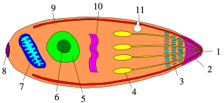
A trophozoite is the activated, feeding stage in the life cycle of certain protozoa such as malaria-causing Plasmodium falciparum and those of the Giardia group. The complementary form of the trophozoite state is the thick-walled cyst form. They are often different from the cyst stage, which is a protective, dormant form of the protozoa. Trophozoites are often found in the host's body fluids and tissues and in many cases, they are the form of the protozoan that causes disease in the host. In the protozoan, Entamoeba histolytica it invades the intestinal mucosa of its host, causing dysentery, which aid in the trophozoites traveling to the liver and leading to the production of hepatic abscesses.

Coccidia (Coccidiasina) are a subclass of microscopic, spore-forming, single-celled obligate intracellular parasites belonging to the apicomplexan class Conoidasida. As obligate intracellular parasites, they must live and reproduce within an animal cell. Coccidian parasites infect the intestinal tracts of animals, and are the largest group of apicomplexan protozoa.

Plasmodium vivax is a protozoal parasite and a human pathogen. This parasite is the most frequent and widely distributed cause of recurring malaria. Although it is less virulent than Plasmodium falciparum, the deadliest of the five human malaria parasites, P. vivax malaria infections can lead to severe disease and death, often due to splenomegaly. P. vivax is carried by the female Anopheles mosquito; the males do not bite.

Plasmodium malariae is a parasitic protozoan that causes malaria in humans. It is one of several species of Plasmodium parasites that infect other organisms as pathogens, also including Plasmodium falciparum and Plasmodium vivax, responsible for most malarial infection. Found worldwide, it causes a so-called "benign malaria", not nearly as dangerous as that produced by P. falciparum or P. vivax. The signs include fevers that recur at approximately three-day intervals – a quartan fever or quartan malaria – longer than the two-day (tertian) intervals of the other malarial parasite.

Eimeria tenella is a species of Eimeria that causes hemorrhagic cecal coccidiosis in young poultry. It is found worldwide.

Eimeria is a genus of apicomplexan parasites that includes various species capable of causing the disease coccidiosis in animals such as cattle, poultry and smaller ruminants including sheep and goats. Eimeria species are considered to be monoxenous because the life cycle is completed within a single host, and stenoxenous because they tend to be host specific, although a number of exceptions have been identified. Species of this genus infect a wide variety of hosts. Thirty-one species are known to occur in bats (Chiroptera), two in turtles, and 130 named species infect fish. Two species infect seals. Five species infect llamas and alpacas: E. alpacae, E. ivitaensis, E. lamae, E. macusaniensis, and E. punonensis. A number of species infect rodents, including E. couesii, E. kinsellai, E. palustris, E. ojastii and E. oryzomysi. Others infect poultry, rabbits and cattle. For full species list, see below.
Eimeria necatrix is a species of Eimeria that causes very severe intestinal coccidiosis in older poultry characterized by congestion, hemorrhage, necrosis of the intestine and bloody feces. Very large schizonts can be seen as white or yellow dots and oocysts can be found occasionally in the ceca.
Megaloschizonts are large schizonts that produce extremely high numbers of merozoites. They are found in various species of the Phylum Apicomplexa. The Apicomplexa phylum contains several parasitic protozoans. They have a very complex life cycle that includes several stages. Megaloschizonts and the smaller schizonts are the part of the life cycle that takes place inside the infected host organism and operates as an asexually reproductive cell. Megaloschizonts appear as grey-white nodules found in the smooth muscle of major organs, such as the heart, liver, lung or spleen.
Karyolysus is a genus of coccidia. With the exception of K. sonomae whose vertebrate host is the yellow-legged frog, species in this genus only infect lizards of the genus Lacerta.
Apicomplexans, a group of intracellular parasites, have life cycle stages that allow them to survive the wide variety of environments they are exposed to during their complex life cycle. Each stage in the life cycle of an apicomplexan organism is typified by a cellular variety with a distinct morphology and biochemistry.
Cytauxzoon is a genus of parasitic alveolates in the phylum Apicomplexa. The name is derived from the Greek meaning an increase in the number of cells in an animal.
Plasmodium coatneyi is a parasitic species that is an agent of malaria in nonhuman primates. P. coatneyi occurs in Southeast Asia. The natural host of this species is the rhesus macaque and crab-eating macaque, but there has been no evidence that zoonosis of P. coatneyi can occur through its vector, the female Anopheles mosquito.

Cystoisospora belli, previously known as Isospora belli, is a parasite that causes an intestinal disease known as cystoisosporiasis. This protozoan parasite is opportunistic in immune suppressed human hosts. It primarily exists in the epithelial cells of the small intestine, and develops in the cell cytoplasm. The distribution of this coccidian parasite is cosmopolitan, but is mainly found in tropical and subtropical areas of the world such as the Caribbean, Central and S. America, India, Africa, and S.E. Asia. In the U.S., it is usually associated with HIV infection and institutional living.
Cystoisospora canis, previously known as Isospora canis, is a microscopic, coccidian parasite that causes an intestinal tract infection in dogs. The intestinal tract infection is coccidiosis caused by a protozoa called coccidia.
Growell India is a multinational feed additive company headquartered in Pune, Maharashtra, India. Growell India manufactures and exports phytogenic feed additives for livestock. The company is a pioneer in phytogenic feed additives and has established a strong presence worldwide since its inception in 1995.

Sarcocystis neurona is primarily a neural parasite of horses and its management is of concern in veterinary medicine. The protozoan Sarcocystis neurona is a protozoan of single celled character and belongs to the family Sarcocystidae, in a group called coccidia. The protozoan, S. neurona, is a member of the genus Sarcocystis, and is most commonly associated with equine protozoal myeloencephalitis (EPM). The protozoan, S. neurona, can be easily cultivated and genetically manipulated, hence its common use as a model to study numerous aspects of cell biology.
Eimeria zuernii is a species of the parasite Eimeria that causes diarrheic disease known as eimeriosis in cattle, and mainly affects younger animals. The disease is also commonly referred to as coccidiosis. The parasite can be found in cattle around the globe.
Eimeria bovis is a parasite belonging to the genus Eimeria and is found globally. The pathogen can cause a diarrheic disease in cattle referred to as either eimeriosis or coccidiosis. The infection predominantly cause disease in younger animals.