Endoreduplication

Last updated

Endoreduplication (also referred to as endoreplication or endocycling) is replication of the nuclear genome in the absence of mitosis, which leads to elevated nuclear gene content and polyploidy. Endoreduplication can be understood simply as a variant form of the mitotic cell cycle (G1-S-G2-M) in which mitosis is circumvented entirely, due to modulation of cyclin-dependent kinase (CDK) activity. [1] [2] [3] [4] Examples of endoreduplication characterised in arthropod, mammalian, and plant species suggest that it is a universal developmental mechanism responsible for the differentiation and morphogenesis of cell types that fulfill an array of biological functions. [1] [2] While endoreduplication is often limited to specific cell types in animals, it is considerably more widespread in plants, such that polyploidy can be detected in the majority of plant tissues. [5] Polyploidy and aneuploidy are common phenomena in cancer cells. [6] Given that oncogenesis and endoreduplication likely involve subversion of common cell cycle regulatory mechanisms, a thorough understanding of endoreduplication may provide important insights for cancer biology.

Contents

Examples in nature

Endoreduplicating cell types that have been studied extensively in model organisms

OrganismNameCell typeBiological functionCitation
fly Drosophilia Melanogaster larval tissues (incl. salivary glands) secretion, embryogenesis [7]
fly ovarian follicle, nurse cells nourishment, protection of oocytes [8]
rodent megakaryocyte platelet formation [9]
rodent hepatocyte regeneration [10]
rodent trophoblast giant cell placental development, nourishment of embryo [11]
plant Arabidopsis Thaliana trichome defense from herbivory, homeostasis [12]
plant leaf epidermal cell leaf size, structure [13]
plant endosperm nourishment of embryo [14]
nematode Caenorhabditis elegans hypodermis secretion, body size [15]
nematode intestine unknown [16]

Endoreduplication, endomitosis and polytenization

Endoreduplication, endomitosis and polytenization are three different processes resulting in polyploidization of a cell in a regulated manner. In endoreduplication cells skip M phase completely by exiting the mitotic cell cycle in the G2 phase after completing the S phase several times, resulting in a mononucleated polyploid cell. The cell ends up with twice as many copies of each chromosome per repeat of the S phase. [17] Endomitosis is a type of cell cycle variation where mitosis is initiated, but stopped during anaphase and thus cytokinesis is not completed. The cell ends up with multiple nuclei in contrast to a cell undergoing endoreduplication. [17] [18] Therefore depending on how far the cell progresses through mitosis, this will give rise to a mononucleated or binucleated polyploid cell. Polytenization arises with under- or overamplification of some genomic regions, creating polytene chromosomes. [3] [4]

Endocycling vs. endomitosis Endocycling vs. endomitosis.png
Endocycling vs. endomitosis

Biological significance

Based on the wide array of cell types in which endoreduplication occurs, a variety of hypotheses have been generated to explain the functional importance of this phenomenon. [1] [2] Unfortunately, experimental evidence to support these conclusions is somewhat limited.

Cell differentiation

In developing plant tissues the transition from mitosis to endoreduplication often coincides with cell differentiation and morphogenesis. [19] However it remains to be determined whether endoreduplication and polyploidy contribute to cell differentiation or vice versa. Targeted inhibition of endoreduplication in trichome progenitors results in the production of multicellular trichomes that exhibit relatively normal morphology, but ultimately dedifferentiate and undergo absorption into the leaf epidermis. [20] This result suggests that endoreduplication and polyploidy may be required for the maintenance of cell identity.

Cell/organism size

Cell ploidy often correlates with cell size, [13] [15] and in some instances, disruption of endoreduplication results in diminished cell and tissue size [21] suggesting that endoreduplication may serve as a mechanism for tissue growth. Relative to mitosis, endoreduplication does not require cytoskeletal rearrangement or the production of new cell membrane and it often occurs in cells that have already differentiated. As such it may represent an energetically efficient alternative to cell proliferation among differentiated cell types that can no longer afford to undergo mitosis. [22] While evidence establishing a connection between ploidy and tissue size is prevalent in the literature, contrary examples also exist. [19]

Oogenesis and embryonic development

Endoreduplication is commonly observed in cells responsible for the nourishment and protection of oocytes and embryos. It has been suggested that increased gene copy number might allow for the mass production of proteins required to meet the metabolic demands of embryogenesis and early development. [1] Consistent with this notion, mutation of the Myc oncogene in Drosophila follicle cells results in reduced endoreduplication and abortive oogenesis. [23] However, reduction of endoreduplication in maize endosperm has limited effect on the accumulation of starch and storage proteins, suggesting that the nutritional requirements of the developing embryo may involve the nucleotides that comprise the polyploid genome rather than the proteins it encodes. [24]

Buffering the genome

Another hypothesis is that endoreduplication buffers against DNA damage and mutation because it provides extra copies of important genes. [1] However, this notion is purely speculative and there is limited evidence to the contrary. For example, analysis of polyploid yeast strains suggests that they are more sensitive to radiation than diploid strains. [25]

Stress response

Research in plants suggests that endoreduplication may also play a role in modulating stress responses. By manipulating expression of E2fe (a repressor of endocycling in plants), researchers were able to demonstrate that increased cell ploidy lessens the negative impact of drought stress on leaf size. [26] Given that the sessile lifestyle of plants necessitates a capacity to adapt to environmental conditions, it is appealing to speculate that widespread polyploidization contributes to their developmental plasticity

Genetic control of endoreplication

The best-studied example of a mitosis-to-endoreduplication transition occurs in Drosophila follicle cells and is activated by Notch signaling. [27] Entry into endoreduplication involves modulation of mitotic and S-phase cyclin-dependent kinase (CDK) activity. [28] Inhibition of M-phase CDK activity is accomplished via transcriptional activation of Cdh/fzr and repression of the G2-M regulator string/cdc25. [28] [29] Cdh/fzr is responsible for activation of the anaphase-promoting complex (APC) and subsequent proteolysis of the mitotic cyclins. String/cdc25 is a phosphatase that stimulates mitotic cyclin-CDK complex activity. Upregulation of S-phase CDK activity is accomplished via transcriptional repression of the inhibitory kinase dacapo. Together, these changes allow for the circumvention of mitotic entry, progression through G1, and entry into S-phase. The induction of endomitosis in mammalian megakaryocytes involves activation of the c-mpl receptor by the thrombopoietin (TPO) cytokine and is mediated by ERK1/2 signaling. [30] As with Drosophila follicle cells, endoreduplication in megakaryocytes results from activation of S-phase cyclin-CDK complexes and inhibition of mitotic cyclin-CDK activity. [31] [32]

Notch regulation of endocycling Notch regulation of endocycling.png
Notch regulation of endocycling

Entry into S-phase during endoreduplication (and mitosis) is regulated through the formation of a prereplicative complex (pre-RC) at replication origins, followed by recruitment and activation of the DNA replication machinery. In the context of endoreduplication these events are facilitated by an oscillation in cyclin E-Cdk2 activity. Cyclin E-Cdk2 activity drives the recruitment and activation of the replication machinery, [33] but it also inhibits pre-RC formation, [34] presumably to ensure that only one round of replication occurs per cycle. Failure to maintain control over pre-RC formation at replication origins results in a phenomenon known as “rereplication” which is common in cancer cells. [2] The mechanism by which cyclin E-Cdk2 inhibits pre-RC formation involves downregulation of APC-Cdh1-mediated proteolysis and accumulation of the protein Geminin, which is responsible for sequestration of the pre-RC component Cdt1. [35] [36]

Oscillations in Cyclin E-Cdk2 activity are modulated via transcriptional and post-transcriptional mechanisms. Expression of cyclin E is activated by E2F transcription factors that were shown to be required for endoreduplication. [37] [38] [39] Recent work suggests that observed oscillations in E2F and cyclin E protein levels result from a negative-feedback loop involving Cul4-dependent ubiquitination and degradation of E2F. [40] Post-transcriptional regulation of cyclin E-Cdk2 activity involves Ago/Fbw7-mediated proteolytic degradation of cyclin E [41] [42] and direct inhibition by factors such as Dacapo and p57. [43] [44]

Premeiotic endomitosis in unisexual vertebrates

The unisexual salamanders (genus Ambystoma ) are the oldest known unisexual vertebrate lineage, having arisen about 5 million years ago. [45] In these polyploid unisexual females, an extra premeiotic endomitotic replication of the genome, doubles the number of chromosomes. [46] As a result, the mature eggs that are produced subsequent to the two meiotic divisions have the same ploidy as the somatic cells of the adult female salamander. Synapsis and recombination during meiotic prophase I in these unisexual females is thought to ordinarily occur between identical sister chromosomes and occasionally between homologous chromosomes. Thus little, if any, genetic variation is produced. Recombination between homeologous chromosomes occurs rarely, if at all. [46]

Related Research Articles

<span class="mw-page-title-main">Cell cycle</span> Series of events and stages that result in cell division

The cell cycle, or cell-division cycle, is the series of events that take place in a cell that causes it to divide into two daughter cells. These events include the duplication of its DNA and some of its organelles, and subsequently the partitioning of its cytoplasm, chromosomes and other components into two daughter cells in a process called cell division.

<span class="mw-page-title-main">Mitosis</span> Process in which chromosomes are replicated and separated into two new identical nuclei

Mitosis is a part of the cell cycle in which replicated chromosomes are separated into two new nuclei. Cell division by mitosis is an equational division which gives rise to genetically identical cells in which the total number of chromosomes is maintained. Mitosis is preceded by the S phase of interphase and is followed by telophase and cytokinesis; which divides the cytoplasm, organelles and cell membrane of one cell into two new cells containing roughly equal shares of these cellular components. The different stages of mitosis altogether define the mitotic phase of a cell cycle—the division of the mother cell into two daughter cells genetically identical to each other.

<span class="mw-page-title-main">Ploidy</span> Number of sets of chromosomes in a cell

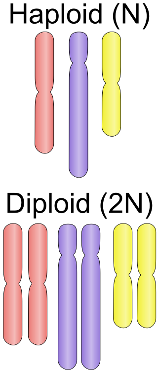
Ploidy is the number of complete sets of chromosomes in a cell, and hence the number of possible alleles for autosomal and pseudoautosomal genes. Sets of chromosomes refer to the number of maternal and paternal chromosome copies, respectively, in each homologous chromosome pair, which chromosomes naturally exist as. Somatic cells, tissues, and individual organisms can be described according to the number of sets of chromosomes present : monoploid, diploid, triploid, tetraploid, pentaploid, hexaploid, heptaploid or septaploid, etc. The generic term polyploid is often used to describe cells with three or more sets of chromosomes.

<span class="mw-page-title-main">Cell division</span> Process by which living cells divide

Cell division is the process by which a parent cell divides into two daughter cells. Cell division usually occurs as part of a larger cell cycle in which the cell grows and replicates its chromosome(s) before dividing. In eukaryotes, there are two distinct types of cell division: a vegetative division (mitosis), producing daughter cells genetically identical to the parent cell, and a cell division that produces haploid gametes for sexual reproduction (meiosis), reducing the number of chromosomes from two of each type in the diploid parent cell to one of each type in the daughter cells. Mitosis is a part of the cell cycle, in which, replicated chromosomes are separated into two new nuclei. Cell division gives rise to genetically identical cells in which the total number of chromosomes is maintained. In general, mitosis is preceded by the S stage of interphase and is followed by telophase and cytokinesis; which divides the cytoplasm, organelles, and cell membrane of one cell into two new cells containing roughly equal shares of these cellular components. The different stages of mitosis all together define the M phase of an animal cell cycle—the division of the mother cell into two genetically identical daughter cells. To ensure proper progression through the cell cycle, DNA damage is detected and repaired at various checkpoints throughout the cycle. These checkpoints can halt progression through the cell cycle by inhibiting certain cyclin-CDK complexes. Meiosis undergoes two divisions resulting in four haploid daughter cells. Homologous chromosomes are separated in the first division of meiosis, such that each daughter cell has one copy of each chromosome. These chromosomes have already been replicated and have two sister chromatids which are then separated during the second division of meiosis. Both of these cell division cycles are used in the process of sexual reproduction at some point in their life cycle. Both are believed to be present in the last eukaryotic common ancestor.

G<sub>1</sub> phase First growth phase in the eukaryotic cell cycle

The G1 phase, gap 1 phase, or growth 1 phase, is the first of four phases of the cell cycle that takes place in eukaryotic cell division. In this part of interphase, the cell synthesizes mRNA and proteins in preparation for subsequent steps leading to mitosis. G1 phase ends when the cell moves into the S phase of interphase. Around 30 to 40 percent of cell cycle time is spent in the G1 phase.

<span class="mw-page-title-main">Telophase</span> Final stage of a cell division for eukaryotic cells both in mitosis and meiosis

Telophase is the final stage in both meiosis and mitosis in a eukaryotic cell. During telophase, the effects of prophase and prometaphase are reversed. As chromosomes reach the cell poles, a nuclear envelope is re-assembled around each set of chromatids, the nucleoli reappear, and chromosomes begin to decondense back into the expanded chromatin that is present during interphase. The mitotic spindle is disassembled and remaining spindle microtubules are depolymerized. Telophase accounts for approximately 2% of the cell cycle's duration.

<span class="mw-page-title-main">Anaphase-promoting complex</span> Cell-cycle regulatory complex

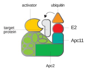
Anaphase-promoting complex is an E3 ubiquitin ligase that marks target cell cycle proteins for degradation by the 26S proteasome. The APC/C is a large complex of 11–13 subunit proteins, including a cullin (Apc2) and RING (Apc11) subunit much like SCF. Other parts of the APC/C have unknown functions but are highly conserved.

<span class="mw-page-title-main">Cyclin</span> Group of proteins

Cyclin is a family of proteins that controls the progression of a cell through the cell cycle by activating cyclin-dependent kinase (CDK) enzymes or group of enzymes required for synthesis of cell cycle.

<span class="mw-page-title-main">S phase</span> DNA replication phase of the cell cycle, between G1 and G2 phase

S phase (Synthesis phase) is the phase of the cell cycle in which DNA is replicated, occurring between G1 phase and G2 phase. Since accurate duplication of the genome is critical to successful cell division, the processes that occur during S-phase are tightly regulated and widely conserved.

G<sub>2</sub> phase Second growth phase in the eukaryotic cell cycle, prior to mitosis

G2 phase, Gap 2 phase, or Growth 2 phase, is the third subphase of interphase in the cell cycle directly preceding mitosis. It follows the successful completion of S phase, during which the cell’s DNA is replicated. G2 phase ends with the onset of prophase, the first phase of mitosis in which the cell’s chromatin condenses into chromosomes.

E2F is a group of genes that encodes a family of transcription factors (TF) in higher eukaryotes. Three of them are activators: E2F1, 2 and E2F3a. Six others act as suppressors: E2F3b, E2F4-8. All of them are involved in the cell cycle regulation and synthesis of DNA in mammalian cells. E2Fs as TFs bind to the TTTCCCGC consensus binding site in the target promoter sequence.

<span class="mw-page-title-main">Cell cycle checkpoint</span> Control mechanism in the eukaryotic cell cycle

Cell cycle checkpoints are control mechanisms in the eukaryotic cell cycle which ensure its proper progression. Each checkpoint serves as a potential termination point along the cell cycle, during which the conditions of the cell are assessed, with progression through the various phases of the cell cycle occurring only when favorable conditions are met. There are many checkpoints in the cell cycle, but the three major ones are: the G1 checkpoint, also known as the Start or restriction checkpoint or Major Checkpoint; the G2/M checkpoint; and the metaphase-to-anaphase transition, also known as the spindle checkpoint. Progression through these checkpoints is largely determined by the activation of cyclin-dependent kinases by regulatory protein subunits called cyclins, different forms of which are produced at each stage of the cell cycle to control the specific events that occur therein.

<span class="mw-page-title-main">G1/S transition</span> Stage in cell cycle

The G1/S transition is a stage in the cell cycle at the boundary between the G1 phase, in which the cell grows, and the S phase, during which DNA is replicated. It is governed by cell cycle checkpoints to ensure cell cycle integrity and the subsequent S phase can pause in response to improperly or partially replicated DNA. During this transition the cell makes decisions to become quiescent, differentiate, make DNA repairs, or proliferate based on environmental cues and molecular signaling inputs. The G1/S transition occurs late in G1 and the absence or improper application of this highly regulated checkpoint can lead to cellular transformation and disease states such as cancer.

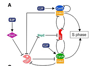
Cyclin A is a member of the cyclin family, a group of proteins that function in regulating progression through the cell cycle. The stages that a cell passes through that culminate in its division and replication are collectively known as the cell cycle Since the successful division and replication of a cell is essential for its survival, the cell cycle is tightly regulated by several components to ensure the efficient and error-free progression through the cell cycle. One such regulatory component is cyclin A which plays a role in the regulation of two different cell cycle stages.

<span class="mw-page-title-main">Cyclin D</span> Member of the cyclin protein family

Cyclin D is a member of the cyclin protein family that is involved in regulating cell cycle progression. The synthesis of cyclin D is initiated during G1 and drives the G1/S phase transition. Cyclin D protein is anywhere from 155 to 477 amino acids in length.

<span class="mw-page-title-main">Cyclin-dependent kinase 2</span> Protein-coding gene in the species Homo sapiens

Cyclin-dependent kinase 2, also known as cell division protein kinase 2, or Cdk2, is an enzyme that in humans is encoded by the CDK2 gene. The protein encoded by this gene is a member of the cyclin-dependent kinase family of Ser/Thr protein kinases. This protein kinase is highly similar to the gene products of S. cerevisiae cdc28, and S. pombe cdc2, also known as Cdk1 in humans. It is a catalytic subunit of the cyclin-dependent kinase complex, whose activity is restricted to the G1-S phase of the cell cycle, where cells make proteins necessary for mitosis and replicate their DNA. This protein associates with and is regulated by the regulatory subunits of the complex including cyclin E or A. Cyclin E binds G1 phase Cdk2, which is required for the transition from G1 to S phase while binding with Cyclin A is required to progress through the S phase. Its activity is also regulated by phosphorylation. Multiple alternatively spliced variants and multiple transcription initiation sites of this gene have been reported. The role of this protein in G1-S transition has been recently questioned as cells lacking Cdk2 are reported to have no problem during this transition.

<span class="mw-page-title-main">CDC25A</span> Protein-coding gene in the species Homo sapiens

M-phase inducer phosphatase 1 also known as dual specificity phosphatase Cdc25A is a protein that in humans is encoded by the cell division cycle 25 homolog A (CDC25A) gene.

<span class="mw-page-title-main">Cyclin A2</span> Protein-coding gene in the species Homo sapiens

Cyclin-A2 is a protein that in humans is encoded by the CCNA2 gene. It is one of the two types of cyclin A: cyclin A1 is expressed during meiosis and embryogenesis while cyclin A2 is expressed in the mitotic division of somatic cells.

A series of biochemical switches control transitions between and within the various phases of the cell cycle. The cell cycle is a series of complex, ordered, sequential events that control how a single cell divides into two cells, and involves several different phases. The phases include the G1 and G2 phases, DNA replication or S phase, and the actual process of cell division, mitosis or M phase. During the M phase, the chromosomes separate and cytokinesis occurs.

<span class="mw-page-title-main">DNA re-replication</span> Undesirable occurrence in eukaryotic cells

DNA re-replication is an undesirable and possibly fatal occurrence in eukaryotic cells in which the genome is replicated more than once per cell cycle. Rereplication is believed to lead to genomic instability and has been implicated in the pathologies of a variety of human cancers. To prevent rereplication, eukaryotic cells have evolved multiple, overlapping mechanisms to inhibit chromosomal DNA from being partially or fully rereplicated in a given cell cycle. These control mechanisms rely on cyclin-dependent kinase (CDK) activity. DNA replication control mechanisms cooperate to prevent the relicensing of replication origins and to activate cell cycle and DNA damage checkpoints. DNA rereplication must be strictly regulated to ensure that genomic information is faithfully transmitted through successive generations.

References

  1. 1 2 3 4 5 Edgar BA, Orr-Weaver TL (2001). "Endoreplication cell cycles: more for less". Cell. 105 (3): 297–306. doi: 10.1016/S0092-8674(01)00334-8 . PMID   11348589.
  2. 1 2 3 4 Lee HO, Davidson JM, Duronio RJ (2008). "Endoreplication: polyploidy with purpose". Genes & Development. 23 (21): 2461–77. doi:10.1101/gad.1829209. PMC   2779750 . PMID   19884253.
  3. 1 2 Edgar BA, Zielke N, Gutierrez C (2014-02-21). "Endocycles: a recurrent evolutionary innovation for post-mitotic cell growth". Nature Reviews Molecular Cell Biology. 15 (3): 197–210. doi:10.1038/nrm3756. ISSN   1471-0080. PMID   24556841. S2CID   641731.
  4. 1 2 Orr-Weaver TL (2015). "When bigger is better: the role of polyploidy in organogenesis". Trends in Genetics. 31 (6): 307–315. doi:10.1016/j.tig.2015.03.011. PMC   4537166 . PMID   25921783.
  5. Galbraith DW, Harkins KR, Knapp S (1991). "Systemic Endopolyploidy in Arabidopsis thaliana". Plant Physiology. 96 (3): 985–9. doi:10.1104/pp.96.3.985. PMC   1080875 . PMID   16668285.
  6. Storchova Z, Pellman D (2004). "From polyploidy to aneuploidy, genome instability and cancer". Nature Reviews Molecular Cell Biology. 5 (1): 45–54. doi:10.1038/nrm1276. PMID   14708009. S2CID   11985415.
  7. Hammond MP, Laird CD (1985). "Control of DNA replication and spatial distribution of defined DNA sequences in salivary gland cells of Drosophila melanogaster". Chromosoma. 91 (3–4): 279–286. doi:10.1007/BF00328223. PMID   3920018. S2CID   1515555.
  8. Hammond MP, Laird CD (1985). "Chromosome structure and DNA replication in nurse and follicle cells of Drosophila melanogaster". Chromosoma. 91 (3–4): 267–278. doi:10.1007/BF00328222. PMID   3920017. S2CID   7919061.
  9. Ravid K, Lu J, Zimmet JM, Jones MR (2002). "Roads to polyploidy: The megakaryocyte example". Journal of Cellular Physiology. 190 (1): 7–20. doi:10.1002/jcp.10035. PMID   11807806. S2CID   37297740.
  10. Wang M, Chen F, Lau JT, Hu Y (2017-05-18). "Hepatocyte polyploidization and its association with pathophysiological processes". Cell Death & Disease. 8 (5): e2805. doi:10.1038/cddis.2017.167. PMC   5520697 . PMID   28518148.
  11. Cross JC (2005). "How to make a placenta: Mechanisms of trophoblast cell differentiation in mice-a review". Placenta. 26: S3–9. doi:10.1016/j.placenta.2005.01.015. PMID   15837063.
  12. Hulskamp M, Schnittger A, Folkers U (1999). Pattern formation and cell differentiation: Trichomes in Arabidopsis as a genetic model system. International Review of Cytology. Vol. 186. pp. 147–178. doi:10.1016/S0074-7696(08)61053-0. ISBN   978-0-12-364590-6. PMID   9770299.
  13. 1 2 Melaragno JE, Mehrotra B, Coleman AW (1993). "Relationship between endopolyploidy and cell size in epidermal tissue of Arabidopsis". The Plant Cell. 5 (11): 1661–8. doi:10.1105/tpc.5.11.1661. JSTOR   3869747. PMC   160394 . PMID   12271050.
  14. Sabelli PA, Larkins BA (2009). "The Development of Endosperm in Grasses". Plant Physiology. 149 (1): 14–26. doi:10.1104/pp.108.129437. PMC   2613697 . PMID   19126691.
  15. 1 2 Flemming AJ, Shen Z, Cunha A, Emmons SW, Leroi AM (2000). "Somatic polyploidization and cellular proliferation drive body size evolution in nematodes". PNAS. 97 (10): 5285–90. Bibcode:2000PNAS...97.5285F. doi: 10.1073/pnas.97.10.5285 . PMC   25820 . PMID   10805788.
  16. Hedgecock EM, White JG (January 1985). "Polyploid tissues in the nematode Caenorhabditis elegans". Developmental Biology. 107 (1): 128–133. doi:10.1016/0012-1606(85)90381-1. ISSN   0012-1606. PMID   2578115.
  17. 1 2 Zielke N, Edgar BA, DePamphilis ML (2013-01-01). "Endoreplication". Cold Spring Harbor Perspectives in Biology. 5 (1): a012948. doi:10.1101/cshperspect.a012948. ISSN   1943-0264. PMC   3579398 . PMID   23284048.
  18. Shu Z, Row S, Deng W (June 2018). "Endoreplication: The Good, the Bad, and the Ugly". Trends in Cell Biology. 28 (6): 465–474. doi:10.1016/j.tcb.2018.02.006. ISSN   0962-8924. PMC   5962415 . PMID   29567370.
  19. 1 2 Inze D, De Veylder L (2006). "Cell cycle regulation in plant development". Annual Review of Genetics. 40: 77–105. doi:10.1146/annurev.genet.40.110405.090431. PMID   17094738.
  20. Bramsiepe J, Wester K, Weinl C, Roodbarkelari F, Kasili R, Larkin JC, Hulskamp M, Schnittger A (2010). Qu L (ed.). "Endoreplication Controls Cell Fate Maintenance". PLOS Genetics. 6 (6): e1000996. doi: 10.1371/journal.pgen.1000996 . PMC   2891705 . PMID   20585618.
  21. Lozano E, Saez AG, Flemming AJ, Cunha A, Leroi AM (2006). "Regulation of growth by ploidy in Caenorhabditis elegans". Current Biology. 16 (5): 493–8. Bibcode:2006CBio...16..493L. doi: 10.1016/j.cub.2006.01.048 . PMID   16527744.
  22. Kondorosi E, Roudier F, Gendreau E (2000). "Plant cell-size control: Growing by ploidy?". Current Opinion in Plant Biology. 3 (6): 488–492. Bibcode:2000COPB....3..488K. doi:10.1016/S1369-5266(00)00118-7. PMID   11074380.
  23. Maines JZ, Stevens LM, Tong X, Stein D (2004). "Drosophila dMyc is required for ovary cell growth and endoreplication". Development. 131 (4): 775–786. doi: 10.1242/dev.00932 . PMID   14724122.
  24. Leiva-Neto JT, Grafi G, Sabelli PA, Dante RA, Woo YM, Maddock S, Gordon-Kamm WJ, Larkins BA (2004). "A Dominant Negative Mutant of Cyclin-Dependent Kinase A Reduces Endoreduplication but Not Cell Size or Gene Expression in Maize Endosperm". The Plant Cell. 16 (7): 1854–69. doi:10.1105/tpc.022178. PMC   514166 . PMID   15208390.
  25. Mortimer RK (1958). "Radiobiological and genetic studies on a polyploid series (haploid to hexaploid) of Saccharomyces cerevisiae". Radiation Research. 9 (3): 312–326. Bibcode:1958RadR....9..312M. doi:10.2307/3570795. JSTOR   3570795. PMID   13579200. S2CID   37053611.
  26. Cookson SJ, Radziejwoski A, Granier C (2006). "Cell and leaf size plasticity in Arabidopsis: what is the role of endoreplication?". Plant, Cell and Environment. 29 (7): 1273–83. doi: 10.1111/j.1365-3040.2006.01506.x . PMID   17080949.
  27. Deng WM, Althauser C, Ruohala-Baker H (2001). "Notch-Delta signaling induces a transition from mitotic cell cycle to endocycle in Drosophila follicle cells". Development. 128 (23): 4737–46. doi:10.1242/dev.128.23.4737. PMID   11731454.
  28. 1 2 Shcherbata HR, Althauser C, Findley SD, Ruohola-Baker H (2004). "The mitotic-to-endocycle switch inDrosophila follicle cells is executed by Notch-dependent regulation of G1/S, G2/M and M/G1 cell-cycle transitions". Development. 131 (13): 3169–81. doi: 10.1242/dev.01172 . PMID   15175253.
  29. Schaeffer V, Althauser C, Shcherbata HR, Deng WM, Ruohola-Baker H (2004). "Notch-dependent Fizzy-related/Hec1/Cdh1 expression is required for the mitotic-to-endocycle transition in Drosophila follicle cells". Current Biology. 14 (7): 630–6. Bibcode:2004CBio...14..630S. doi:10.1016/j.cub.2004.03.040. hdl: 11858/00-001M-0000-002D-1B8D-3 . PMID   15062106. S2CID   18877076.
  30. Kaushansky K (2005). "The molecular mechanisms that control thrombopoiesis". The Journal of Clinical Investigation. 115 (12): 3339–47. doi:10.1172/JCI26674. PMC   1297257 . PMID   16322778.
  31. Garcia P, Cales C (1996). "Endoreplication in megakaryoblastic cell lines is accompanied by sustained expression of G1/S cyclins and downregulation of cdc25c". Oncogene. 13 (4): 695–703. PMID   8761290.
  32. Zhang Y, Wang Z, Ravid K (1996). "The cell cycle in polyploid megakaryocytes is associated with reduced activity of cyclin B1-dependent cdc2 kinase". Journal of Biological Chemistry. 271 (8): 4266–72. doi: 10.1074/jbc.271.8.4266 . PMID   8626773.
  33. Su TT, O'Farrell PH (1998). "Chromosome Association of Minichromosome Maintenance Proteins in Drosophila Endoreplication Cycles". Journal of Cell Biology. 140 (3): 451–460. doi:10.1083/jcb.140.3.451. PMC   2140170 . PMID   9456309.
  34. Arias EE, Walter JC (2004). "Strength in numbers: Preventing rereplication via multiple mechanisms in eukaryotic cells". Genes & Development. 21 (5): 497–518. doi: 10.1101/gad.1508907 . PMID   17344412.
  35. Narbonne-Reveau K, Senger S, Pal M, Herr A, Richardson HE, Asano M, Deak P, Lilly MA (2008). "APC/CFzr/Cdh1 promotes cell cycle progression during the Drosophila endocycle". Development. 135 (8): 1451–61. doi: 10.1242/dev.016295 . PMID   18321983.
  36. Zielke N, Querings S, Rottig C, Lehner C, Sprenger F (2008). "The anaphase-promoting complex/cyclosome (APC/C) is required for rereplication control in endoreplication cycles". Genes & Development. 22 (12): 1690–1703. doi:10.1101/gad.469108. PMC   2428065 . PMID   18559483.
  37. Duronio RJ, O'Farrell PH (1995). "Developmental control of the G1 to S transition in Drosophila: Cyclin E is a limiting downstream target of E2F". Genes & Development. 9 (12): 1456–68. doi: 10.1101/gad.9.12.1456 . PMID   7601350.
  38. Duronio RJ, O'Farrell PH, Xie JE, Brook A, Dyson N (1995). "The transcription factor E2F is required for S phase during Drosophila embryogenesis". Genes & Development. 9 (12): 1445–55. doi: 10.1101/gad.9.12.1445 . PMID   7601349.
  39. Duronio RJ, Bonnette PC, O'Farrell PH (1998). "Mutations of the Drosophila dDP, dE2F, and cyclin E Genes Reveal Distinct Roles for the E2F-DP Transcription Factor and Cyclin E during the G1-S Transition". Molecular and Cellular Biology. 18 (1): 141–151. doi:10.1128/MCB.18.1.141. PMC   121467 . PMID   9418862.
  40. Shibutani ST, de la Cruz AF, Tran V, Turbyfill WJ, Reis T, Edgar BA, Duronio RJ (2008). "Intrinsic negative cell cycle regulation provided by PIP box- and Cul4Cdt2-mediated destruction of E2f1 during S phase". Developmental Cell. 15 (6): 890–900. doi:10.1016/j.devcel.2008.10.003. PMC   2644461 . PMID   19081076.
  41. Koepp DM, Schaefer LK, Ye X, Keyomarsi K, Chu C, Harper JW, Elledge SJ (2001). "Phosphorylation-dependent ubiquitination of cyclin E by the SCFFbw7 ubiquitin ligase". Science. 294 (5540): 173–7. Bibcode:2001Sci...294..173K. doi: 10.1126/science.1065203 . PMID   11533444. S2CID   23404627.
  42. Moberg KH, Bell DW, Wahrer DC, Haber DA, Hariharan IK (2001). "Archipelago regulates cyclin E levels in Drosophila and is mutated in human cancer lines". Nature. 413 (6853): 311–6. doi:10.1038/35095068. PMID   11565033. S2CID   4372821.
  43. de Nooij JC, Graber KH, Hariharan IK (2001). "Expression of cyclin-dependent kinase inhibitor Dacapo is regulated by cyclin E". Mechanisms of Development. 97 (1–2): 73–83. doi: 10.1016/S0925-4773(00)00435-4 . PMID   11025208.
  44. Ullah Z, Kohn MJ, Yagi R, Vassilev LT, DePamphilis ML (2008). "Differentiation of trophoblast stem cells into giant cells is triggered by p57/Kip2 inhibition of CDK1 activity". Genes & Development. 22 (21): 3024–36. doi:10.1101/gad.1718108. PMC   2577795 . PMID   18981479.
  45. Bi K, Bogart JP (2010). "Time and time again: unisexual salamanders (genus Ambystoma) are the oldest unisexual vertebrates". BMC Evol. Biol. 10 (1): 238. Bibcode:2010BMCEE..10..238B. doi: 10.1186/1471-2148-10-238 . PMC   3020632 . PMID   20682056.
  46. 1 2 Bi K, Bogart JP (2010). "Probing the meiotic mechanism of intergenomic exchanges by genomic in situ hybridization on lampbrush chromosomes of unisexual Ambystoma (Amphibia: Caudata)". Chromosome Res. 18 (3): 371–82. doi:10.1007/s10577-010-9121-3. PMID   20358399. S2CID   2015354.