Related Research Articles

A cell wall is a structural layer surrounding some types of cells, just outside the cell membrane. It can be tough, flexible, and sometimes rigid. It provides the cell with both structural support and protection, and also acts as a filtering mechanism. Cell walls are absent in many eukaryotes, including animals, but are present in some other ones like fungi, algae and plants, and in most prokaryotes. A major function is to act as pressure vessels, preventing over-expansion of the cell when water enters.

Fibrils are structural biological materials found in nearly all living organisms. Not to be confused with fibers or filaments, fibrils tend to have diameters ranging from 10–100 nanometers. Fibrils are not usually found alone but rather are parts of greater hierarchical structures commonly found in biological systems. Due to the prevalence of fibrils in biological systems, their study is of great importance in the fields of microbiology, biomechanics, and materials science.
Profilin is an actin-binding protein involved in the dynamic turnover and reconstruction of the actin cytoskeleton. It is found in most eukaryotic organisms. Profilin is important for spatially and temporally controlled growth of actin microfilaments, which is an essential process in cellular locomotion and cell shape changes. This restructuring of the actin cytoskeleton is essential for processes such as organ development, wound healing, and the hunting down of infectious intruders by cells of the immune system.

Hexosaminidase is an enzyme involved in the hydrolysis of terminal N-acetyl-D-hexosamine residues in N-acetyl-β-D-hexosaminides.

The liver X receptor (LXR) is a member of the nuclear receptor family of transcription factors and is closely related to nuclear receptors such as the PPARs, FXR and RXR. Liver X receptors (LXRs) are important regulators of cholesterol, fatty acid, and glucose homeostasis. LXRs were earlier classified as orphan nuclear receptors, however, upon discovery of endogenous oxysterols as ligands they were subsequently deorphanized.
Xyloglucan is a hemicellulose that occurs in the primary cell wall of all vascular plants; however, all enzymes responsible for xyloglucan metabolism are found in Charophyceae algae. In many dicotyledonous plants, it is the most abundant hemicellulose in the primary cell wall. Xyloglucan binds to the surface of cellulose microfibrils and may link them together. It is the substrate of xyloglucan endotransglycosylase, which cuts and ligates xyloglucans, as a means of integrating new xyloglucans into the cell wall. It is also thought to be the substrate of alpha-expansin, which promotes cell wall enlargement.

Brassinolide is a plant hormone. The first isolated brassinosteroid, it was discovered when it was shown that pollen from rapeseed could promote stem elongation and cell division. The biologically active component was isolated and named brassinolide.
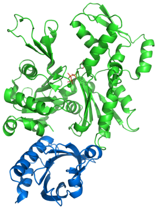
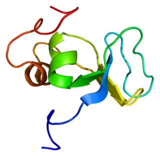
Acid growth refers to the ability of plant cells and plant cell walls to elongate or expand quickly at low (acidic) pH. The cell wall needs to be modified in order to maintain the turgor pressure. This modification is controlled by plant hormones like auxin. Auxin also controls the expression of some cell wall genes. This form of growth does not involve an increase in cell number. During acid growth, plant cells enlarge rapidly because the cell walls are made more extensible by expansin, a pH-dependent wall-loosening protein. Expansin loosens the network-like connections between cellulose microfibrils within the cell wall, which allows the cell volume to increase by turgor and osmosis. A typical sequence leading up to this would involve the introduction of a plant hormone (auxin, for example) that causes protons (H+ ions) to be pumped out of the cell into the cell wall. As a result, the cell wall solution becomes more acidic. It was suggested by different scientist that the epidermis is a unique target of the auxin but this theory has been disapproved over time. This activates expansin activity, causing the wall to become more extensible and to undergo wall stress relaxation, which enables the cell to take up water and to expand. The acid growth theory has been very controversial in the past.
Dystrobrevin is a protein that binds to dystrophin in the costamere of skeletal muscle cells. In humans, there are at least two isoforms of dystrobrevin, dystrobrevin alpha and dystrobrevin beta.

PDX1, also known as insulin promoter factor 1, is a transcription factor in the ParaHox gene cluster. In vertebrates, Pdx1 is necessary for pancreatic development, including β-cell maturation, and duodenal differentiation. In humans this protein is encoded by the PDX1 gene, which was formerly known as IPF1. The gene was originally identified in the clawed frog Xenopus laevis and is present widely across the evolutionary diversity of bilaterian animals, although it has been lost in evolution in arthropods and nematodes. Despite the gene name being Pdx1, there is no Pdx2 gene in most animals; single-copy Pdx1 orthologs have been identified in all mammals. Coelacanth and cartilaginous fish are, so far, the only vertebrates shown to have two Pdx genes, Pdx1 and Pdx2.

Filamin A, alpha (FLNA) is a protein that in humans is encoded by the FLNA gene.
The plant cell wall is made up of hydrated polymetric material, allowing it to have viscoelastic properties. The primary cell wall of a plant consists of cellulose fibers, hemicellulose, and xyloglucans. This load bearing network is also surrounded by pectins and glycoproteins.

The UDP-forming form of cellulose synthase is the main enzyme that produces cellulose. Systematically, it is known as UDP-glucose:(1→4)-β-D-glucan 4-β-D-glucosyltransferase in enzymology. It catalyzes the chemical reaction:
Latent-transforming growth factor beta-binding protein 1 is a protein that in humans is encoded by the LTBP1 gene.

Organic solute transporter alpha, also known as OST-alpha, is a protein which in humans is encoded by the SLC51A gene.

αE-catenin, also known as Catenin alpha-1 is a protein that in humans is encoded by the CTNNA1 gene. αE-catenin is highly expressed in cardiac muscle and localizes to adherens junctions at intercalated disc structures where it functions to mediate the anchorage of actin filaments to the sarcolemma. αE-catenin also plays a role in tumor metastasis and skin cell function.
Arabinogalactan-proteins (AGPs) are highly glycosylated proteins (glycoproteins) found in the cell walls of plants. Each one consists of a protein with sugar molecules attached. They are members of the wider class of hydroxyproline (Hyp)-rich cell wall glycoproteins, a large and diverse group of glycosylated wall proteins.

Endo-polygalacturonase (EC 3.2.1.15, pectin depolymerase, pectolase, pectin hydrolase, and poly-α-1,4-galacturonide glycanohydrolase; systematic name (1→4)-α-D-galacturonan glycanohydrolase (endo-cleaving)) is an enzyme that hydrolyzes the α-1,4 glycosidic bonds between galacturonic acid residues:
The acid-growth hypothesis is a theory that explains the expansion dynamics of cells and organs in plants. It was originally proposed by Achim Hager and Robert Cleland in 1971. They hypothesized that the naturally occurring plant hormone, auxin (indole-3-acetic acid, IAA), induces H+ proton extrusion into the apoplast. Such derived apoplastic acidification then activates a range of enzymatic reactions which modifies the extensibility of plant cell walls. Since its formulation in 1971, the hypothesis has stimulated much research and debate. Most debates have concerned the signalling role of auxin and the molecular nature of cell wall modification. The current version holds that auxin activates small auxin-up RNA (SAUR) proteins, which in turn regulate protein phosphatases that modulate proton-pump activity. Acid growth is responsible for short-term (seconds to minutes) variation in growth rate, but many other mechanisms influence longer-term growth.

Mei Hong is a Chinese-American biophysical chemist and professor of chemistry at the Massachusetts Institute of Technology. She is known for her creative development and application of solid-state nuclear magnetic resonance (ssNMR) spectroscopy to elucidate the structures and mechanisms of membrane proteins, plant cell walls, and amyloid proteins. She has received a number of recognitions for her work, including the American Chemical Society Nakanishi Prize in 2021, Günther Laukien Prize in 2014, the Protein Society Young Investigator award in 2012, and the American Chemical Society’s Pure Chemistry award in 2003.
References
- ↑ Cosgrove DJ (September 2000). "Loosening of plant cell walls by expansins" (PDF). Nature. 407 (6802): 321–6. Bibcode:2000Natur.407..321C. doi:10.1038/35030000. PMID 11014181. S2CID 4358466. Archived from the original (PDF) on 2006-09-08. Retrieved 2009-01-09.
- 1 2 McQueen-Mason S, Durachko DM, Cosgrove DJ (November 1992). "Two endogenous proteins that induce cell wall extension in plants". Plant Cell. 4 (11): 1425–33. doi:10.1105/tpc.4.11.1425. JSTOR 3869513. PMC 160229 . PMID 11538167.
- ↑ Cho HT, Kende H (September 1997). "Expression of expansin genes is correlated with growth in deepwater rice". Plant Cell. 9 (9): 1661–71. doi:10.1105/tpc.9.9.1661. PMC 157041 . PMID 9338967.
- ↑ Downes BP, Crowell DN (June 1998). "Cytokinin regulates the expression of a soybean β-expansin gene by a post-transcriptional mechanism" (PDF). Plant Mol. Biol. 37 (3): 437–44. doi:10.1023/A:1005920732211. PMID 9617811. S2CID 1815686.
- ↑ Cho HT, Cosgrove DJ (December 2002). "Regulation of root hair initiation and expansin gene expression in Arabidopsis". Plant Cell. 14 (12): 3237–53. doi:10.1105/tpc.006437. PMC 151215 . PMID 12468740.
- ↑ Sun Y, Veerabomma S, Abdel-Mageed HA, et al. (August 2005). "Brassinosteroid regulates fiber development on cultured cotton ovules". Plant Cell Physiol. 46 (8): 1384–91. doi: 10.1093/pcp/pci150 . PMID 15958497.
- 1 2 3 4 Cosgrove DJ, Bedinger P, Durachko DM (June 1997). "Group I allergens of grass pollen as cell wall-loosening agents". Proc. Natl. Acad. Sci. U.S.A. 94 (12): 6559–64. Bibcode:1997PNAS...94.6559C. doi: 10.1073/pnas.94.12.6559 . PMC 21089 . PMID 9177257.
- 1 2 3 Sampedro, J.; Cosgrove, D.J. (2005). "The expansin superfamily". Genome Biol. 6 (12): 242. doi: 10.1186/gb-2005-6-12-242 . PMC 1414085 . PMID 16356276.
- 1 2 3 Li Y, Darley CP, Ongaro V, et al. (March 2002). "Plant expansins are a complex multigene family with an ancient evolutionary origin". Plant Physiol. 128 (3): 854–64. doi:10.1104/pp.010658. PMC 152199 . PMID 11891242.
- ↑ Saloheimo M, Paloheimo M, Hakola S, et al. (September 2002). "Swollenin, a Trichoderma reesei protein with sequence similarity to the plant expansins, exhibits disruption activity on cellulosic materials". Eur. J. Biochem. 269 (17): 4202–11. doi:10.1046/j.1432-1033.2002.03095.x. PMID 12199698.
- ↑ Laine MJ, Haapalainen M, Wahlroos T, Kankare K, Nissinen R, Kassuwi S, Metzler MC (November 2000). "The cellulase encoded by the native plasmid of Clavibacter michiganensis ssp. sepedonicus plays a role in virulence and contains an expansin-like domain". Physiological and Molecular Plant Pathology. 57 (5): 221–233. doi:10.1006/pmpp.2000.0301.
- 1 2 Kerff F, Amoroso A, Herman R, et al. (November 2008). "Crystal structure and activity of Bacillus subtilis YoaJ (EXLX1), a bacterial expansin that promotes root colonization". Proc. Natl. Acad. Sci. U.S.A. 105 (44): 16876–81. Bibcode:2008PNAS..10516876K. doi: 10.1073/pnas.0809382105 . PMC 2579346 . PMID 18971341.
- ↑ Qin L, Kudla U, Roze EH, et al. (January 2004). "Plant degradation: a nematode expansin acting on plants". Nature. 427 (6969): 30. Bibcode:2004Natur.427...30Q. doi: 10.1038/427030a . PMID 14702076. S2CID 4414051.
- ↑ Kende H, Bradford K, Brummell D (May 2004). "Nomenclature for members of the expansin superfamily of genes and proteins". Plant Mol. Biol. 55 (3): 311–4. doi:10.1007/s11103-004-0158-6. hdl: 1874/19664 . PMID 15604683. S2CID 21108229.
- 1 2 McQueen-Mason SJ, Cosgrove DJ (January 1995). "Expansin mode of action on cell walls. Analysis of wall hydrolysis, stress relaxation, and binding". Plant Physiol. 107 (1): 87–100. doi:10.1104/pp.107.1.87. PMC 161171 . PMID 11536663.
- ↑ Rose JK, Lee HH, Bennett AB (May 1997). "Expression of a divergent expansin gene is fruit-specific and ripening-regulated". Proc. Natl. Acad. Sci. U.S.A. 94 (11): 5955–60. Bibcode:1997PNAS...94.5955R. doi: 10.1073/pnas.94.11.5955 . PMC 20888 . PMID 9159182.
- ↑ Chen F, Bradford KJ (November 2000). "Expression of an expansin is associated with endosperm weakening during tomato seed germination". Plant Physiol. 124 (3): 1265–74. doi:10.1104/pp.124.3.1265. PMC 59224 . PMID 11080302.
- 1 2 Lee Y, Choi D, Kende H (December 2001). "Expansins: ever-expanding numbers and functions". Curr. Opin. Plant Biol. 4 (6): 527–32. doi:10.1016/S1369-5266(00)00211-9. PMID 11641069.
- ↑ Cosgrove, D.J. (November 2005). "Growth of the plant cell wall" (PDF). Nat. Rev. Mol. Cell Biol. 6 (11): 850–61. doi:10.1038/nrm1746. PMID 16261190. S2CID 25196267. Archived from the original (PDF) on 2006-09-08. Retrieved 2009-01-10.
- ↑ Yennawar NH, Li LC, Dudzinski DM, Tabuchi A, Cosgrove DJ (October 2006). "Crystal structure and activities of EXPB1 (Zea m 1), a β-expansin and group-1 pollen allergen from maize". Proc. Natl. Acad. Sci. U.S.A. 103 (40): 14664–71. Bibcode:2006PNAS..10314664Y. doi: 10.1073/pnas.0605979103 . PMC 1595409 . PMID 16984999.
- ↑ Shani N, Shani Z, Shoseyov O, Mruwat R, Shoseyov D (January 2011). "Oxidized cellulose binding to allergens with a carbohydrate-binding module attenuates allergic reactions". J. Immunol. 186 (2): 1240–7. doi: 10.4049/jimmunol.1000640 . PMID 21169552.
- ↑ Valdivia ER, Wu Y, Li LC, Cosgrove DJ, Stephenson AG (2007). "A group-1 grass pollen allergen influences the outcome of pollen competition in maize". PLOS ONE. 2 (1): e154. Bibcode:2007PLoSO...2..154V. doi: 10.1371/journal.pone.0000154 . PMC 1764715 . PMID 17225858.