FADD

Last updated
FADD
FADD based on pdb file 2GF5.gif
Available structures
PDB Ortholog search: PDBe RCSB
Identifiers
Aliases FADD , GIG3, MORT1, Fas associated via death domain, IMD90
External IDs OMIM: 602457 MGI: 109324 HomoloGene: 2836 GeneCards: FADD
Orthologs
SpeciesHumanMouse
Entrez
Ensembl
UniProt
RefSeq (mRNA)

NM_003824

NM_010175

RefSeq (protein)

NP_003815

NP_034305

Location (UCSC) Chr 11: 70.2 – 70.21 Mb n/a
PubMed search [2] [3]
Wikidata
View/Edit Human View/Edit Mouse

FAS-associated death domain protein, also called MORT1, is encoded by the FADD gene on the 11q13.3 region of chromosome 11 in humans. [4]

Contents

FADD is an adaptor protein that bridges members of the tumor necrosis factor receptor superfamily, such as the Fas-receptor, to procaspases 8 and 10 to form the death-inducing signaling complex (DISC) during apoptosis. As well as its most well known role in apoptosis, FADD has also been seen to play a role in other processes including proliferation, cell cycle regulation and development.

Structure

FADD is a 23 kDa protein, made up of 208 amino acids. It contains two main domains: a C terminal death domain (DD) and an N terminal death effector domain (DED). Each domain, although sharing very little sequence similarity, are structurally similar to one another, with each consisting of 6 α helices. [5] [6] The DD of FADD binds to receptors such as the Fas receptor at the plasma membrane via their DD. [7] The interaction between the death domains are electrostatic interactions involving α helices 2 and 3 of the 6 helix domain. [8] The DED binds to the DED of intracellular molecules such as procaspase 8. [9] It is thought that this interaction occurs through hydrophobic interactions. [6]

Functions

Extrinsic apoptosis

Upon stimulation by the Fas ligand, the Fas receptor trimerises. Many receptors, including Fas, contain a cytoplasmic DD and are therefore named death receptors. FADD binds to the DD of this trimeric structure via its death domain [7] resulting in unmasking of FADD's DED and subsequent recruitment of procaspase 8 and 10 via an interaction between the DEDs of both FADD and the procaspases. [10] This generates a complex known as the death inducing signalling complex (DISC). [11] Procaspase 8 and 10 are known as initiator caspases. These are inactive molecules, but when bought into close proximity with other procaspases of the same type, autocatalytic cleavage occurs at an aspartate residue within their own structures, resulting in an activated protein. This activated protein can then go on to cleave and activate further caspases, initiating the caspase cascade. [12] The activated caspases can go on to cleave intracellular proteins such as inhibitor of caspase-activated DNase (ICAD), which ultimately leads to apoptosis of the cell. [13]

Binding of TRAIL to death receptors four and five (DR4 and DR5) can lead to apoptosis by the same mechanism. [14]

Apoptosis can also be triggered by binding of a ligand to tumor necrosis factor receptor 1 (TNFR1); however, the mechanism by which this occurs is slightly more complex. Another DD-containing adaptor protein named TRADD, along with other proteins, binds to activated TNF1R, forming what is known as complex I. This results in activation of the NFκB pathway, which promotes cell survival. This complex is then internalised, and FADD binds to TRADD via an interaction of the DD’s of the two adapter proteins, forming what is known as complex II. FADD again recruits procaspase 8, which initiates the caspase cascade leading to apoptosis. [15]

Extrinsic apoptosis pathway: The Fas receptor (FasR) is stimulated by Fas ligand (FasL), recruiting FADD to the FasR via an interaction between the death domains (DD) of both molecules. Procaspase 8 is recruited to FADD and interacts via the death effector domains (DED) of both molecules. This results in the cleavage and activation of procaspase 8, forming caspase 8, which goes on to cleave and activate other caspases such as procaspase 3 to initiate the caspase cascade which leads to cell death. Extrinsic apoptosis.jpg
Extrinsic apoptosis pathway: The Fas receptor (FasR) is stimulated by Fas ligand (FasL), recruiting FADD to the FasR via an interaction between the death domains (DD) of both molecules. Procaspase 8 is recruited to FADD and interacts via the death effector domains (DED) of both molecules. This results in the cleavage and activation of procaspase 8, forming caspase 8, which goes on to cleave and activate other caspases such as procaspase 3 to initiate the caspase cascade which leads to cell death.

Necroptosis

FADD also plays a role in regulating necroptosis, a process requiring the serine/threonine kinases, RIPK1 and RIPK3. Activated caspase 8 cleaves these kinases, inhibiting necroptosis. Since activation of caspase 8 requires FADD in order to bring the procaspase 8 molecules into close proximity to one another to facilitate their activation, FADD is required for negatively regulating necroptosis. In accordance, cells deficient in FADD induce necroptosis as they are unable to recruit and activate procaspase 8. FADD can also bind to RIPK1 and RIPK3 directly, however the significance of this interaction is currently unclear. [13]

Autophagic cell death

Autophagy is a process which allows cell survival under stressed conditions but can also lead to cell death.

Using its DD, FADD interacts with ATG5, a protein involved in autophagy. This interaction has been shown to be essential for autophagic cell death, which is induced by IFN-γ. [16]

In contrast, it has also been found to inhibit autophagic cell death and therefore promote cell survival. FADD binds to ATG5 in a complex which also contains ATG12, Caspase 8 and RIPK1. The formation of this complex is stimulated by autophagic signalling. Caspase 8 then cleaves RIPK1, leading to inhibition of this signalling, inhibiting cell death. [17]

Development

FADD knockout in mouse embryos is lethal, showing a role for FADD in embryonic development. This is thought to be due to abnormal development of the heart. [18] This abnormal heart development may be due to FADD dependent regulation of the NFκB pathway. [19]

FADD also plays a role in the development of the eyes of zebrafish. [20]

Cell cycle regulation

FADD is thought to have a role in regulating the cell cycle of T lymphocytes. This regulation is dependent on phosphorylation of FADD on Serine 194, which is carried out by Casein Kinase 1a (CKIα). This phosphorylated form of FADD is found mainly in the nucleus and the abundance of phosphorylated FADD increases significantly in the G2 phase of the cell cycle compared to the G1 phase where only very little can be detected. As it is found at the mitotic spindle during G2, it has been proposed to mediate the G2/M transition, however, the mechanism by which it does this it not yet known. [21]

Lymphocyte proliferation

FADD is essential for T cell proliferation when the T cell receptor is stimulated by antigen. [22] In contrast, FADD has no effect on the proliferation of B cells induced by stimulation of the B cell receptor. However, it is required for B cell proliferation induced by stimulation of TLR3 and TLR4. [23]

Inflammation

Activation of nuclear factor kappa B (NFκB) signalling leads to transcription of various proinflammatory cytokines as well as anti-apoptotic genes. It was found that NFκB signalling was inhibited in FADD-deficient cells after stimulation of the TNF-R1 or Fas receptors. This suggests a role of FADD in activation of the NFκB pathway. Conversely, FADD also has a role in inhibition of this pathway. Normally, upon stimulation of the receptors TL4 or IL-1R1, the adaptor protein, MyD88, is recruited to the plasma membrane where is binds to IL-1 receptor associated Kinase (IRAK) via a DD-DD interaction. This activates a signalling pathway which results in translocation of NFκB to the nucleus, where it induces the transcription of the inflammatory cytokines. FADD can interfere with the interaction between MyD88 and IRAK, by binding to MyD88 via its DD and therefore this disrupts the cascade which would lead to NFκB translocation and inflammation. [24] [25]

Other

FADD is required for an efficient antiviral response. Upon viral infection, FADD is needed to increase the levels of Irf7 a molecule which is needed for the production of IFN-α. IFN-α is a key molecule involved in the response against viruses. [26]

FADD is involved in the activation of the phosphatases which dephosphorylate and deactivate Protein Kinase C (PKC). Without FADD, PKC remains active and is able to continue signalling cascades leading to processes including cytoskeletal rearrangements and cell motility. [27]

Recent research has also shown that it may have a role in regulating glucose levels and the phosphorylated form of FADD is important for this function. [28]

Regulation

Regulation of FADD by MKRN1: MKRN1 ubiquitinylates FADD targeting it for degradation by the 26S proteosome. As it is degraded, FADD can no longer bind to the Fas receptor (Fas R) to induce apoptosis. Regulation of FADD by MKRN1.png
Regulation of FADD by MKRN1: MKRN1 ubiquitinylates FADD targeting it for degradation by the 26S proteosome. As it is degraded, FADD can no longer bind to the Fas receptor (Fas R) to induce apoptosis.

Subcellular localisation

FADD can be found in both the nucleus and cytoplasm of cells. Phosphorylation of Ser194 of FADD in humans (or Ser191 in mice) is thought to regulate its subcellular localisation. A nuclear localization sequence and nuclear export signal, both located in the DED of FADD, are also required for it to enter and exit the nucleus. Depending on its subcellular localisation, FADD can have different roles. In the cytoplasm, its main function is to induce apoptosis. However, in the nucleus, it can have the opposite effect and instead promote survival. [25] [29]

c-FLIP

Cellular FLICE inhibitory protein (c-FLIP) is a regulatory protein which contains two DEDs. There are two isoforms of C-FLIP: C-FLIPS and FLIPL. It was originally thought to act as a negative regulator of apoptosis by binding to the DED of FADD and therefore preventing procaspase 8 from binding and inhibiting formation of the DISC. [30] However, it has been seen that both c-FLIP and procaspase 8 can be found at the same DISC. [31] Therefore, it has been proposed that the presence of c-FLIP inhibits the close interaction of the procaspases to one another. Without this close proximity, the procaspases cannot be completely cleaved and remain in an inactive state. [30]

PKC

The activity of protein kinase C has a negative effect on Fas receptor mediated apoptosis. This is because it inhibits the recruitment of FADD to the receptor and so a DISC is not formed. It has been shown that by either increasing or decreasing the amount of PKC in T cells, more or less FADD is recruited to FasR respectively, when the FasR is stimulated. [32]

MKRN1

MKRN1 is an E3 ubiquitin ligase which negatively regulates FADD by targeting it for ubiquitin mediated degradation. In doing so, MKRN1 is able to control the level of apoptosis. [33]

Roles in inflammatory diseases

Increased levels of FADD were found in the leukocytes of patients with relapsing remitting multiple sclerosis, contributing to inflammation. [34] In rheumatoid arthritis, it is thought that stimulation of Fas receptors on macrophages, leads to formation of the FADD containing DISCs. Formation of these sequesters FADD away from MyD88 allowing MyD88 to interact with IRAK and induce the enhanced inflammation associated with this disease. [35]

Roles in cancer

As FADD has such an important role in apoptosis, loss of FADD can give cancer cells a proliferative advantage as apoptosis would no longer be induced when the Fas receptors are stimulated. [25]

However, there is significant upregulation of FADD in ovarian cancer [36] and head and neck squamous cell carcinoma. It is not yet clear what advantage this has on the cancer cells, but given FADDs roles in cell cycle regulation and cell survival, it likely that it may be related to this. [37] There are also elevated levels of FADD in non small cell lung cancer. FADD can be used as a prognosis marker for both of these diseases, with high levels of FADD being correlated with poor outcome. [38]

Therapeutic target

Taxol is a drug used in anticancer therapies due to its ability to interfere with microtubule assembly, which leads to cell cycle arrest. FADD phosphorylated at Ser194 makes cells more sensitive to cell cycle arrest induced by taxol. [21] Taxol can also cause apoptosis of cells and this requires procaspase 10, which is activated by recruitment to FADD. [39]

It has been shown that the activation of JNK leads to the phosphorylation of FADD. Phosphorylated FADD can induce G2/M cell cycle arrest, potentially by increasing the stability of p53. Therefore, drugs which can activate this pathway may have a therapeutic potential. [40] However, high levels of phosphorylated FADD have been correlated with a poor prognosis in many cancers such as that of the head and neck. This is likely to be due to its activation of the NF-κB pathway, which is antiapoptotic. Therefore, inhibition of FADD phosphorylation may be developed as a potential anti cancer strategy. [41] For example, It has been suggested that inhibition of FADD might work as a potential targeted therapy for drug-resistant ovarian cancer. [36]

Interactions

FADD has been seen to interact with Fas receptor,: [7]

See also

Related Research Articles

<span class="mw-page-title-main">Fas ligand</span> Protein-coding gene in the species Homo sapiens

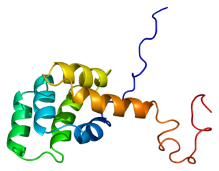
Fas ligand is a type-II transmembrane protein expressed on cytotoxic T lymphocytes and natural killer (NK) cells. Its binding with Fas receptor (FasR) induces programmed cell death in the FasR-carrying target cell. Fas ligand/receptor interactions play an important role in the regulation of the immune system and the progression of cancer.

<span class="mw-page-title-main">Death effector domain</span> InterPro Domain

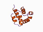
The death-effector domain (DED) is a protein interaction domain found only in eukaryotes that regulates a variety of cellular signalling pathways. The DED domain is found in inactive procaspases and proteins that regulate caspase activation in the apoptosis cascade such as FAS-associating death domain-containing protein (FADD). FADD recruits procaspase 8 and procaspase 10 into a death induced signaling complex (DISC). This recruitment is mediated by a homotypic interaction between the procaspase DED and a second DED that is death effector domain in an adaptor protein that is directly associated with activated TNF receptors. Complex formation allows proteolytic activation of procaspase into the active caspase form which results in the initiation of apoptosis. Structurally the DED domain are a subclass of protein motif known as the death fold and contains 6 alpha helices, that closely resemble the structure of the Death domain (DD).

<span class="mw-page-title-main">Death fold</span> Tertiary protein structure motif

The death fold is a tertiary structure motif commonly found in proteins involved in apoptosis or inflammation-related processes. This motif is commonly found in domains that participate in protein–protein interactions leading to the formation of large functional complexes. Examples of death fold domains include the death domain (DD), death effector domain (DED), caspase recruitment domain (CARD), and pyrin domain (PYD).

<span class="mw-page-title-main">Fas receptor</span> Protein found in humans

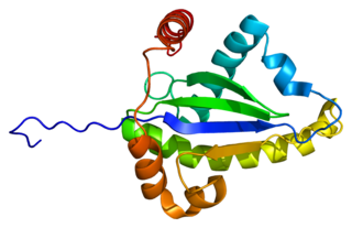
The Fas receptor, also known as Fas, FasR, apoptosis antigen 1, cluster of differentiation 95 (CD95) or tumor necrosis factor receptor superfamily member 6 (TNFRSF6), is a protein that in humans is encoded by the FAS gene. Fas was first identified using a monoclonal antibody generated by immunizing mice with the FS-7 cell line. Thus, the name Fas is derived from FS-7-associated surface antigen.

<span class="mw-page-title-main">Death-inducing signaling complex</span>

The death-inducing signaling complex or DISC is a multi-protein complex formed by members of the death receptor family of apoptosis-inducing cellular receptors. A typical example is FasR, which forms the DISC upon trimerization as a result of its ligand (FasL) binding. The DISC is composed of the death receptor, FADD, and caspase 8. It transduces a downstream signal cascade resulting in apoptosis.

<span class="mw-page-title-main">Caspase 8</span> Protein-coding gene in the species Homo sapiens

Caspase-8 is a caspase protein, encoded by the CASP8 gene. It most likely acts upon caspase-3. CASP8 orthologs have been identified in numerous mammals for which complete genome data are available. These unique orthologs are also present in birds.

<span class="mw-page-title-main">TRAF2</span> Protein-coding gene in humans

TNF receptor-associated factor 2 is a protein that in humans is encoded by the TRAF2 gene.

<span class="mw-page-title-main">Caspase 3</span> Protein-coding gene in the species Homo sapiens

Caspase-3 is a caspase protein that interacts with caspase-8 and caspase-9. It is encoded by the CASP3 gene. CASP3 orthologs have been identified in numerous mammals for which complete genome data are available. Unique orthologs are also present in birds, lizards, lissamphibians, and teleosts.

<span class="mw-page-title-main">TRADD</span> Protein-coding gene in the species Homo sapiens

Tumor necrosis factor receptor type 1-associated DEATH domain protein is a protein that in humans is encoded by the TRADD gene.

<span class="mw-page-title-main">Death receptor 4</span> Protein found in humans

Death receptor 4 (DR4), also known as TRAIL receptor 1 (TRAILR1) and tumor necrosis factor receptor superfamily member 10A (TNFRSF10A), is a cell surface receptor of the TNF-receptor superfamily that binds TRAIL and mediates apoptosis.

<span class="mw-page-title-main">TRAF1</span> Protein-coding gene in the species Homo sapiens

TNF receptor-associated factor 1 is a protein that in humans is encoded by the TRAF1 gene.

<span class="mw-page-title-main">Caspase 10</span> Protein-coding gene in the species Homo sapiens

Caspase-10 is an enzyme that, in humans, is encoded by the CASP10 gene.

<span class="mw-page-title-main">Death receptor 5</span> Protein found in humans

Death receptor 5 (DR5), also known as TRAIL receptor 2 (TRAILR2) and tumor necrosis factor receptor superfamily member 10B (TNFRSF10B), is a cell surface receptor of the TNF-receptor superfamily that binds TRAIL and mediates apoptosis.

<span class="mw-page-title-main">RIPK1</span> Enzyme found in humans

Receptor-interacting serine/threonine-protein kinase 1 (RIPK1) functions in a variety of cellular pathways related to both cell survival and death. In terms of cell death, RIPK1 plays a role in apoptosis and necroptosis. Some of the cell survival pathways RIPK1 participates in include NF-κB, Akt, and JNK.

<span class="mw-page-title-main">RIPK2</span> Protein-coding gene in humans

Receptor-interacting serine/threonine-protein kinase 2 is an enzyme that in humans is encoded by the RIPK2 gene.

<span class="mw-page-title-main">PEA15</span> Protein-coding gene in the species Homo sapiens

Astrocytic phosphoprotein PEA-15 is a protein that in humans is encoded by the PEA15 gene.

<span class="mw-page-title-main">CASP8AP2</span> Protein-coding gene in the species Homo sapiens

CASP8-associated protein 2 is a protein, that in humans is encoded by the CASP8AP2 gene.

<span class="mw-page-title-main">Death domain</span>

The death domain (DD) is a protein interaction module composed of a bundle of six alpha-helices. DD is a subclass of protein motif known as the death fold and is related in sequence and structure to the death effector domain (DED) and the caspase recruitment domain (CARD), which work in similar pathways and show similar interaction properties. DD bind each other forming oligomers. Mammals have numerous and diverse DD-containing proteins. Within these proteins, the DD domains can be found in combination with other domains, including: CARDs, DEDs, ankyrin repeats, caspase-like folds, kinase domains, leucine zippers, leucine-rich repeats (LRR), TIR domains, and ZU5 domains.

The Death Domain database is a secondary database of protein-protein interactions (PPI) of the death domain superfamily. Members of this superfamily are key players in apoptosis, inflammation, necrosis, and immune cell signaling pathways. Negative death domain superfamily-mediated signaling events result in various human diseases which include, cancers, neurodegenerative diseases, and immunological disorders. Creating death domain databases are of particular interest to researchers in the biomedical field as it enables a further understanding of the molecular mechanisms involved in death domain interactions while also providing easy access to tools such as an interaction map that illustrates the protein-protein interaction network and information. There is currently only one database that exclusively looks at death domains but there are other databases and resources that have information on this superfamily. According to PubMed, this database has been cited by seven peer-reviewed articles to date because of its extensive and specific information on the death domains and their PPI summaries.

<span class="mw-page-title-main">Activation-induced cell death</span>

AICD is programmed cell death caused by the interaction of Fas receptors and Fas ligands. AICD is a negative regulator of activated T lymphocytes that results from repeated stimulation of their T-cell receptors (TCR) and helps to maintain peripheral immune tolerance. Alteration of the process may lead to autoimmune diseases.

References

  1. 1 2 3 GRCh38: Ensembl release 89: ENSG00000168040 - Ensembl, May 2017
  2. "Human PubMed Reference:". National Center for Biotechnology Information, U.S. National Library of Medicine.
  3. "Mouse PubMed Reference:". National Center for Biotechnology Information, U.S. National Library of Medicine.
  4. Kim, P.K.M., Dutra, A.S., Chandrasekharappa, S.C., PUCK, J.M (1996). "Genomic structure and mapping of human FADD, an intracellular mediator of lymphocyte apoptosis". Journal of Immunology. 157 (12): 5461–5466. doi: 10.4049/jimmunol.157.12.5461 . PMID   8955195.
  5. Huang B, Eberstadt M, Olejniczak ET, Meadows RP, Fesik SW (1996). "NMR structure and mutagenesis of the Fas (APO-1/CD95) death domain". Nature. 384 (6610): 638–641. Bibcode:1996Natur.384..638H. doi:10.1038/384638a0. PMID   8967952. S2CID   2492303.
  6. 1 2 Eberstadt M, Huang B, Chen Z, Meadows RP, Ng SC, Zheng L, Lenardo MJ, Fesik SW (1998). "NMR structure and mutagenesis of the FADD (Mort1) death-effector domain". Nature. 392 (6679): 941–945. Bibcode:1998Natur.392..941E. doi:10.1038/31972. PMID   9582077. S2CID   4370202.
  7. 1 2 3 Boldin, M. P., Varfolomeev, E. E., Pancer, Z., Mett, I. L., Camonis, J. H. & Wallach, D. (1995). "A Novel Protein That Interacts with the Death Domain of Fas/APO1 Contains a Sequence Motif Related to the Death Domain". Journal of Biological Chemistry. 270 (14): 7795–7798. doi: 10.1074/jbc.270.14.7795 . PMID   7536190.{{cite journal}}: CS1 maint: multiple names: authors list (link)
  8. Jeong, E. J., Bang, S., Lee, T. H., Park, Y. I., Sim, W. S., Kim, K. S. (1999). "The solution structure of FADD death domain - Structural basis of death domain interactions of Fas and FADD". Journal of Biological Chemistry. 274 (23): 16337–16342. doi: 10.1074/jbc.274.23.16337 . PMID   10347191.
  9. Boldin, M. P., Goncharov, T. M., Goltsev, Y. V., wallach, D. (1996). "Involvement of MACH, a novel MORT1/FADD-interacting protease, in Fas/APO-1- and TNF receptor-induced cell death". Cell. 85 (6): 803–815. doi: 10.1016/s0092-8674(00)81265-9 . PMID   8681376. S2CID   7415784.
  10. 1 2 3 Kischkel FC, Lawrence DA, Tinel A, LeBlanc H, Virmani A, Schow P, Gazdar A, Blenis J, Arnott D, Ashkenazi A (2001). "Death receptor recruitment of endogenous caspase-10 and apoptosis initiation in the absence of caspase-8". Journal of Biological Chemistry. 276 (49): 46639–46646. doi: 10.1074/jbc.M105102200 . PMID   11583996.
  11. Pruul H, McDonald PJ (1995). "Cytotoxicity-dependent APO-1 (Fas/CD95)-associated proteins form a death-inducing signalling complex (DISC) with the receptor". EMBO Journal. 14 (22): 5579–5588. doi:10.1002/j.1460-2075.1995.tb00245.x. PMC   394672 . PMID   8521815.
  12. Weinlich R, Dillon CP, Green DR (2011). "Ripped to death". Trends in Cell Biology. 21 (11): 630–637. doi:10.1016/j.tcb.2011.09.002. PMC   3205316 . PMID   21978761.
  13. 1 2 3 4 Lee, E.-W., Seo, J., Jeong, M., Lee, S., Song, J (2012). "The roles of FADD in extrinsic apoptosis and necroptosis". BMB Reports. 45 (9): 496–508. doi: 10.5483/BMBRep.2012.45.9.186 . PMID   23010170.
  14. 1 2 Bodmer JL, Holler N, Reynard S, Vinciguerra P, Schneider P, Juo P, Blenis J, Tschopp J (2000). "TRAIL receptor-2 signals apoptosis through FADD and caspase-8". Nature Cell Biology. 2 (4): 241–243. doi:10.1038/35008667. PMID   10783243. S2CID   13547815.
  15. 1 2 Micheau, O., Tschopp, J. (2003). "Induction of TNF receptor I-mediated apoptosis via two sequential signaling complexes" (PDF). Cell. 114 (2): 181–190. doi:10.1016/s0092-8674(03)00521-x. PMID   12887920. S2CID   17145731.
  16. 1 2 Pyo JO, Jang MH, Kwon YK, Lee HJ, Jun JI, Woo HN, Cho DH, Choi B, Lee H, Kim JH, Mizushima N, Oshumi Y, Jung YK (2005). "Essential roles of Atg5 and FADD in autophagic cell death - Dissection of autophagic cell death into vacuole formation and cell death". Journal of Biological Chemistry. 280 (21): 20722–20729. doi: 10.1074/jbc.M413934200 . PMID   15778222.
  17. Bell BD, Leverrier S, Weist BM, Newton RH, Arechiga AF, Luhrs KA, Morrissette NS, Walsh CM (2008). ". FADD and caspase-8 control the outcome of autophagic signaling in proliferating T cells". Proceedings of the National Academy of Sciences of the United States of America. 105 (43): 16677–16682. Bibcode:2008PNAS..10516677B. doi: 10.1073/pnas.0808597105 . PMC   2575479 . PMID   18946037.
  18. Yeh, W. C., De La Pompa, J. L., Mccurach, M. E., Shu, H. B., Elia, A. J., Shahinian, A., Ng, M., Wakeham, A., Khoo, W., Mitchell, K., El-Deiry, W. S., Lowe, S. W., Goeddel, D. V., Mak, T. W. (1998). "FADD: Essential for embryo development and signaling from some, but not all, inducers of apoptosis". Science. 279 (5358): 1954–1958. Bibcode:1998Sci...279.1954Y. doi:10.1126/science.279.5358.1954. PMID   9506948.
  19. Sakamaki K, Takagi C, Kitayama A, Kurata T, Yamamoto TS, Chiba K, Kominami K, Jung SK, Okawa K, Nozaki M, Kubota HY, Ueno N (2012). "Multiple functions of FADD in apoptosis, NF-kappa B-related signaling, and heart development in Xenopus embryos". Genes to Cells. 17 (11): 875–896. doi: 10.1111/gtc.12004 . PMID   23025414. S2CID   23264540.
  20. Gregory-Evans CY, Moosajee M, Hodges MD, Mackay DS, Game L, Vargesson N, Bloch-Zupan A, Rüschendorf F, Santos-Pinto L, Wackens G, Gregory-Evans K (2007). "SNP genome scanning localizes oto-dental syndrome to chromosome 11q13 and microdeletions at this locus implicate FGF3 in dental and inner-ear disease and FADD in ocular coloboma". Human Molecular Genetics. 16 (20): 2482–2493. doi: 10.1093/hmg/ddm204 . PMID   17656375.
  21. 1 2 3 Alappat EC, Feig C, Boyerinas B, Volkland J, Samuels M, Murmann AE, Thorburn A, Kidd VJ, Slaughter CA, Osborn SL, Winoto A, Tang WJ, Peter ME (2005). "Phosphorylation of FADD at serine 194 by CKI alpha regulates its nonapoptotic activities". Molecular Cell. 19 (3): 321–332. doi: 10.1016/j.molcel.2005.06.024 . PMID   16061179.
  22. Zhang J, Cado D, Chen A, Kabra NH, Winoto A (1998). "Fas-mediated apoptosis and activation-induced T-cell proliferation are defective in mice lacking FADD/Mort1". Nature. 392 (6673): 296–300. Bibcode:1998Natur.392..296Z. doi:10.1038/32681. PMID   9521326. S2CID   4420585.
  23. Vander Elst P, van den Berg E, Pepermans H, vander Auwera L, Zeeuws R, Tourwe D, van Binst G (2006). "The Fas-associated death domain protein is required in apoptosis and TLR-induced proliferative responses in B cells". Journal of Immunology. 176 (11): 6852–6861. doi:10.4049/jimmunol.176.11.6852. PMC   3110081 . PMID   16709845.
  24. Wajant H, Haas E, Schwenzer R, Muhlenbeck F, Kreuz S, Schubert G, Grell M, Smith C, Scheurich P (2000). "Inhibition of death receptor-mediated gene induction by a cycloheximide-sensitive factor occurs at the level of or upstream of Fas-associated death domain protein (FADD)". Journal of Biological Chemistry. 275 (32): 24357–24366. doi: 10.1074/jbc.M000811200 . PMID   10823821.
  25. 1 2 3 4 Tourneur L, Chiocchia G (2010). "FADD: a regulator of life and death". Trends in Immunology. 31 (7): 260–269. doi:10.1016/j.it.2010.05.005. PMID   20576468.
  26. Balachandran, S., Venkataraman, T., Fisher, P. B., Barber, G. N (2007). "Fas-associated death domain-containing protein-mediated antiviral innate immune signaling involves the regulation of Irf7". Journal of Immunology. 178 (4): 2429–2439. doi: 10.4049/jimmunol.178.4.2429 . PMID   17277150.
  27. Cheng W, Wang L, Zhang R, Du P, Yang B, Zhuang H, Tang B, Yao C, Yu M, Wang Y, Zhang J, Yin W, Li J, Zheng W, Lu M, Hua Z (2012). "Regulation of Protein Kinase C Inactivation by Fas-associated Protein with Death Domain". Journal of Biological Chemistry. 287 (31): 26126–26135. doi: 10.1074/jbc.M112.342170 . PMC   3406696 . PMID   22582393.
  28. Yao C, Zhuang H, Du P, Cheng W, Yang B, Guan S, Hu Y, Zhu D, Christine M, Shi L, Hua ZC (2013). "Domain-containing Protein (FADD) Phosphorylation in Regulating Glucose Homeostasis: from Proteomic Discovery to Physiological Validation". Molecular & Cellular Proteomics. 12 (10): 2689–2700. doi: 10.1074/mcp.M113.029306 . PMC   3790283 . PMID   23828893.
  29. Gómez-Angelats M, Cidlowski JA (2003). "Molecular evidence for the nuclear localization of FADD". Cell Death and Differentiation. 10 (7): 791–797. doi: 10.1038/sj.cdd.4401237 . PMID   12815462.
  30. 1 2 Krueger A, Baumann S, Krammer PH, Kirchhoff S (2001). "FLICE-inhibitory proteins: Regulators of death receptor-mediated apoptosis". Molecular and Cellular Biology. 21 (24): 8247–8254. doi:10.1128/mcb.21.24.8247-8254.2001. PMC   99990 . PMID   11713262.
  31. 1 2 Scaffidi, C., Schmitz, I., Krammer, P. H., Peter, M. E. (1999). "The role of c-FLIP in modulation of CD95-induced apoptosis". Journal of Biological Chemistry. 274 (3): 1541–1548. doi: 10.1074/jbc.274.3.1541 . PMID   9880531.
  32. Gómez-Angelats M, Cidlowski JA (2001). "Protein kinase C regulates FADD recruitment and death-inducing signaling complex formation in Fas/CD95-induced apoptosis". Journal of Biological Chemistry. 276 (48): 44944–44952. doi: 10.1074/jbc.M104919200 . PMID   11581255.
  33. 1 2 Lee EW, Kim JH, Ahn YH, Seo J, Ko A, Jeong M, Kim SJ, Ro JY, Park KM, Lee HW, Park EJ, Chun KH, Song J (2012). "Ubiquitination and degradation of the FADD adaptor protein regulate death receptor-mediated apoptosis and necroptosis". Nature Communications. 3: 978. Bibcode:2012NatCo...3..978L. doi: 10.1038/ncomms1981 . PMID   22864571.
  34. Reuss R, Mistarz M, Mirau A, Kraus J, Bödeker RH, Oschmann P (2014). "FADD is upregulated in relapsing remitting multiple". Neuroimmunomodulation. 21 (5): 221–225. doi:10.1159/000356522. PMID   24603611. S2CID   207652468.
  35. Ma Y, Liu H, Tu-Rapp H, Thiesen HJ, Ibrahim SM, Cole SM, Pope RM (2004). "Fas ligation on macrophages enhances IL-1R1-Toll-like receptor 4 signaling and promotes chronic inflammation". Nature Immunology. 5 (4): 380–387. doi:10.1038/ni1054. PMID   15004557. S2CID   39471972.
  36. 1 2 Razaghi A, Villacrés C, Jung V, Mashkour N, Butler M, Owens L, Heimann K (2017). "Improved therapeutic efficacy of mammalian expressed-recombinant interferon gamma against ovarian cancer cells". Experimental Cell Research. 359 (1): 20–29. doi:10.1016/j.yexcr.2017.08.014. PMID   28803068. S2CID   12800448.
  37. Pattje WJ, Melchers LJ, Slagter-Menkema L, Mastik MF, Schrijvers ML, Gibcus JH, Kluin PM, Hoegen-Chouvalova O, van der Laan BF, Roodenburg JL, van der Wal JE, Schuuring E, Langendijk JA (2013). "FADD expression is associated with regional and distant metastasis in squamous cell carcinoma of the head and neck". Histopathology. 63 (2): 263–270. doi:10.1111/his.12174. PMID   23763459. S2CID   36206578.
  38. Cimino Y, Costes A, Damotte D, Validire P, Mistou S, Cagnard N, Alifano M, Régnard JF, Chiocchia G, Sautès-Fridman C, Tourneur L (2012). "FADD protein release mirrors the development and aggressiveness of human non-small cell lung cancer". British Journal of Cancer. 106 (12): 1989–1996. doi:10.1038/bjc.2012.196. PMC   3388563 . PMID   22669160.
  39. Park SJ, Wu CH, Gordon JD, Zhong X, Emami A, Safa AR (2004). "Taxol induces caspase-10-dependent apoptosis". Journal of Biological Chemistry. 279 (49): 51057–51067. doi: 10.1074/jbc.M406543200 . PMID   15452117.
  40. Matsuyoshi S, Shimada K, Nakamura M, Ishida E, Konishi N (2006). "FADD phosphorylation is critical for cell cycle regulation in breast cancer cells". British Journal of Cancer. 94 (4): 532–539. doi:10.1038/sj.bjc.6602955. PMC   2361184 . PMID   16450001.
  41. Schinske KA, Nyati S, Khan AP, Williams TM, Johnson TD, Ross BD, Tomás RP, Rehemtulla A (2011). "A Novel Kinase Inhibitor of FADD Phosphorylation Chemosensitizes through the Inhibition of NF-kappa B". Molecular Cancer Therapeutics. 10 (10): 1807–1817. doi:10.1158/1535-7163.mct-11-0362. PMC   3191281 . PMID   21859840.
  42. Buechler C, Bared SM, Aslanidis C, Ritter M, Drobnik W, Schmitz G (Nov 2002). "Molecular and functional interaction of the ATP-binding cassette transporter A1 with Fas-associated death domain protein". J. Biol. Chem. 277 (44): 41307–10. doi: 10.1074/jbc.C200436200 . PMID   12235128.
  43. Roth W, Stenner-Liewen F, Pawlowski K, Godzik A, Reed JC (Mar 2002). "Identification and characterization of DEDD2, a death effector domain-containing protein". J. Biol. Chem. 277 (9): 7501–8. doi: 10.1074/jbc.M110749200 . PMID   11741985.
  44. Screaton RA, Kiessling S, Sansom OJ, Millar CB, Maddison K, Bird A, Clarke AR, Frisch SM (Apr 2003). "Fas-associated death domain protein interacts with methyl-CpG binding domain protein 4: a potential link between genome surveillance and apoptosis". Proc. Natl. Acad. Sci. U.S.A. 100 (9): 5211–6. Bibcode:2003PNAS..100.5211S. doi: 10.1073/pnas.0431215100 . PMC   154324 . PMID   12702765.
  45. Stilo R, Liguoro D, di Jeso B, Leonardi A, Vito P (Apr 2003). "The alpha-chain of the nascent polypeptide-associated complex binds to and regulates FADD function". Biochem. Biophys. Res. Commun. 303 (4): 1034–41. doi:10.1016/s0006-291x(03)00487-x. PMID   12684039.
  46. Condorelli G, Vigliotta G, Cafieri A, Trencia A, Andalò P, Oriente F, Miele C, Caruso M, Formisano P, Beguinot F (Aug 1999). "PED/PEA-15: an anti-apoptotic molecule that regulates FAS/TNFR1-induced apoptosis". Oncogene. 18 (31): 4409–15. doi:10.1038/sj.onc.1202831. PMID   10442631. S2CID   20510429.

Further reading