Fluorescence in the life sciences

Last updated
Distribution of fluorescent proteins in animals. Distribution of fluorescent proteins in metazoans - 40851 2020 161 Fig2 HTML.png
Distribution of fluorescent proteins in animals.
The hippocampus of a mouse imaged via fluorescence microscopy. Neuronal explosion.jpg
The hippocampus of a mouse imaged via fluorescence microscopy.
Biofluorescent emission spectra from amphibians Amphibian biofluorescent emission spectra - 41598 2020 59528 Fig2-top.png
Biofluorescent emission spectra from amphibians
Example uses of fluorescent proteins for imaging in the life sciences Imaging Life with Fluorescent Proteins (10690274384) (2).jpg
Example uses of fluorescent proteins for imaging in the life sciences

Fluorescence is used in the life sciences generally as a non-destructive way of tracking or analysing biological molecules. Some proteins or small molecules in cells are naturally fluorescent, which is called intrinsic fluorescence or autofluorescence (such as NADH, tryptophan or endogenous chlorophyll, phycoerythrin or green fluorescent protein). Alternatively, specific or general proteins, nucleic acids, lipids or small molecules can be "labelled" with an extrinsic fluorophore, a fluorescent dye which can be a small molecule, protein or quantum dot. Several techniques exist to exploit additional properties of fluorophores, such as fluorescence resonance energy transfer, where the energy is passed non-radiatively to a particular neighbouring dye, allowing proximity or protein activation to be detected; another is the change in properties, such as intensity, of certain dyes depending on their environment allowing their use in structural studies. [1] [2] [3]

Contents

Fluorescence

A simplified Jablonski diagram illustrating the change of energy levels. Jablonskidiagram.svg
A simplified Jablonski diagram illustrating the change of energy levels.

The principle behind fluorescence is that the fluorescent moiety contains electrons which can absorb a photon and briefly enter an excited state before either dispersing the energy non-radiatively or emitting it as a photon, but with a lower energy, i.e., at a longer wavelength (wavelength and energy are inversely proportional). [4] The difference in the excitation and emission wavelengths is called the Stokes shift, and the time that an excited electron takes to emit the photon is called a lifetime. The quantum yield is an indicator of the efficiency of the dye (it is the ratio of emitted photons per absorbed photon), and the extinction coefficient is the amount of light that can be absorbed by a fluorophore. Both the quantum yield and extinction coefficient are specific for each fluorophore and multiplied together calculates the brightness of the fluorescent molecule. [5]

Labelling

Reactive dyes

Fluorophores can be attached to proteins via specific functional groups, such as:

or non-specificately (glutaraldehyde) or non-covalently (e.g. via hydrophobicity, etc.).

These fluorophores are either small molecules, protein or quantum dots.

Organic fluorophores fluoresce thanks to delocalized electrons which can jump a band and stabilize the energy absorbed, hence most fluorophores are conjugated systems. Several families exist and their excitations range from the infrared to the ultraviolet.
Lanthanides (chelated) are uniquely fluorescent metals, which emit thanks to transitions involving 4f orbits, which are forbidden, hence they have very low absorption coefficients and slow emissions, requiring excitation through fluorescent organic chelators (e.g. dipicolinate-based Terbium (III) chelators [6] ).
A third class of small molecule fluorophore is that of the transition metal-ligand complexes, which display molecular fluorescence from a metal-to-ligand charge transfer state which is partially forbidden, these are generally complexes of Ruthenium, Rhenium or Osmium.

Quantum dots

Quantum dots are fluorescent semiconductor nanoparticles that typically brighter than conventional stains. They are generally more expensive, toxic, do not permeate cell membranes, and cannot be manufactured by the cell.

Fluorescent proteins

Several fluorescent protein exist in nature[ citation needed ], but the most important one as a research tool is Green Fluorescent Protein (GFP) from the jellyfish Aequorea victoria , [7] which spontaneously fluoresces upon folding via specific serine-tyrosine-glycine residues. The benefit that GFP and other fluorescent proteins have over organic dyes or quantum dots is that they can be expressed exogenously in cells alone or as a fusion protein, a protein that is created by ligating the fluorescent gene (e.g., GFP) to another gene and whose expression is driven by a housekeeping gene promoter or another specific promoter. This approach allows fluorescent proteins to be used as reporters for any number of biological events, such as sub-cellular localization and expression patterns. A variant of GFP is naturally found in corals, specifically the Anthozoa, and several mutants have been created to span the visible spectra and fluoresce longer and more stably. Other proteins are fluorescent but require a fluorophore cofactor, and hence can only be used in vitro; these are often found in plants and algae (phytofluors, phycobiliprotein such as allophycocyanin).

Computational techniques

The above techniques can be combined with computational methods to estimate staining levels without staining the cell. These approaches, generally, rely on training a deep-convolutional neural network to perform imaging remapping, converting the bright-field or phase image into a fluorescent image. [8] [9] By decoupling the training corpus from the cells under investigation, these approaches provide an avenue for using stains that are otherwise incompatible with live cell imaging, such as anti-body staining. [10]

Bioluminescence and fluorescence

Fluorescence, chemiluminescence and phosphorescence are 3 different types of luminescence properties, i.e. emission of light from a substance. Fluorescence is a property where light is absorbed and remitted within a few nanoseconds (approx. 10ns) at a lower energy (=higher wavelength), while bioluminescence is biological chemiluminescence, a property where light is generated by a chemical reaction of an enzyme on a substrate. Phosphorescence is a property of materials to absorb light and emit the energy several milliseconds or more later (due to forbidden transitions to the ground state of a triplet state, while fluorescence occurs in excited singlet states). Until recently, this was not applicable to life science research due to the size of the inorganic particles. However the boundary between the fluorescence and phosphorescence is not clean cut as transition metal-ligand complexes, which combine a metal and several organic moieties, have long lifetimes, up to several microseconds (as they display mixed singlet-triplet states).

Comparison with radioactivity

Prior to its widespread use in the past three decades radioactivity was the most common label.

The advantages of fluorescence over radioactive labels are as follows:

Note: a channel is similar to "colour" but distinct, it is the pair of excitation and emission filters specific for a dye, e.g. agilent microarrays are dual channel, working on cy3 and cy5, these are colloquially referred to as green and red.

Fluorescence is not necessarily more convenient to use because it requires specialized detection equipment of its own. For non-quantitative or relative quantification applications it can be useful but it is poorly suited for making absolute measurement because of fluorescence quenching, whereas measuring radioactively labeled molecules is always direct and highly sensitive.

Disadvantages of fluorophores include:

Additional useful properties

The basic property of fluorescence are extensively used, such as a marker of labelled components in cells (fluorescence microscopy) or as an indicator in solution (Fluorescence spectroscopy), but other additional properties, not found with radioactivity, make it even more extensively used.

FRET

Cartoon of FRET between two protein interacting protein, labelled with fluorescein and tetramethylrhodamine Squid's fret.svg
Cartoon of FRET between two protein interacting protein, labelled with fluorescein and tetramethylrhodamine

FRET (Förster resonance energy transfer) is a property in which the energy of the excited electron of one fluorphore, called the donor, is passed on to a nearby acceptor dye, either a dark quencher or another fluorophore, which has an excitation spectrum which overlaps with the emission spectrum of the donor dye resulting in a reduced fluorescence. This can be used to:

Sensitivity to environment

Example of an environmentally sensitive dye: Badan exhibits a large change in dipole moment upon excitation (due to internal charge transfer between the tertiary amine and ketone). This results in a significant lowering of the energy from solvent relaxation. Badan.svg
Example of an environmentally sensitive dye: Badan exhibits a large change in dipole moment upon excitation (due to internal charge transfer between the tertiary amine and ketone). This results in a significant lowering of the energy from solvent relaxation.

Environment-sensitive dyes change their properties (intensity, half-life, and excitation and emission spectra) depending on the polarity (hydrophobicity and charge) of their environments. Examples include: Indole, Cascade Yellow, prodan, Dansyl, Dapoxyl, NBD, PyMPO, Pyrene and diethylaminocumarin.
This change is most pronounced when electron-donating and electron-withdrawing groups are placed at opposite ends of an aromatic ring system, [12] as this results in a large change in dipole moment when excited.

When a fluorophore is excited, it generally has a larger dipole moment (μE) than in the ground state (μG). Absorption of a photon by a fluorophore takes a few picoseconds. Before this energy is released (emission: 1–10 ns), the solvent molecules surrounding the fluorophore reorient (10–100 ps) due to the change in polarity in the excited singlet state; this process is called solvent relaxation. As a result of this relaxation, the energy of the excited state of the fluorophore is lowered (longer wavelength), hence fluorophores that have a large change in dipole moment have larger stokes shift changes in different solvents. The difference between the energy levels can be roughly determined with the Lipper-Mataga equation.

A hydrophobic dye is a dye which is insoluble in water, a property independent of solvatochromism.
Additionally, The term environment-sensitive in chemistry actually describes changes due to one of a variety of different environmental factors, such as pH or temperature, not just polarity; however, in biochemistry environment-sensitive fluorphore and solvatochromic fluorophore are used interchangeably: this convention is so widespread that suppliers describe them as environment-sensitive over solvatochromic.

Fluorescence lifetime

Fluorescent moieties emit photons several nanoseconds after absorption following an exponential decay curve, which differs between dyes and depends on the surrounding solvent. When the dye is attached to a macromolecules the decay curve becomes multiexponential. Conjugated dyes generally have a lifetime between 1–10 ns, a small amount of longer lived exceptions exist, notably pyrene with a lifetime of 400ns in degassed solvents or 100ns in lipids and coronene with 200ns. On a different category of fluorphores are the fluorescent organometals (lanthanides and transition metal-ligand complexes) which have been previously described, which have much longer lifetimes due to the restricted states: lanthanides have lifetimes of 0.5 to 3 ms, while transition metal-ligand complexes have lifetimes of 10 ns to 10 µs. Note that fluorescent lifetime should not be confused with the photodestruction lifetime or the "shelf-life" of a dye.

Multiphoton excitation

Multiphoton excitation is a way of focusing the viewing plane of the microscope by taking advantage of the phenomenon where two simultaneous low energy photons are absorbed by a fluorescent moiety which normally absorbs one photon with double their individual energy: say two NIR photons (800 nm) to excite a UV dye (400 nm).

Fluorescence anisotropy

A perfectly immobile fluorescent moiety when exited with polarized light will emit light which is also polarized. However, if a molecule is moving, it will tend to "scramble" the polarization of the light by radiating at a different direction from the incident light.

Fluorescent thermometry

Some fluorescent chemicals exhibit significant fluorescent quenching when exposed to increasing temperatures. This effect has been used to measure and examine the thermogenic properties of mitochondria. This involves placing mitochondria-targeting thermosensitive fluorophores inside cells, which naturally localise inside the mitochondria due to the inner mitochondrial membrane matrix-face's negative charge (as the fluorophores are cationic). [13] The temperature of these fluorophores is inversely proportional to their fluorescence emission, and thus by measuring the fluorescent output, the temperature of actively-respiring mitochondria can be deduced. The fluorophores used are typically lipophilic cations derived from Rhodamine-B, [13] such as ThermoFisher's MitoTracker probes. [14] This technique has contributed significantly to the general scientific consensus that mitochondria are physiologically maintained at close to 50 ˚C, more than 10˚C above the rest of the cell. [15]

Structure of MitoThermo Yellow Mitothermoyellow.png
Structure of MitoThermo Yellow

The inverse relationship between fluorescence and temperature can be explained by the change in the number of atomic collisions in the fluorophore's environment, depending on the kinetic energy. Collisions promote radiationless decay and loss of extra energy as heat, so more collisions or more forceful collisions will promote radiationless decay and reduce fluorescence emission. [16]

This temperature-measurement technique is, however, limited. These cationic fluorophores are heavily influenced by the charge of the inner mitochondrial membrane matrix-face, dependent on the cell type. For example, the thermosensitive fluorophore MTY (MitoTracker Yellow) shows a sudden and drastic drop in fluorescence after the addition of oligomycin (an ATP synthase inhibitor) to the mitochondria of human primary fibroblasts. This would suggest a sharp increase in mitochondrial temperature but is, in reality, explained by the hyperpolarisation of the mitochondrial inner membrane by oligomycin - leading to the breakdown of the positively-charged MTY fluorophore. [13]

Methods

Ethidium bromide stained agarose gel. Ethidium bromide fluoresces orange when intercalating DNA and when exposed to UV light. AgarosegelUV.jpg
Ethidium bromide stained agarose gel. Ethidium bromide fluoresces orange when intercalating DNA and when exposed to UV light.

Also, many biological molecules have an intrinsic fluorescence that can sometimes be used without the need to attach a chemical tag. Sometimes this intrinsic fluorescence changes when the molecule is in a specific environment, so the distribution or binding of the molecule can be measured. Bilirubin, for instance, is highly fluorescent when bound to a specific site on serum albumin. Zinc protoporphyrin, formed in developing red blood cells instead of hemoglobin when iron is unavailable or lead is present, has a bright fluorescence and can be used to detect these problems.

The number of fluorescence applications in the biomedical, biological and related sciences continuously expands. Methods of analysis in these fields are also growing, often with nomenclature in the form of acronyms such as: FLIM, FLI, FLIP, CALI, FLIE, FRET, FRAP, FCS, PFRAP, smFRET, FIONA, FRIPS, SHREK, SHRIMP or TIRF. Most of these techniques rely on fluorescence microscopes, which use high intensity light sources, usually mercury or xenon lamps, LEDs, or lasers, to excite fluorescence in the samples under observation. Optical filters then separate excitation light from emitted fluorescence to be detected by eye or with a (CCD) camera or other light detector (e.g., photomultiplier tubes, spectrographs). Considerable research is underway to improve the capabilities of such microscopes, the fluorescent probes used, and the applications they are applied to. Of particular note are confocal microscopes, which use a pinhole to achieve optical sectioning, which affords a quantitative, 3D view of the sample.

See also

Related Research Articles

<span class="mw-page-title-main">Fluorescence</span> Emission of light by a substance that has absorbed light

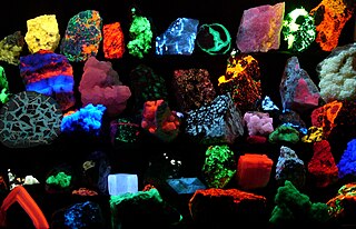
Fluorescence is the emission of light by a substance that has absorbed light or other electromagnetic radiation. It is a form of luminescence. In most cases, the emitted light has a longer wavelength, and therefore a lower photon energy, than the absorbed radiation. A perceptible example of fluorescence occurs when the absorbed radiation is in the ultraviolet region of the electromagnetic spectrum, while the emitted light is in the visible region; this gives the fluorescent substance a distinct color that can only be seen when the substance has been exposed to UV light. Fluorescent materials cease to glow nearly immediately when the radiation source stops, unlike phosphorescent materials, which continue to emit light for some time after.

<span class="mw-page-title-main">Fluorescent tag</span>

In molecular biology and biotechnology, a fluorescent tag, also known as a fluorescent label or fluorescent probe, is a molecule that is attached chemically to aid in the detection of a biomolecule such as a protein, antibody, or amino acid. Generally, fluorescent tagging, or labeling, uses a reactive derivative of a fluorescent molecule known as a fluorophore. The fluorophore selectively binds to a specific region or functional group on the target molecule and can be attached chemically or biologically. Various labeling techniques such as enzymatic labeling, protein labeling, and genetic labeling are widely utilized. Ethidium bromide, fluorescein and green fluorescent protein are common tags. The most commonly labelled molecules are antibodies, proteins, amino acids and peptides which are then used as specific probes for detection of a particular target.

<span class="mw-page-title-main">Fluorescence spectroscopy</span> Type of electromagnetic spectroscopy

Fluorescence spectroscopy is a type of electromagnetic spectroscopy that analyzes fluorescence from a sample. It involves using a beam of light, usually ultraviolet light, that excites the electrons in molecules of certain compounds and causes them to emit light; typically, but not necessarily, visible light. A complementary technique is absorption spectroscopy. In the special case of single molecule fluorescence spectroscopy, intensity fluctuations from the emitted light are measured from either single fluorophores, or pairs of fluorophores.

<span class="mw-page-title-main">Fluorophore</span> Agents that emit light after excitation by light

A fluorophore is a fluorescent chemical compound that can re-emit light upon light excitation. Fluorophores typically contain several combined aromatic groups, or planar or cyclic molecules with several π bonds.

Plate readers, also known as microplate readers or microplate photometers, are instruments which are used to detect biological, chemical or physical events of samples in microtiter plates. They are widely used in research, drug discovery, bioassay validation, quality control and manufacturing processes in the pharmaceutical and biotechnological industry and academic organizations. Sample reactions can be assayed in 1-1536 well format microtiter plates. The most common microplate format used in academic research laboratories or clinical diagnostic laboratories is 96-well with a typical reaction volume between 100 and 200 µL per well. Higher density microplates are typically used for screening applications, when throughput and assay cost per sample become critical parameters, with a typical assay volume between 5 and 50 µL per well. Common detection modes for microplate assays are absorbance, fluorescence intensity, luminescence, time-resolved fluorescence, and fluorescence polarization.

<span class="mw-page-title-main">Förster resonance energy transfer</span> Photochemical energy transfer mechanism

Förster resonance energy transfer (FRET), fluorescence resonance energy transfer, resonance energy transfer (RET) or electronic energy transfer (EET) is a mechanism describing energy transfer between two light-sensitive molecules (chromophores). A donor chromophore, initially in its electronic excited state, may transfer energy to an acceptor chromophore through nonradiative dipole–dipole coupling. The efficiency of this energy transfer is inversely proportional to the sixth power of the distance between donor and acceptor, making FRET extremely sensitive to small changes in distance.

A total internal reflection fluorescence microscope (TIRFM) is a type of microscope with which a thin region of a specimen, usually less than 200 nanometers can be observed.

<span class="mw-page-title-main">Fluorescence microscope</span> Optical microscope that uses fluorescence and phosphorescence

A fluorescence microscope is an optical microscope that uses fluorescence instead of, or in addition to, scattering, reflection, and attenuation or absorption, to study the properties of organic or inorganic substances. "Fluorescence microscope" refers to any microscope that uses fluorescence to generate an image, whether it is a simple set up like an epifluorescence microscope or a more complicated design such as a confocal microscope, which uses optical sectioning to get better resolution of the fluorescence image.

<span class="mw-page-title-main">Two-photon excitation microscopy</span> Fluorescence imaging technique

Two-photon excitation microscopy is a fluorescence imaging technique that is particularly well-suited to image scattering living tissue of up to about one millimeter in thickness. Unlike traditional fluorescence microscopy, where the excitation wavelength is shorter than the emission wavelength, two-photon excitation requires simultaneous excitation by two photons with longer wavelength than the emitted light. The laser is focused onto a specific location in the tissue and scanned across the sample to sequentially produce the image. Due to the non-linearity of two-photon excitation, mainly fluorophores in the micrometer-sized focus of the laser beam are excited, which results in the spatial resolution of the image. This contrasts with confocal microscopy, where the spatial resolution is produced by the interaction of excitation focus and the confined detection with a pinhole.

In particle physics, the quantum yield of a radiation-induced process is the number of times a specific event occurs per photon absorbed by the system.

<span class="mw-page-title-main">Texas Red</span> Chemical compound

Texas Red or sulforhodamine 101 acid chloride is a red fluorescent dye, used in histology for staining cell specimens, for sorting cells with fluorescent-activated cell sorting machines, in fluorescence microscopy applications, and in immunohistochemistry. Texas Red fluoresces at about 615 nm, and the peak of its absorption spectrum is at 589 nm. The powder is dark purple. Solutions can be excited by a dye laser tuned to 595-605 nm, or less efficiently a krypton laser at 567 nm. The absorption extinction coefficient at 596 nm is about 85,000 M−1cm−1.

<span class="mw-page-title-main">Autofluorescence</span>

Autofluorescence is the natural emission of light by biological structures such as mitochondria and lysosomes when they have absorbed light, and is used to distinguish the light originating from artificially added fluorescent markers (fluorophores).

<span class="mw-page-title-main">Photobleaching</span> Loss of colour by a pigment when illuminated

In optics, photobleaching is the photochemical alteration of a dye or a fluorophore molecule such that it is permanently unable to fluoresce. This is caused by cleaving of covalent bonds or non-specific reactions between the fluorophore and surrounding molecules. Such irreversible modifications in covalent bonds are caused by transition from a singlet state to the triplet state of the fluorophores. The number of excitation cycles to achieve full bleaching varies. In microscopy, photobleaching may complicate the observation of fluorescent molecules, since they will eventually be destroyed by the light exposure necessary to stimulate them into fluorescing. This is especially problematic in time-lapse microscopy.

<span class="mw-page-title-main">STED microscopy</span> Technique in fluorescence microscopy

Stimulated emission depletion (STED) microscopy is one of the techniques that make up super-resolution microscopy. It creates super-resolution images by the selective deactivation of fluorophores, minimizing the area of illumination at the focal point, and thus enhancing the achievable resolution for a given system. It was developed by Stefan W. Hell and Jan Wichmann in 1994, and was first experimentally demonstrated by Hell and Thomas Klar in 1999. Hell was awarded the Nobel Prize in Chemistry in 2014 for its development. In 1986, V.A. Okhonin had patented the STED idea. This patent was unknown to Hell and Wichmann in 1994.

Fluorescence anisotropy or fluorescence polarization is the phenomenon where the light emitted by a fluorophore has unequal intensities along different axes of polarization. Early pioneers in the field include Aleksander Jablonski, Gregorio Weber, and Andreas Albrecht. The principles of fluorescence polarization and some applications of the method are presented in Lakowicz's book.

In chemistry, a dark quencher is a substance that absorbs excitation energy from a fluorophore and dissipates the energy as heat; while a typical (fluorescent) quencher re-emits much of this energy as light. Dark quenchers are used in molecular biology in conjunction with fluorophores. When the two are close together, such as in a molecule or protein, the fluorophore's emission is suppressed. This effect can be used to study molecular geometry and motion.

Super-resolution microscopy is a series of techniques in optical microscopy that allow such images to have resolutions higher than those imposed by the diffraction limit, which is due to the diffraction of light. Super-resolution imaging techniques rely on the near-field or on the far-field. Among techniques that rely on the latter are those that improve the resolution only modestly beyond the diffraction-limit, such as confocal microscopy with closed pinhole or aided by computational methods such as deconvolution or detector-based pixel reassignment, the 4Pi microscope, and structured-illumination microscopy technologies such as SIM and SMI.

Photo-activated localization microscopy and stochastic optical reconstruction microscopy (STORM) are widefield fluorescence microscopy imaging methods that allow obtaining images with a resolution beyond the diffraction limit. The methods were proposed in 2006 in the wake of a general emergence of optical super-resolution microscopy methods, and were featured as Methods of the Year for 2008 by the Nature Methods journal. The development of PALM as a targeted biophysical imaging method was largely prompted by the discovery of new species and the engineering of mutants of fluorescent proteins displaying a controllable photochromism, such as photo-activatible GFP. However, the concomitant development of STORM, sharing the same fundamental principle, originally made use of paired cyanine dyes. One molecule of the pair, when excited near its absorption maximum, serves to reactivate the other molecule to the fluorescent state.

Time-resolved fluorescence energy transfer (TR-FRET) is the practical combination of time-resolved fluorometry (TRF) with Förster resonance energy transfer (FRET) that offers a powerful tool for drug discovery researchers. TR-FRET combines the low background aspect of TRF with the homogeneous assay format of FRET. The resulting assay provides an increase in flexibility, reliability and sensitivity in addition to higher throughput and fewer false positive/false negative results. FRET involves two fluorophores, a donor and an acceptor. Excitation of the donor by an energy source produces an energy transfer to the acceptor if the two are within a given proximity to each other. The acceptor in turn emits light at its characteristic wavelength.

<span class="mw-page-title-main">Fluorescence imaging</span> Type of non-invasive imaging technique

Fluorescence imaging is a type of non-invasive imaging technique that can help visualize biological processes taking place in a living organism. Images can be produced from a variety of methods including: microscopy, imaging probes, and spectroscopy.

References

  1. Joseph R. Lakowicz (2006). Principles of fluorescence spectroscopy. Springer. pp. 26–. ISBN   978-0-387-31278-1 . Retrieved 25 June 2011.
  2. Fluorescence Fundamentals. Invitrogen.com. Retrieved on 2011-06-25.
  3. Juan Carlos Stockert, Alfonso Blázquez-Castro (2017). Fluorescence Microscopy in Life Sciences. Bentham Science Publishers. ISBN   978-1-68108-519-7 . Retrieved 17 December 2017.
  4. Animation for the principle of fluorescence and UV-visible absorbance
  5. Au-Yeung, Ho Yu; Tong, Ka Yan (2021). "Chapter 16. Transition Metals and Imaging Probes in Neurobiology and Neurodegenerative Diseases". Metal Ions in Bio-Imaging Techniques. Springer. pp. 437–456. doi:10.1515/9783110685701-022. S2CID   233678495.
  6. Lamture, JB; Wensel, TG (1995). "Intensely luminescent immunoreactive conjugates of proteins and dipicolinate-based polymeric Tb (III) chelates". Bioconjugate Chemistry. 6 (1): 88–92. doi:10.1021/bc00031a010. PMID   7711110.
  7. Chalfie, M; Tu, Y; Euskirchen, G; Ward, WW; Prasher, DC (1994). "Green fluorescent protein as a marker for gene expression". Science. 263 (5148): 802–5. Bibcode:1994Sci...263..802C. doi:10.1126/science.8303295. PMID   8303295.
  8. Christiansen, Eric M.; Yang, Samuel J.; Ando, D. Michael; Javaherian, Ashkan; Skibinski, Gaia; Lipnick, Scott; Mount, Elliot; O’Neil, Alison; Shah, Kevan; Lee, Alicia K.; Goyal, Piyush; Fedus, William; Poplin, Ryan; Esteva, Andre; Berndl, Marc; Rubin, Lee L.; Nelson, Philip; Finkbeiner, Steven (April 2018). "In Silico Labeling: Predicting Fluorescent Labels in Unlabeled Images". Cell. 173 (3): 792–803.e19. doi:10.1016/j.cell.2018.03.040. PMC   6309178 . PMID   29656897.
  9. Kandel, Mikhail E.; He, Yuchen R.; Lee, Young Jae; Chen, Taylor Hsuan-Yu; Sullivan, Kathryn Michele; Aydin, Onur; Saif, M. Taher A.; Kong, Hyunjoon; Sobh, Nahil; Popescu, Gabriel (7 December 2020). "Phase imaging with computational specificity (PICS) for measuring dry mass changes in sub-cellular compartments". Nature Communications. 11 (1): 6256. arXiv: 2002.08361 . Bibcode:2020NatCo..11.6256K. doi:10.1038/s41467-020-20062-x. PMC   7721808 . PMID   33288761.
  10. Kandel, Mikhail E.; Kim, Eunjae; Lee, Young Jae; Tracy, Gregory; Chung, Hee Jung; Popescu, Gabriel (28 May 2021). "Multiscale Assay of Unlabeled Neurite Dynamics Using Phase Imaging with Computational Specificity". ACS Sensors. 6 (5): 1864–1874. doi:10.1021/acssensors.1c00100. PMC   8815662 . PMID   33882232. S2CID   220936922.
  11. "The Micro Imager by Biospace Lab". Biospacelab.com. Archived from the original on 2009-01-05. Retrieved 2011-06-25.
  12. Evanko, Daniel (2005). "A 'flaky' but useful fluorophore". Nature Methods. 2 (3): 160–161. doi:10.1038/nmeth0305-160b. S2CID   33954899.
  13. 1 2 3 Chrétien, Dominique; Bénit, Paule; Leroy, Christine; El-Khoury, Riyad; Park, Sunyou; Lee, Jung Yeol; Chang, Young-Tae; Lenaers, Guy; Rustin, Pierre; Rak, Malgorzata (2020-12-02). "Pitfalls in Monitoring Mitochondrial Temperature Using Charged Thermosensitive Fluorophores". Chemosensors. 8 (4): 124. doi: 10.3390/chemosensors8040124 . ISSN   2227-9040.
  14. Arai, Satoshi; Suzuki, Madoka; Park, Sung-Jin; Yoo, Jung Sun; Wang, Lu; Kang, Nam-Young; Ha, Hyung-Ho; Chang, Young-Tae (2015). "Mitochondria-targeted fluorescent thermometer monitors intracellular temperature gradient". Chemical Communications. 51 (38): 8044–8047. doi:10.1039/c5cc01088h. ISSN   1359-7345. PMID   25865069.
  15. Chrétien, Dominique; Bénit, Paule; Ha, Hyung-Ho; Keipert, Susanne; El-Khoury, Riyad; Chang, Young-Tae; Jastroch, Martin; Jacobs, Howard T.; Rustin, Pierre; Rak, Malgorzata (January 2018). "Mitochondria are physiologically maintained at close to 50 °C". PLOS Biology. 16 (1): e2003992. doi: 10.1371/journal.pbio.2003992 . ISSN   1545-7885. PMC   5784887 . PMID   29370167.
  16. "3.6: Variables that Influence Fluorescence Measurements". Chemistry LibreTexts. 2018-10-26. Retrieved 2022-06-08.
  17. Zinchuk, Grossenbacher-Zinchuk (2009). "Recent advances in quantitative colocalization analysis: Focus on neuroscience". Prog Histochem Cytochem. 44 (3): 125–172. doi:10.1016/j.proghi.2009.03.001. PMID   19822255.