Gemmata obscuriglobus

Last updated

Gemmata obscuriglobus
Santarella plosbio 2010 gemmata obscuriglobus fig3.png
Electron micrographs of representative examples illustrating G. obscuriglobus internal morphology. Scale bar = 500nm. [1]
Scientific classification
Domain:
Phylum:
Class:
Order:
Family:
Genus:
Gemmata

Franzmann and Skerman 1985 [2]
Species:
G. obscuriglobus
Binomial name
Gemmata obscuriglobus
Franzmann and Skerman 1985 [2]

Gemmata obscuriglobus is a species of Gram-negative, aerobic, heterotrophic bacteria of the phylum Planctomycetota. G. obscuriglobus occur in freshwater habitats [2] and was first described in 1984, [2] [3] and is the only described species in its genus.

Contents

G. obscuriglobus is exceptional for its unusual morphology and for the unusual features in its genome, often considered to represent large differences in internal organization compared with most prokaryotes [4]

Structure and Morphology

Cell shape and appendages

G. obscuriglobus is a large, roughly spherical bacterium with a cell diameter of 1–2μm. It is motile and possesses multiple flagella per cell.

Internal cell composition

Dense, compact DNA and a deeply invaginated membrane are characteristics of the species. [2] [4]

Membrane structure

Santarella plosbio 2013 gemmata obscuriglobus reconstruction fig1A fig1E.png
Electron micrograph (top) and whole-cell three-dimensional reconstruction (bottom) of Gemmata obscuriglobus. This reconstruction suggests continuous, non-enclosed membranes. The outer membrane is shown in green, the inner membrane in cyan, the DNA in yellow, a poly-phosphate granule in blue, and membrane cavitation in pink. Scale bar = 500nm. [5]
Sagulenko plosone 2014 gemmata obscuriglobus reconstruction fig1A fig1B.png
Electron micrograph (top) and three-dimensional reconstruction of the nuclear body (bottom) of Gemmata obscuriglobus. This reconstruction suggests a closed membrane around the nuclear body. The top panel labels the nuclear body (NB), nuclear DNA (N), and riboplasm (R). The nuclear body appears surrounded by a membrane that is a single layer in some places (arrowheads) and a double membrane in others (arrows). Top panel scale bar = 1µm, bottom panel scale bar = 500nm. [6]

Among the most notable features of G. obscuriglobus is its highly complex and morphologically distinctive cell membrane system, including deep invaginations of its membrane that historically have been described as closed internal membranes that may surround the bacterium's DNA by analogy to a eukaryotic cell nucleus. [7] [8] The concept of membrane-bound genetic material has been described as a "cell plan" unique to a proposed PVC superphylum composed of the Planctomycetota, Verrucomicrobiota, and Chlamydiota and distinct from the rest of the Gram-negative bacteria. The question of whether G. obscuriglobus and other members of the PVC group possess closed internal membranes and therefore have a unique "cell plan" is considered important in understanding the evolution of membrane-bound compartments, which are often considered a distinguishing feature between eukaryotes and prokaryotes; the lineage that gave rise to the PVC superphylum is speculated to be related to an "intermediate" state between prokaryotes and eukaryotes. The question of how the PVC superfamily membranes are organized and how they relate to eukaryotes is an active and controversial area of research. [4] [9] [10] [11] [12] [13]

Three-dimensional tomogram reconstructions of whole cells reported in 2013 suggest that contrary to historical belief, G. obscuriglobus membranes are continuous and do not enclose distinct cellular compartments. [5] However, this study has been criticized for not detecting or modeling some commonly reported structural features, [14] and a 2014 study using similar methodology was interpreted as supporting the earlier hypothesis of closed internal compartments. [6]

Membrane and cell wall composition

Early characterization of G. obscuriglobus found that it lacked a traditional Gram-negative peptidoglycan (PG) cell wall and instead possessed a proteinaceous exterior layer, [15] later described as possibly analogous to an archaeal S-layer. [13] More recent reports have found the exterior to more closely chemically resemble typical Gram-negative features. Compositional analysis of the membrane has been reported to find lipopolysaccharide in G. obscuriglobus, consistent with typical features of Gram-negative outer membranes. [16] A 2015 study of several Planctomycetota including G. obscuriglobus identified the presence of a PG cell wall following the typical Gram-negative structure by both biochemical and bioinformatic analysis. [17]

Reproduction

Electron micrograph of G. obscuriglobus in the process of dividing by budding. Labels indicate the nucleoid (N) of the mother (larger) and daughter (smaller) cells, and the nucleoid envelope (NE) of the daughter cell, which is described as not yet fully formed. Scale bar = 1mm. Lee bmccellbio 2009 gemmata obscuriglobus budding fig4D.png
Electron micrograph of G. obscuriglobus in the process of dividing by budding. Labels indicate the nucleoid (N) of the mother (larger) and daughter (smaller) cells, and the nucleoid envelope (NE) of the daughter cell, which is described as not yet fully formed. Scale bar = 1μm.

Like most Planctomycetota, G. obscuriglobus reproduces by budding rather than the fission more commonly observed in bacterial species. It is relatively slow-growing, with an estimated generation time of around 13 hours based on bulk cell culture. [2] Its life cycle consists of a motile or "swarmer" phase and a sessile phase during which budding occurs, although these are less distinct than in other Planctomycetota whose life cycles have been studied. Observations of individual cells in culture found that approximately 12 hours were required for bud maturation and separation, followed by an asymmetrical lag phase in which mother cells were quicker to begin a new budding cycle than were newly budded daughter cells. [18]

It has been reported that the budding process involves transfer of naked DNA to the daughter cell, after which it is then surrounded by a nucleoid membrane. [18] However, three-dimensional reconstructions indicate that DNA is never surrounded by a closed membrane in a newly created bud, but instead is free to diffuse from the mother to daughter cell cytoplasm after the "neck" between the mother and daughter cell membranes, initially as narrow as 30 nanometers, widens sufficiently to accommodate condensed DNA. [5]

Genetic Characteristics

Genomic content and organization

The G. obscuriglobus genome was sequenced by the J. Craig Venter Institute. The bacterium has a large genome by the standards of other PVC bacteria, around 9 megabases, and contains about 8,000 genes. It has 67% GC content. It possesses unusual genetic infrastructure, lacking a key component of most bacterial cell division processes, the protein FtsZ. [4] A study of indels in protein-coding genes of the PVC grouping identified a number of biochemical pathways with unusually high numbers of indels in the G. obscuriglobus genome, including ribosomal proteins. [19]

Nucleoids

One or more nucleoid-like regions of densely compact DNA is commonly observed in G. obscuriglobus cells. Complex internal structure resembling a liquid crystal has been reported, with some structural similarities to the chromatin of eukaryotes such as dinoflagellates. [20] The structure of the nucleoid has been implicated in the unusual radiation tolerance of G. obscuriglobus. [21]

Transcription and translation of genes have been reported to occur in spatially segregated locations within the cell, which is otherwise characteristic of eukaryotic but not prokaryotic cells. [22]

Physiology

Sterol synthesis

G. obscuriglobus is one of the few prokaryotes known to synthesize sterols, [23] a process critical to the maintenance of eukaryotic cell membranes and ubiquitous in eukaryotes. [24] The sterols identified in the bacterium, lanosterol and parkeol, are relatively simple compared to eukaryotic sterols; as indicated by phylogenetic analysis, the G. obscuriglobus sterol biosynthetic pathway was among the most primitive known at the time it was identified. [23]

Endocytosis

G. obscuriglobus was the first bacterium shown to possess a mechanism for protein import into the cell, analogous to eukaryotic endocytosis. Active, ATP-dependent, likely receptor-mediated import of extracellular proteins has been observed under laboratory conditions, although it is of unknown functional significance. This may suggest that Planctomycetota and eukaryote endocytosis mechanism share a common evolutionary origin, that the two processes may be an example of convergent evolution, or that G. obscuriglobus acquired its endocytotic infrastructure through horizontal gene transfer; of the three possibilities, the latter is considered unlikely due to statistical features of the bacterial genes associated with the process. [25] [26] There is disagreement over the possibility that proteins with homology to clathrins are represented in the G. obscuriglobus proteome. [1] [14] The bacterium's ability to synthesize sterols may also be involved in its capacity for membrane invaginations and endocytosis because sterols are known to facilitate membrane deformation. [5] [14]

Related Research Articles

<span class="mw-page-title-main">Cell (biology)</span> Basic unit of many life forms

The cell is the basic structural and functional unit of all forms of life. Every cell consists of cytoplasm enclosed within a membrane, and contains many macromolecules such as proteins, DNA and RNA, as well as many small molecules of nutrients and metabolites. The term comes from the Latin word cellula meaning 'small room'.

<span class="mw-page-title-main">Gram-negative bacteria</span> Group of bacteria that do not retain the Gram stain used in bacterial differentiation

Gram-negative bacteria are bacteria that do not retain the crystal violet stain used in the Gram staining method of bacterial differentiation. Their defining characteristic is their cell envelopes, which consists of a thin peptidoglycan cell wall sandwiched between an inner (cytoplasmic) membrane and an outer membrane. These bacteria are found in all environments that support life on Earth.

In cell biology, an organelle is a specialized subunit, usually within a cell, that has a specific function. The name organelle comes from the idea that these structures are parts of cells, as organs are to the body, hence organelle, the suffix -elle being a diminutive. Organelles are either separately enclosed within their own lipid bilayers or are spatially distinct functional units without a surrounding lipid bilayer. Although most organelles are functional units within cells, some function units that extend outside of cells are often termed organelles, such as cilia, the flagellum and archaellum, and the trichocyst.

<span class="mw-page-title-main">Three-domain system</span> Hypothesis for classification of life

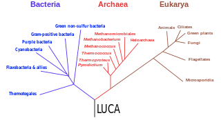
The three-domain system is a biological classification introduced by Carl Woese, Otto Kandler, and Mark Wheelis in 1990 that divides cellular life forms into three domains, namely Archaea, Bacteria, and Eukarya. The key difference from earlier classifications such as the two-empire system and the five-kingdom classification is the splitting of Archaea from Bacteria as completely different organisms. It has been challenged by the two-domain system that divides organisms into Bacteria and Archaea only, as Eukaryotes are considered as one group of Archaea.

The periplasm is a concentrated gel-like matrix in the space between the inner cytoplasmic membrane and the bacterial outer membrane called the periplasmic space in gram-negative bacteria. Using cryo-electron microscopy it has been found that a much smaller periplasmic space is also present in gram-positive bacteria, between cell wall and the plasma membrane. The periplasm may constitute up to 40% of the total cell volume of gram-negative bacteria, but is a much smaller percentage in gram-positive bacteria.

<span class="mw-page-title-main">Planctomycetota</span> Phylum of aquatic bacteria

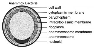
The Planctomycetota are a phylum of widely distributed bacteria, occurring in both aquatic and terrestrial habitats. They play a considerable role in global carbon and nitrogen cycles, with many species of this phylum capable of anaerobic ammonium oxidation, also known as anammox. Many Planctomycetota occur in relatively high abundance as biofilms, often associating with other organisms such as macroalgae and marine sponges.

The bacterium, despite its simplicity, contains a well-developed cell structure which is responsible for some of its unique biological structures and pathogenicity. Many structural features are unique to bacteria and are not found among archaea or eukaryotes. Because of the simplicity of bacteria relative to larger organisms and the ease with which they can be manipulated experimentally, the cell structure of bacteria has been well studied, revealing many biochemical principles that have been subsequently applied to other organisms.

<span class="mw-page-title-main">Alpha solenoid</span>

An alpha solenoid is a protein fold composed of repeating alpha helix subunits, commonly helix-turn-helix motifs, arranged in antiparallel fashion to form a superhelix. Alpha solenoids are known for their flexibility and plasticity. Like beta propellers, alpha solenoids are a form of solenoid protein domain commonly found in the proteins comprising the nuclear pore complex. They are also common in membrane coat proteins known as coatomers, such as clathrin, and in regulatory proteins that form extensive protein-protein interactions with their binding partners. Examples of alpha solenoid structures binding RNA and lipids have also been described.

<span class="mw-page-title-main">PVC superphylum</span> Superphylum of bacteria

The PVC superphylum is a superphylum of bacteria named after its three important members, Planctomycetota, Verrucomicrobiota, and Chlamydiota. Cavalier-Smith postulated that the PVC bacteria probably lost or reduced their peptidoglycan cell wall twice. It has been hypothesised that a member of the PVC clade might have been the host cell in the endosymbiotic event that gave rise to the first proto-eukaryotic cell.

<span class="mw-page-title-main">Prokaryote</span> Unicellular organism lacking a membrane-bound nucleus

A prokaryote is a single-cell organism whose cell lacks a nucleus and other membrane-bound organelles. The word prokaryote comes from the Ancient Greek πρό 'before' and κάρυον 'nut, kernel'. In the two-empire system arising from the work of Édouard Chatton, prokaryotes were classified within the empire Prokaryota. But in the three-domain system, based upon molecular analysis, prokaryotes are divided into two domains: Bacteria and Archaea. Organisms with nuclei are placed in a third domain, Eukaryota.

<span class="mw-page-title-main">Eukaryote</span> Domain of life whose cells have nuclei

The eukaryotes constitute the domain of Eukarya, organisms whose cells have a membrane-bound nucleus. All animals, plants, fungi, and many unicellular organisms are eukaryotes. They constitute a major group of life forms alongside the two groups of prokaryotes: the Bacteria and the Archaea. Eukaryotes represent a small minority of the number of organisms, but given their generally much larger size, their collective global biomass is much larger than that of prokaryotes.

<span class="mw-page-title-main">Cell membrane</span> Biological membrane that separates the interior of a cell from its outside environment

The cell membrane is a biological membrane that separates and protects the interior of a cell from the outside environment. The cell membrane consists of a lipid bilayer, made up of two layers of phospholipids with cholesterols interspersed between them, maintaining appropriate membrane fluidity at various temperatures. The membrane also contains membrane proteins, including integral proteins that span the membrane and serve as membrane transporters, and peripheral proteins that loosely attach to the outer (peripheral) side of the cell membrane, acting as enzymes to facilitate interaction with the cell's environment. Glycolipids embedded in the outer lipid layer serve a similar purpose. The cell membrane controls the movement of substances in and out of a cell, being selectively permeable to ions and organic molecules. In addition, cell membranes are involved in a variety of cellular processes such as cell adhesion, ion conductivity, and cell signalling and serve as the attachment surface for several extracellular structures, including the cell wall and the carbohydrate layer called the glycocalyx, as well as the intracellular network of protein fibers called the cytoskeleton. In the field of synthetic biology, cell membranes can be artificially reassembled.

The following outline is provided as an overview of and topical guide to life forms:

Membrane vesicle trafficking in eukaryotic animal cells involves movement of biochemical signal molecules from synthesis-and-packaging locations in the Golgi body to specific release locations on the inside of the plasma membrane of the secretory cell. It takes place in the form of Golgi membrane-bound micro-sized vesicles, termed membrane vesicles (MVs).

<span class="mw-page-title-main">Poribacteria</span> Phylum of bacteria

Poribacteria are a candidate phylum of bacteria originally discovered in the microbiome of marine sponges (Porifera). Poribacteria are Gram-negative primarily aerobic mixotrophs with the ability for oxidative phosphorylation, glycolysis, and autotrophic carbon fixation via the Wood – Ljungdahl pathway. Poribacterial heterotrophy is characterised by an enriched set of glycoside hydrolases, uronic acid degradation, as well as several specific sulfatases. This heterotrophic repertoire of poribacteria was suggested to be involved in the degradation of the extracellular sponge host matrix.

<span class="mw-page-title-main">Lokiarchaeota</span> Phylum of archaea

Lokiarchaeota is a proposed phylum of the Archaea. The phylum includes all members of the group previously named Deep Sea Archaeal Group, also known as Marine Benthic Group B. Lokiarchaeota is part of the superphylum Asgard containing the phyla: Lokiarchaeota, Thorarchaeota, Odinarchaeota, Heimdallarchaeota, and Helarchaeota. A phylogenetic analysis disclosed a monophyletic grouping of the Lokiarchaeota with the eukaryotes. The analysis revealed several genes with cell membrane-related functions. The presence of such genes support the hypothesis of an archaeal host for the emergence of the eukaryotes; the eocyte-like scenarios.

<span class="mw-page-title-main">Parkeol</span> Chemical compound

Parkeol is a relatively uncommon sterol secondary metabolite found mostly in plants, particularly noted in Butyrospermum parkii. It can be synthesized as a minor product by several oxidosqualene cyclase enzymes, and is the sole product of the enzyme parkeol synthase.

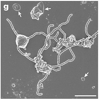
Plesiocystis pacifica is a species of marine myxobacteria. Like other members of this order, P. pacifica is a rod-shaped Gram-negative bacterium that can move by gliding and can form aggregates of cells called fruiting bodies. The species was first described in 2003, based on two strains isolated from samples collected from the Pacific coast of Japan.

<span class="mw-page-title-main">Bacterial secretion system</span> Protein complexes present on the cell membranes of bacteria for secretion of substances

Bacterial secretion systems are protein complexes present on the cell membranes of bacteria for secretion of substances. Specifically, they are the cellular devices used by pathogenic bacteria to secrete their virulence factors to invade the host cells. They can be classified into different types based on their specific structure, composition and activity. Generally, proteins can be secreted through two different processes. One process is a one-step mechanism in which proteins from the cytoplasm of bacteria are transported and delivered directly through the cell membrane into the host cell. Another involves a two-step activity in which the proteins are first transported out of the inner cell membrane, then deposited in the periplasm, and finally through the outer cell membrane into the host cell.

"Candidatus Brocadia" is a candidatus genus of bacteria, meaning that while it is well-characterized, it has not been grown as a pure culture yet. Due to this, much of what is known about Candidatus species has been discovered using culture-independent techniques such as metagenomic sequence analysis.

References

  1. 1 2 Santarella-Mellwig, Rachel; Franke, Josef; Jaedicke, Andreas; Gorjanacz, Matyas; Bauer, Ulrike; Budd, Aidan; Mattaj, Iain W.; Devos, Damien P.; Schmid, Sandra L. (19 January 2010). "The Compartmentalized Bacteria of the Planctomycetes-Verrucomicrobia-Chlamydiae Superphylum Have Membrane Coat-Like Proteins". PLOS Biology. 8 (1): e1000281. doi: 10.1371/journal.pbio.1000281 . PMC   2799638 . PMID   20087413.
  2. 1 2 3 4 5 6 Franzmann, P. D.; Skerman, V. B. D. (May 1984). "Gemmata obscuriglobus, a new genus and species of the budding bacteria". Antonie van Leeuwenhoek. 50 (3): 261–268. doi:10.1007/BF02342136. PMID   6486770. S2CID   24707205.
  3. "Gemmata obscuriglobus". Integrated Taxonomic Information System . Retrieved 30 October 2015.
  4. 1 2 3 4 Devos, Damien P. (September 2013). "Gemmata obscuriglobus". Current Biology. 23 (17): R705–R707. doi: 10.1016/j.cub.2013.07.013 . PMID   24028944.
  5. 1 2 3 4 Santarella-Mellwig, R; Pruggnaller, S; Roos, N; Mattaj, IW; Devos, DP (2013). "Three-dimensional reconstruction of bacteria with a complex endomembrane system". PLOS Biology. 11 (5): e1001565. doi: 10.1371/journal.pbio.1001565 . PMC   3660258 . PMID   23700385.
  6. 1 2 Sagulenko, E; Morgan, GP; Webb, RI; Yee, B; Lee, KC; Fuerst, JA (2014). "Structural studies of planctomycete Gemmata obscuriglobus support cell compartmentalisation in a bacterium". PLOS ONE. 9 (3): e91344. Bibcode:2014PLoSO...991344S. doi: 10.1371/journal.pone.0091344 . PMC   3954628 . PMID   24632833.
  7. Fuerst, JA; Webb, RI (15 September 1991). "Membrane-bounded nucleoid in the eubacterium Gemmata obscuriglobus". Proceedings of the National Academy of Sciences of the United States of America. 88 (18): 8184–8. Bibcode:1991PNAS...88.8184F. doi: 10.1073/pnas.88.18.8184 . PMC   52471 . PMID   11607213.
  8. Fuerst, JA (July 1995). "The planctomycetes: emerging models for microbial ecology, evolution and cell biology" (PDF). Microbiology. 141 (7): 1493–506. doi:10.1099/13500872-141-7-1493. PMID   7551018.
  9. Fuerst, John A. (2010). "Beyond Prokaryotes and Eukaryotes: Planctomycetes and Cell Organization". Nature Education. 3 (9): 44.
  10. McInerney, JO; Martin, WF; Koonin, EV; Allen, JF; Galperin, MY; Lane, N; Archibald, JM; Embley, TM (November 2011). "Planctomycetes and eukaryotes: a case of analogy not homology". BioEssays. 33 (11): 810–7. doi:10.1002/bies.201100045. PMC   3795523 . PMID   21858844.
  11. Fuerst, JA (October 2013). "The PVC superphylum: exceptions to the bacterial definition?". Antonie van Leeuwenhoek. 104 (4): 451–66. doi:10.1007/s10482-013-9986-1. PMID   23912444. S2CID   14283647.
  12. Devos, DP (February 2014). "Re-interpretation of the evidence for the PVC cell plan supports a Gram-negative origin". Antonie van Leeuwenhoek. 105 (2): 271–4. doi:10.1007/s10482-013-0087-y. hdl: 10261/129395 . PMID   24292377. S2CID   16557669.
  13. 1 2 Devos, Damien P. (January 2014). "PVC bacteria: variation of, but not exception to, the Gram-negative cell plan". Trends in Microbiology. 22 (1): 14–20. doi:10.1016/j.tim.2013.10.008. hdl: 10261/129431 . PMID   24286661.
  14. 1 2 3 Fuerst, John A.; Sagulenko, Evgeny (August 2014). "Towards understanding the molecular mechanism of the endocytosis-like process in the bacterium Gemmata obscuriglobus". Biochimica et Biophysica Acta (BBA) - Molecular Cell Research. 1843 (8): 1732–1738. doi: 10.1016/j.bbamcr.2013.10.002 . PMID   24144586.
  15. Stackebrandt, Erko; Wehmeyer, Uta; Liesack, Werner (December 1986). "16S ribosomal RNA- and cell wall analysis of Gemmata obscuriglobus, a new member of the order Planctomycetales". FEMS Microbiology Letters. 37 (3): 289–292. doi: 10.1111/j.1574-6968.1986.tb01810.x .
  16. Mahat, Rajendra; Seebart, Corrine; Basile, Franco; Ward, Naomi L. (19 October 2015). "Global and targeted lipid analysis of Gemmata obscuriglobus reveals the presence of lipopolysaccharide, a signature of the classical Gram-negative outer membrane". Journal of Bacteriology. 198 (2): JB.00517–15. doi:10.1128/JB.00517-15. PMC   4751799 . PMID   26483522.
  17. Jeske, O; Schüler, M; Schumann, P; Schneider, A; Boedeker, C; Jogler, M; Bollschweiler, D; Rohde, M; Mayer, C; Engelhardt, H; Spring, S; Jogler, C (12 May 2015). "Planctomycetes do possess a peptidoglycan cell wall". Nature Communications. 6: 7116. Bibcode:2015NatCo...6.7116J. doi:10.1038/ncomms8116. PMC   4432640 . PMID   25964217.
  18. 1 2 3 Lee, Kuo-Chang; Webb, Rick I; Fuerst, John A (2009). "The cell cycle of the planctomycete Gemmata obscuriglobus with respect to cell compartmentalization". BMC Cell Biology. 10 (1): 4. doi: 10.1186/1471-2121-10-4 . PMC   2656463 . PMID   19144151.
  19. Kamneva, O. K.; Liberles, D. A.; Ward, N. L. (3 November 2010). "Genome-Wide Influence of Indel Substitutions on Evolution of Bacteria of the PVC Superphylum, Revealed Using a Novel Computational Method". Genome Biology and Evolution. 2: 870–886. doi:10.1093/gbe/evq071. PMC   3000692 . PMID   21048002.
  20. Yee, Benjamin; Sagulenko, Evgeny; Morgan, Garry P.; Webb, Richard I.; Fuerst, John A. (2012). "Electron tomography of the nucleoid of Gemmata obscuriglobus reveals complex liquid crystalline cholesteric structure". Frontiers in Microbiology. 3: 326. doi: 10.3389/fmicb.2012.00326 . PMC   3440768 . PMID   22993511.
  21. Lieber, A; Leis, A; Kushmaro, A; Minsky, A; Medalia, O (March 2009). "Chromatin organization and radio resistance in the bacterium Gemmata obscuriglobus". Journal of Bacteriology. 191 (5): 1439–45. doi:10.1128/jb.01513-08. PMC   2648202 . PMID   19074379.
  22. Gottshall, E. Y.; Seebart, C.; Gatlin, J. C.; Ward, N. L. (14 July 2014). "Spatially segregated transcription and translation in cells of the endomembrane-containing bacterium Gemmata obscuriglobus". Proceedings of the National Academy of Sciences. 111 (30): 11067–11072. Bibcode:2014PNAS..11111067G. doi: 10.1073/pnas.1409187111 . PMC   4121771 . PMID   25024214.
  23. 1 2 Pearson, A.; Budin, M.; Brocks, J. J. (5 December 2003). "Phylogenetic and biochemical evidence for sterol synthesis in the bacterium Gemmata obscuriglobus". Proceedings of the National Academy of Sciences. 100 (26): 15352–15357. Bibcode:2003PNAS..10015352P. doi: 10.1073/pnas.2536559100 . PMC   307571 . PMID   14660793.
  24. Desmond, E.; Gribaldo, S. (10 September 2009). "Phylogenomics of Sterol Synthesis: Insights into the Origin, Evolution, and Diversity of a Key Eukaryotic Feature". Genome Biology and Evolution. 1: 364–381. doi:10.1093/gbe/evp036. PMC   2817430 . PMID   20333205.
  25. Lonhienne, T. G. A.; Sagulenko, E.; Webb, R. I.; Lee, K.-C.; Franke, J.; Devos, D. P.; Nouwens, A.; Carroll, B. J.; Fuerst, J. A. (21 June 2010). "Endocytosis-like protein uptake in the bacterium Gemmata obscuriglobus". Proceedings of the National Academy of Sciences. 107 (29): 12883–12888. doi: 10.1073/pnas.1001085107 . PMC   2919973 . PMID   20566852.
  26. Jermy, Andrew (August 2010). "Evolution: Bacterial endocytosis uncovered". Nature Reviews Microbiology. 8 (8): 534–535. doi:10.1038/nrmicro2408. PMID   20665955. S2CID   12013205.