Related Research Articles

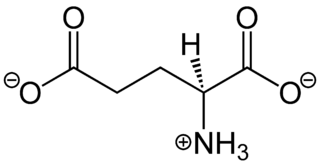
Glutamic acid is an α-amino acid that is used by almost all living beings in the biosynthesis of proteins. It is non-essential in humans, meaning the body can synthesize it. It is also an excitatory neurotransmitter, in fact the most abundant one, in the vertebrate nervous system. It serves as the precursor for the synthesis of the inhibitory gamma-aminobutyric acid (GABA) in GABA-ergic neurons.

Drosophila melanogaster is a species of fly in the family Drosophilidae. The species is known generally as the common fruit fly or vinegar fly. Starting with Charles W. Woodworth's proposal of the use of this species as a model organism, D. melanogaster continues to be widely used for biological research in genetics, physiology, microbial pathogenesis, and life history evolution. As of 2017, six Nobel prizes had been awarded for research using Drosophila.
Neurotoxicity is a form of toxicity in which a biological, chemical, or physical agent produces an adverse effect on the structure or function of the central and/or peripheral nervous system. It occurs when exposure to a substance – specifically, a neurotoxin or neurotoxicant– alters the normal activity of the nervous system in such a way as to cause permanent or reversible damage to nervous tissue. This can eventually disrupt or even kill neurons, which are cells that transmit and process signals in the brain and other parts of the nervous system. Neurotoxicity can result from organ transplants, radiation treatment, certain drug therapies, recreational drug use, and exposure to heavy metals, bites from certain species of venomous snakes, pesticides, certain industrial cleaning solvents, fuels and certain naturally occurring substances. Symptoms may appear immediately after exposure or be delayed. They may include limb weakness or numbness, loss of memory, vision, and/or intellect, uncontrollable obsessive and/or compulsive behaviors, delusions, headache, cognitive and behavioral problems and sexual dysfunction. Chronic mold exposure in homes can lead to neurotoxicity which may not appear for months to years of exposure. All symptoms listed above are consistent with mold mycotoxin accumulation.

Astrocytes, also known collectively as astroglia, are characteristic star-shaped glial cells in the brain and spinal cord. They perform many functions, including biochemical support of endothelial cells that form the blood–brain barrier, provision of nutrients to the nervous tissue, maintenance of extracellular ion balance and a role in the repair and scarring process of the brain and spinal cord following traumatic injuries. The proportion of astrocytes in the brain is not well defined; depending on the counting technique used, studies have found that the astrocyte proportion varies by region and ranges from 20% to 40% of all glia.
Molecular neuroscience is a branch of neuroscience that observes concepts in molecular biology applied to the nervous systems of animals. The scope of this subject covers topics such as molecular neuroanatomy, mechanisms of molecular signaling in the nervous system, the effects of genetics and epigenetics on neuronal development, and the molecular basis for neuroplasticity and neurodegenerative diseases. As with molecular biology, molecular neuroscience is a relatively new field that is considerably dynamic.

Glutamate receptors are synaptic and non synaptic receptors located primarily on the membranes of neuronal and glial cells. Glutamate is abundant in the human body, but particularly in the nervous system and especially prominent in the human brain where it is the body's most prominent neurotransmitter, the brain's main excitatory neurotransmitter, and also the precursor for GABA, the brain's main inhibitory neurotransmitter. Glutamate receptors are responsible for the glutamate-mediated postsynaptic excitation of neural cells, and are important for neural communication, memory formation, learning, and regulation.

Spectrin is a cytoskeletal protein that lines the intracellular side of the plasma membrane in eukaryotic cells. Spectrin forms pentagonal or hexagonal arrangements, forming a scaffold and playing an important role in maintenance of plasma membrane integrity and cytoskeletal structure. The hexagonal arrangements are formed by tetramers of spectrin subunits associating with short actin filaments at either end of the tetramer. These short actin filaments act as junctional complexes allowing the formation of the hexagonal mesh. The protein is named spectrin since it was first isolated as a major protein component of human red blood cells which had been treated with mild detergents; the detergents lysed the cells and the hemoglobin and other cytoplasmic components were washed out. In the light microscope the basic shape of the red blood cell could still be seen as the spectrin-containing submembranous cytoskeleton preserved the shape of the cell in outline. This became known as a red blood cell "ghost" (spectre), and so the major protein of the ghost was named spectrin.
Glutamate transporters are a family of neurotransmitter transporter proteins that move glutamate – the principal excitatory neurotransmitter – across a membrane. The family of glutamate transporters is composed of two primary subclasses: the excitatory amino acid transporter (EAAT) family and vesicular glutamate transporter (VGLUT) family. In the brain, EAATs remove glutamate from the synaptic cleft and extrasynaptic sites via glutamate reuptake into glial cells and neurons, while VGLUTs move glutamate from the cell cytoplasm into synaptic vesicles. Glutamate transporters also transport aspartate and are present in virtually all peripheral tissues, including the heart, liver, testes, and bone. They exhibit stereoselectivity for L-glutamate but transport both L-aspartate and D-aspartate.
The fruitless gene (fru) is a Drosophila melanogaster gene that encodes several variants of a putative transcription factor protein. Normal fruitless function is required for proper development of several anatomical structures necessary for courtship, including motor neurons which innervate muscles needed for fly sexual behaviors. The gene does not have an obvious mammalian homolog, but appears to function in sex determination in species as distant as the mosquito Anopheles gambiae.

Satellite glial cells(or satellite cells) are glial cells that cover the surface of neuron cell bodies in ganglia of the peripheral nervous system. Thus, they are found in sensory, sympathetic, and parasympathetic ganglia. Both satellite glial cells (SGCs) and Schwann cells are derived from the neural crest of the embryo during development. SGCs have been found to play a variety of roles, including control over the microenvironment of sympathetic ganglia. They are thought to have a similar role to astrocytes in the central nervous system (CNS). They supply nutrients to the surrounding neurons and also have some structural function. Satellite cells also act as protective, cushioning cells. Additionally, they express a variety of receptors that allow for a range of interactions with neuroactive chemicals. Many of these receptors and other ion channels have recently been implicated in health issues including chronic pain and herpes simplex. There is much more to be learned about these cells, and research surrounding additional properties and roles of the SGCs is ongoing.

Excitatory amino acid transporter 1 (EAAT1) is a protein that, in humans, is encoded by the SLC1A3 gene. EAAT1 is also often called the GLutamate ASpartate Transporter 1 (GLAST-1).

The GAL4-UAS system is a biochemical method used to study gene expression and function in organisms such as the fruit fly. It has also been adapted to study receptor chemical-binding functions in vitro in cell culture. It was developed by Hitoshi Kakidani and Mark Ptashne, and Nicholas Webster and Pierre Chambon in 1988, then adapted by Andrea Brand and Norbert Perrimon in 1993 and is considered a powerful technique for studying the expression of genes. The system has two parts: the Gal4 gene, encoding the yeast transcription activator protein Gal4, and the UAS, an enhancer to which GAL4 specifically binds to activate gene transcription.

Excitatory amino acid transporter 2 (EAAT2) also known as solute carrier family 1 member 2 (SLC1A2) and glutamate transporter 1 (GLT-1) is a protein that in humans is encoded by the SLC1A2 gene. Alternatively spliced transcript variants of this gene have been described, but their full-length nature is not known.

Excitatory amino acid transporter 3 (EAAT3), is a protein that in humans is encoded by the SLC1A1 gene.

Cystine/glutamate transporter is an antiporter that in humans is encoded by the SLC7A11 gene.

Notch proteins are a family of type-1 transmembrane proteins that form a core component of the Notch signaling pathway, which is highly conserved in metazoans. The Notch extracellular domain (NECD) mediates interactions with DSL family ligands, allowing it to participate in juxtacrine signaling.The Notch intracellular domain (NICD) acts as a transcriptional activator when in complex with CSL family transcription factors. Members of this Type 1 transmembrane protein family share several core structures, including an extracellular domain consisting of multiple epidermal growth factor (EGF)-like repeats and an intracellular domain transcriptional activation domain (TAD). Notch family members operate in a variety of different tissues and play a role in a variety of developmental processes by controlling cell fate decisions. Much of what is known about Notch function comes from studies done in Caenorhabditis elegans (C.elegans) and Drosophila melanogaster. Human homologs have also been identified, but details of Notch function and interactions with its ligands are not well known in this context.
Pigment dispersing factor (pdf) is a gene that encodes the protein PDF, which is part of a large family of neuropeptides. Its hormonal product, pigment dispersing hormone (PDH), was named for the diurnal pigment movement effect it has in crustacean retinal cells upon its initial discovery in the central nervous system of arthropods. The movement and aggregation of pigments in retina cells and extra-retinal cells is hypothesized to be under a split hormonal control mechanism. One hormonal set is responsible for concentrating chromatophoral pigment by responding to changes in the organism's exposure time to darkness. Another hormonal set is responsible for dispersion and responds to the light cycle. However, insect pdf genes do not function in such pigment migration since they lack the chromatophore.
Doubletime (dbt) also known as discs overgrown (dco) is a gene that encodes the double-time protein (DBT) in Drosophila melanogaster. The double-time protein is a kinase that phosphorylates PER protein that regulates the molecularly-driven, biological clock controlling circadian rhythm. The mammalian homolog of doubletime is casein kinase I epsilon. Different mutations in the dbt gene have been shown to cause lengthening, shortening, or complete loss in period of locomotor activity in flies. Drosophila and certain vertebrate Casein Kinase Id shows circadian function that has been evolutionary conserved over long time spans.

Michael Morris Rosbash is an American geneticist and chronobiologist. Rosbash is a professor and researcher at Brandeis University and investigator at the Howard Hughes Medical Institute. Rosbash's research group cloned the Drosophila period gene in 1984 and proposed the Transcription Translation Negative Feedback Loop for circadian clocks in 1990. In 1998, they discovered the cycle gene, clock gene, and cryptochrome photoreceptor in Drosophila through the use of forward genetics, by first identifying the phenotype of a mutant and then determining the genetics behind the mutation. Rosbash was elected to the National Academy of Sciences in 2003. Along with Michael W. Young and Jeffrey C. Hall, he was awarded the 2017 Nobel Prize in Physiology or Medicine "for their discoveries of molecular mechanisms controlling the circadian rhythm".
In neuroscience, glutamate refers to the anion of glutamic acid in its role as a neurotransmitter: a chemical that nerve cells use to send signals to other cells. It is by a wide margin the most abundant excitatory neurotransmitter in the vertebrate nervous system. It is used by every major excitatory function in the vertebrate brain, accounting in total for well over 90% of the synaptic connections in the human brain. It also serves as the primary neurotransmitter for some localized brain regions, such as cerebellum granule cells.
References
- 1 2 3 "Homosexuality Turned On and Off in Fruit Flies", LiveScience, 9 December 2007, accessed 10 December 2007
- ↑ Augustin H, Grosjean Y, Chen K, Sheng Q, Featherstone DE (2007). "Nonvesicular release of glutamate by glial xCT transporters suppresses glutamate receptor clustering in vivo". J. Neurosci. 27 (1): 111–23. doi:10.1523/JNEUROSCI.4770-06.2007. PMC 2193629 . PMID 17202478.
| This gene article is a stub. You can help Wikipedia by expanding it. |