Glutamate receptor

Last updated
The AMPA receptor bound to a glutamate antagonist showing the amino terminal, ligand binding, and transmembrane domain, PDB 3KG2 AMPA receptor.png
The AMPA receptor bound to a glutamate antagonist showing the amino terminal, ligand binding, and transmembrane domain, PDB 3KG2
Glutamic acid L-glutamic-acid-skeletal.png
Glutamic acid

Glutamate receptors are synaptic and non synaptic receptors located primarily on the membranes of neuronal and glial cells. [1] Glutamate (the conjugate base of glutamic acid) is abundant in the human body, but particularly in the nervous system and especially prominent in the human brain where it is the body's most prominent neurotransmitter, the brain's main excitatory neurotransmitter, and also the precursor for GABA, the brain's main inhibitory neurotransmitter. [2] Glutamate receptors are responsible for the glutamate-mediated postsynaptic excitation of neural cells, and are important for neural communication, memory formation, learning, and regulation.

Contents

Glutamate receptors are implicated in a number of neurological conditions. Their central role in excitotoxicity and prevalence in the central nervous system has been linked or speculated to be linked to many neurodegenerative diseases, and several other conditions have been further linked to glutamate receptor gene mutations or receptor autoantigen/antibody activity.

Function

Glutamate

Glutamate is the most prominent neurotransmitter in the body, and is the main excitatory neurotransmitter, being present in over 50% of nervous tissue. [2] [3] Glutamate was initially discovered to be a neurotransmitter in insect studies in the early 1960s.

Glutamate is also used by the brain to synthesize GABA (γ-Aminobutyric acid), the main inhibitory neurotransmitter of the mammalian central nervous system. GABA plays a role in regulating neuronal excitability throughout the nervous system and is also directly responsible for the regulation of muscle tone in humans. [2] [4]

Glutamate receptors

Mammalian glutamate receptors are classified based on their pharmacology. [5] However, glutamate receptors in other organisms have different pharmacology, and therefore these classifications do not hold. One of the major functions of glutamate receptors appears to be the modulation of synaptic plasticity, a property of the brain thought to be vital for memory and learning. Both metabotropic and ionotropic glutamate receptors have been shown to have an effect on synaptic plasticity. [6] An increase or decrease in the number of ionotropic glutamate receptors on a postsynaptic cell may lead to long-term potentiation or long-term depression of that cell, respectively. [7] [8] [9] Additionally, metabotropic glutamate receptors may modulate synaptic plasticity by regulating postsynaptic protein synthesis through second messenger systems. [10] Research shows that glutamate receptors are present in CNS glial cells as well as neurons. [11] These glutamate receptors are suggested to play a role in modulating gene expression in glial cells, both during the proliferation and differentiation of glial precursor cells in brain development and in mature glial cells. [12]

Glutamate receptors serve to facilitate the impact of the neurotransmitter glutamate in the central nervous system. These receptors are pivotal in excitatory synaptic transmission, synaptic plasticity, and neuronal development. They are vital for functions like learning, memory, and neuronal communication. [13] Various subtypes of glutamate receptors, such as NMDA (N-methyl-D-aspartate), AMPA (α-amino-3-hydroxy-5-methyl-4-isoxazolepropionic acid), and kainate receptors, have distinct roles in synaptic transmission and plasticity. [13] [14]

1. NMDA (N-methyl-D-aspartate) receptors: These receptors are involved in synaptic plasticity, learning, and memory. They are unique in that they require both glutamate and the co-agonist glycine to activate, and they are also voltage-dependent, meaning they only open when the postsynaptic membrane is depolarized. NMDA receptors are permeable to calcium ions, which can trigger intracellular signaling pathways that lead to changes in synaptic strength. [14]

2. AMPA (α-amino-3-hydroxy-5-methyl-4-isoxazolepropionic acid) receptors: These receptors mediate the majority of fast excitatory synaptic transmission in the brain. They are permeable to sodium and potassium ions and are responsible for the rapid depolarization of the postsynaptic membrane that underlies the excitatory postsynaptic potential (EPSP). AMPA receptors are also involved in synaptic plasticity, particularly in the early stages of long-term potentiation (LTP).

3. Kainate receptors: These receptors are involved in both pre- and postsynaptic signaling and are thought to play a role in regulating synaptic transmission and plasticity. They are activated by the neurotransmitter kainate and are permeable to both sodium and potassium ions. Kainate receptors are expressed in a variety of brain regions and are involved in processes such as sensory processing, motor control, and learning and memory.

Each subtype of glutamate receptor has a unique function and plays a crucial role in neuronal communication and plasticity. [14]

Types

Ionotropic glutamate receptors (iGluRs) form the ion channel pore that activates when glutamate binds to the receptor. Metabotropic glutamate receptors (mGluRs) affect the cell through a signal transduction cascade, and they may be primarily activating (mGlur1/5) or primarily inhibitory (mGlur2/3 and mGlur4/6/7/8). Ionotropic receptors tend to be quicker in relaying information, but metabotropic ones are associated with a more prolonged stimulus. The signalling cascade induced by metabotropic receptor activation means that even a relatively brief or small synaptic signal can have large and long-lasting effects, i.e. the system can have high "gain." NMDA receptor activation is particularly complex, as channel opening requires not only glutamate binding but also glycine or serine binding simultaneously at a separate site, and it also displays a degree of voltage dependence due to Zn2+ or Mg2+ binding in the pore. [15] Furthermore, Ca2+ currents through the NMDA receptor modulate not just the membrane potential but act as an important second messenger system. The particular dynamics of the NMDAR allow it to function as a neural coincidence detector, and the NMDAR Ca2+ currents are critical in synaptic plasticity (LTP and LTD) and learning and memory in general. [15]

Of the many specific subtypes of glutamate receptors, it is customary to refer to primary subtypes by a chemical that binds to it more selectively than glutamate. The research, however, is ongoing, as subtypes are identified and chemical affinities measured. Several compounds are routinely used in glutamate receptor research and associated with receptor subtypes:

TypeNameAgonist(s)Antagonists
ionotropic NMDA receptor NMDA Ketamine
Kainate receptor Kainate UBP-302
AMPA receptor AMPA Perampanel
Group 1 metabotropic mGluR1, mGluR5 DHPG LY-344,545
Group 2 metabotropic mGluR2, mGluR3 DCG-IV LY-341,495
Group 3 metabotropic mGluR4, mGluR6, mGluR7, mGluR8 L-AP4 MMPIP (mGlur7)

Due to the diversity of glutamate receptors, their subunits are encoded by numerous gene families. Sequence similarities between mammals show a common evolutionary origin for many mGluR and all iGluR genes. [16] Conservation of reading frames and splice sites of GluR genes between chimpanzees and humans is complete, suggesting no gross structural changes after humans diverged from the human-chimpanzee common ancestor. However, there is a possibility that two human-specific "fixed" amino acid substitutions, D71G in GRIN3A and R727H in GRIN3B, are specifically associated with human brain function. [17]

Ionotropic

Mammalian ionotropic glutamate receptor subunits and their genes: [18] [19]

Mammalian receptor familySubunit

(Old nomenclature)

GeneChromosome
(human)
AMPA GluA1 (GluR1) GRIA1 5q33
GluA2 (GluR2) GRIA2 4q32-33
GluA3 (GluR3) GRIA3 Xq25-26
GluA4 (GluR4) GRIA4 11q22-23
Kainate GluK1 (GluR5) GRIK1 21q21.1-22.1
GluK2 (GluR6) GRIK2 6q16.3-q21
GluK3 (GluR7) GRIK3 1p34-p33
GluK4 (KA-1) GRIK4 11q22.3
GluK5 (KA-2) GRIK5 19q13.2
NMDA GluN1(NR1) GRIN1 9q34.3
GluN2A (NR2A) GRIN2A 16p13.2
GluN2B (NR2B) GRIN2B 12p12
GluN2C (NR2C) GRIN2C 17q24-q25
GluN2D (NR2D) GRIN2D 19q13.1qter
GluN3A (NR3A) GRIN3A 9q31.1
GluN3B (NR3B) GRIN3B 19p13.3

Metabotropic

Mammalian metabotropic glutamate receptors are all named mGluR# and are further broken down into three groups:

GroupReceptorGeneChromosome
(human)
Effect
1mGluR1 GRM1 6q24Increase in Ca2+ concentration in the cytoplasm.
mGluR5 GRM5 11q14.3Release of K+ from the cell by activating K+ ionic channels
2mGluR2 GRM2 3p21.2Inhibition of adenylyl cyclase causing shutdown of the cAMP-dependent pathway
And therefore decreasing amount of cAMP
mGluR3 GRM3 7q21.1-q21.2
3mGluR4 GRM4 6p21.3Activation of Ca2+ channels, allowing more Ca2+ to enter the cell [20]
mGluR6 GRM6 5q35
mGluR7 GRM7 3p26-p25
mGluR8 GRM8 7q31.3-q32.1

In other (non mammalian) organisms, the classification and subunit composition of glutamate receptors is different.

Structure, mechanism and function

Glutamate receptors exist primarily in the central nervous system. These receptors can be found on the dendrites of postsynaptic cells and bind to glutamate released into the synaptic cleft by presynaptic cells. They are also present on both astrocytes and oligodendrocytes. [12] Ionotropic and metabotropic glutamate receptors, with the exception of NMDA, are found on cultured glial cells, which can open in response to glutamate and cause cells to activate second messengers to regulate gene expression and release neuroactive compounds. [11] Furthermore, brain slices show glutamate receptors are ubiquitously expressed in both developing and mature astrocytes and oligodendrocytes in vivo. Because of this, glial glutamate receptors are thought to be vital for glial cell development. [12]

Ionotropic

Ionotropic glutamate receptors, by definition, are ligand-gated nonselective cation channels that allow the flow of K+, Na+ and sometimes Ca2+ in response to glutamate binding. (In C. elegans and Drosophila , invertebrate-specific subunits enable the flow of negative chloride ions rather than cations.) Upon binding, the agonist will stimulate direct action of the central pore of the receptor, an ion channel, allowing ion flow and causing excitatory postsynaptic current (EPSC). This current is depolarizing and, if enough glutamate receptors are activated, may trigger an action potential in the postsynaptic neuron. All produce excitatory postsynaptic current, but the speed and duration of the current is different for each type. NMDA receptors have an internal binding site for an Mg2+ ion, creating a voltage-dependent block, which is removed by outward flow of positive current. [21] Since the block must be removed by outward current flow, NMDA receptors rely on the EPSC produced by AMPA receptors to open. NMDA receptors are permeable to Ca2+, [22] which is an important cation in the nervous system [23] and has been linked to gene regulation. [24] The flow of Ca2+ through NMDA receptors is thought to cause both long-term potentiation (LTP, of synapse efficacy) and long-term depression (LTD) by transducing signaling cascades and regulating gene expression.

Metabotropic

Metabotropic glutamate receptors, which belong to subfamily C of G protein-coupled receptors are divided into three groups, with a total of eight subtypes (in mammals; this is not necessarily the case for most organisms). The mGluRs are composed of three distinct regions: the extracellular region, the transmembrane region, and the intracellular region. [25] The extracellular region is composed of a venus flytrap (VFT) module that binds glutamate, [26] and a cysteine-rich domain that is thought to play a role in transmitting the conformational change induced by ligand binding from in the VFT module to the transmembrane region. [25] The transmembrane region consists of seven transmembrane domains and connects the extracellular region to the intracellular region where G protein coupling occurs. [26] Glutamate binding to the extracellular region of an mGluR causes G proteins bound to the intracellular region to be phosphorylated, affecting multiple biochemical pathways and ion channels in the cell. [27] Because of this, mGluRs can both increase or decrease the excitability of the postsynaptic cell, thereby causing a wide range of physiological effects.

Outside the central nervous system

Glutamate receptors are thought to be responsible for the reception and transduction of umami taste stimuli. Taste receptors of the T1R family, belonging to the same class of GPCR as metabotropic glutamate receptors are involved. Additionally, the mGluRs, as well as ionotropic glutamate receptors in neural cells, have been found in taste buds and may contribute to the umami taste. [28] Numerous ionotropic glutamate receptor subunits are expressed by heart tissue, but their specific function is still unknown. Western blots and northern blots confirmed the presence of iGluRs in cardiac tissue. Immunohistochemistry localized the iGluRs to cardiac nerve terminals, ganglia, conducting fibers, and some myocardiocytes. [29] Glutamate receptors are (as mentioned above) also expressed in pancreatic islet cells. [30] AMPA iGluRs modulate the secretion of insulin and glucagon in the pancreas, opening the possibility of treatment of diabetes via glutamate receptor antagonists. [31] [32] Small unmyelinated sensory nerve terminals in the skin also express NMDA and non-NMDA receptors. Subcutaneous injections of receptor blockers in rats successfully analgesized skin from formalin-induced inflammation, raising possibilities of targeting peripheral glutamate receptors in the skin for pain treatment. [33]

General clinical implications

Specific medical conditions and symptoms are discussed below.

Autoimmunity and antibody interactions with glutamate receptors and their subunit genes

Various neurological disorders are accompanied by antibody or autoantigen activity associated with glutamate receptors or their subunit genes (e.g. GluR3 in Rasmussen's encephalitis, [34] and GluR2 in nonfamilial olivopontocerebellar degeneration). [35] In 1994 GluR3 was shown to act as an autoantigen in Rasmussen's encephalitis, leading to speculation that autoimmune activity might underlie the condition. [36] Such findings "suggest" links between glutamate receptors and autoimmune interactions are possible and may be significant in some degenerative diseases, [35] however the exact role of such antibodies in disease manifestation is still not entirely known. [37]

Excitotoxicity

Overstimulation of glutamate receptors causes neurodegeneration and neuronal damage through a process called excitotoxicity. Excessive glutamate, or excitotoxins acting on the same glutamate receptors, overactivate glutamate receptors (specifically NMDARs), causing high levels of calcium ions (Ca2+) to influx into the postsynaptic cell. [38]

High Ca2+ concentrations activate a cascade of cell degradation processes involving proteases, lipases, nitric oxide synthase, and a number of enzymes that damage cell structures often to the point of cell death. [39] Ingestion of or exposure to excitotoxins that act on glutamate receptors can induce excitotoxicity and cause toxic effects on the central nervous system. [40] This becomes a problem for cells, as it feeds into a cycle of positive feedback cell death.

Glutamate excitotoxicity triggered by overstimulation of glutamate receptors also contributes to intracellular oxidative stress. Proximal glial cells use a cystine/glutamate antiporter (xCT) to transport cystine into the cell and glutamate out. Excessive extracellular glutamate concentrations reverse xCT, so glial cells no longer have enough cystine to synthesize glutathione (GSH), an antioxidant. [41] Lack of GSH leads to more reactive oxygen species (ROSs) that damage and kill the glial cell, which then cannot reuptake and process extracellular glutamate. [42] This is another positive feedback in glutamate excitotoxicity. In addition, increased Ca2+ concentrations activate nitric oxide synthase (NOS) and the over-synthesis of nitric oxide (NO). High NO concentration damages mitochondria, leading to more energy depletion, and adds oxidative stress to the neuron as NO is a ROS. [43]

Neurodegeneration

In the case of traumatic brain injury or cerebral ischemia (e.g., cerebral infarction or hemorrhage), acute neurodegeneration caused by excitotoxicity may spread to proximal neurons through two processes. Hypoxia and hypoglycemia trigger bioenergetic failure; mitochondria stop producing ATP energy. Na+/K+-ATPase can no longer maintain sodium/potassium ion concentration gradients across the plasma membrane. Glutamate transporters (EAATs), which use the Na+/K+ gradient, reverse glutamate transport (efflux) in affected neurons and astrocytes, and depolarization increases downstream synaptic release of glutamate. [44] In addition, cell death via lysis or apoptosis releases cytoplasmic glutamate outside of the ruptured cell. [45] These two forms of glutamate release cause a continual cascade of excitotoxic cell death and further increased extracellular glutamate concentrations.

Glutamate receptors' significance in excitotoxicity also links it to many neurogenerative diseases. Conditions such as exposure to excitotoxins, old age, congenital predisposition, and brain trauma can trigger glutamate receptor activation and ensuing excitotoxic neurodegeneration. This damage to the central nervous system propagates symptoms associated with a number of diseases. [46]

Conditions with demonstrated associations to glutamate receptors

A number of diseases in humans have a proven association with genetic mutations of glutamate receptor genes, or autoantigen/antibody interactions with glutamate receptors or their genes. Glutamate receptors and impaired regulation (in particular, those resulting in excessive glutamate levels) are also one cause of excitotoxicity (described above), which itself has been implicated or associated with a number of specific neurodegenerative conditions where neural cell death or degradation within the brain occurs over time. [42] [46]

Excessive synaptic receptor stimulation by glutamate is directly related to many conditions. Magnesium is one of many antagonists at the glutamate receptor, and magnesium deficiencies have demonstrated relationships with many glutamate receptor-related conditions. [47]

Glutamate receptors have been found to have an influence in ischemia/stroke, seizures, Parkinson's disease, Huntington's disease, and aching, [48] addiction [49] and an association with both ADHD [50] and autism. [51]

In most cases these are areas of ongoing research.

Aching

Hyperalgesia is directly involved with spinal NMDA receptors. Administered NMDA antagonists in a clinical setting produce significant side effects, although more research is being done in intrathecal administration. [40] Since spinal NMDA receptors link the area of pain to the brain's pain processing center, the thalamus, these glutamate receptors are a prime target for treatment. One proposed way to cope with the pain is subconsciously through the visualization technique. [52] [ unreliable source? ]

Attention deficit hyperactivity disorder (ADHD)

In 2006 the glutamate receptor subunit gene GRIN2B (responsible for key functions in memory and learning) was associated with ADHD. [53] This followed earlier studies showing a link between glutamate modulation and hyperactivity (2001), [54] and then between the SLC1A3 solute carrier gene-encoding part of the glutamate transporter process that mapped to chromosome 5 (5p12) noted in multiple ADHD genome scans. [55]

Further mutations to four different metabotropic glutamate receptor genes were identified in a study of 1013 children with ADHD compared to 4105 controls with non-ADHD, replicated in a subsequent study of 2500 more patients. Deletions and duplications affected GRM1, GRM5, GRM7 and GRM8. The study concluded that "CNVs affecting metabotropic glutamate receptor genes were enriched across all cohorts (P = 2.1 × 10⁻⁹)", "over 200 genes interacting with glutamate receptors […] were collectively affected by CNVs", "major hubs of the (affected genes') network include TNIK50, GNAQ51, and CALM", and "the fact that children with ADHD are more likely to have alterations in these genes reinforces previous evidence that the GRM pathway is important in ADHD". [50]

A SciBX article in January 2012 commented that "UPenn and MIT teams have independently converged on mGluRs as players in ADHD and autism. The findings suggest agonizing mGluRs in patients with ADHD." [56]

Autism

The etiology of autism may include excessive glutamatergic mechanisms. In small studies, memantine has been shown to significantly improve language function and social behavior in children with autism. [57] [58] Research is underway on the effects of memantine in adults with autism spectrum disorders. [59]

A link between glutamate receptors and autism was also identified via the structural protein ProSAP1 SHANK2 and potentially ProSAP2 SHANK3. The study authors concluded that the study "illustrates the significant role glutamatergic systems play in autism" and "By comparing the data on ProSAP1/Shank2−/− mutants with ProSAP2/Shank3αβ−/− mice, we show that different abnormalities in synaptic glutamate receptor expression can cause alterations in social interactions and communication. Accordingly, we propose that appropriate therapies for autism spectrum disorders are to be carefully matched to the underlying synaptopathic phenotype." [51]

Diabetes

Diabetes is a peculiar case because it is influenced by glutamate receptors present outside of the central nervous system, and it also influences glutamate receptors in the central nervous system.

Diabetes mellitus, an endocrine disorder, induces cognitive impairment and defects of long-term potential in the hippocampus, interfering with synaptic plasticity. Defects of long-term potential in the hippocampus are due to abnormal glutamate receptors, to be specific the malfunctioning NMDA glutamate receptors during early stages of the disease. [60]

Research is being done to address the possibility of using hyperglycemia and insulin to regulate these receptors and restore cognitive functions. Pancreatic islets regulating insulin and glucagon levels also express glutamate receptors. [30] Treating diabetes via glutamate receptor antagonists is possible, but not much research has been done. The difficulty of modifying peripheral GluR without having detrimental effects on the central nervous system, which is saturated with GluR, may be the cause of this.

Huntington's disease

In 2004, a specific genotype of human GluR6 was discovered to have a slight influence on the age of onset of Huntington's disease. [61]

In addition to similar mechanisms causing Parkinson's disease with respect to NMDA or AMPA receptors, Huntington's disease was also proposed to exhibit metabolic and mitochondrial deficiency, which exposes striatal neurons to the over activation of NMDA receptors. [40] Using folic acid has been proposed as a possible treatment for Huntington's due to the inhibition it exhibits on homocysteine, which increases vulnerability of nerve cells to glutamate. [62] This could decrease the effect glutamate has on glutamate receptors and reduce cell response to a safer level, not reaching excitotoxicity.

Ischemia

During ischemia, the brain has been observed to have an unnaturally high concentration of extracellular glutamate. [63] This is linked to an inadequate supply of ATP, which drives the glutamate transport levels that keep the concentrations of glutamate in balance. [64] This usually leads to an excessive activation of glutamate receptors, which may lead to neuronal injury. After this overexposure, the postsynaptic terminals tend to keep glutamate around for long periods of time, which results in a difficulty in depolarization. [64] Antagonists for NMDA and AMPA receptors seem to have a large benefit, with more aid the sooner it is administered after onset of the neural ischemia. [65]

Multiple sclerosis

Inducing experimental autoimmune encephalomyelitis in animals as a model for multiple sclerosis(MS) has targeted some glutamate receptors as a pathway for potential therapeutic applications. [66] This research has found that a group of drugs interact with the NMDA, AMPA, and kainate glutamate receptor to control neurovascular permeability, inflammatory mediator synthesis, and resident glial cell functions including CNS myelination. Oligodendrocytes in the CNS myelinate axons; the myelination dysfunction in MS is partly due to the excitotoxicity of those cells. By regulating the drugs which interact with those glutamate receptors, regulating glutamate binding may be possible, and thereby reduce the levels of Ca2+ influx. The experiments showed improved oligodendrocyte survival, and remyelination increased. Furthermore, CNS inflammation, apoptosis, and axonal damage were reduced. [66]

Parkinson's disease (Parkinsonism)

Late onset neurological disorders, such as Parkinson's disease, may be partially due to glutamate binding NMDA and AMPA glutamate receptors. [40] In vitro spinal cord cultures with glutamate transport inhibitors led to degeneration of motor neurons, which was counteracted by some AMPA receptor antagonists such as GYKI 52466. [40] Research also suggests that the metabotropic glutamate receptor mGlu4 is directly involved in movement disorders associated with the basal ganglia through selectively modulating glutamate in the striatum. [67]

Rasmussen's encephalitis

In 1994, GluR3 was shown to act as an autoantigen in Rasmussen's encephalitis, leading to speculation that autoimmune activity might underlie the condition. [36]

Schizophrenia

In schizophrenia, the expression of the mRNA for the NR2A subunit of the NMDA glutamate receptor was found to be decreased in a subset of inhibitory interneurons in the cerebral cortex. [68] This is suggested by upregulation of GABA, an inhibitory neurotransmitter. In schizophrenia, the expression of the NR2A subunit of NDMA receptors in mRNA was experimentally undetectable in 49-73% in GABA neurons that usually express it. These are mainly in GABA cells expressing the calcium-buffering protein parvalbumin (PV), which exhibits fast-spiking firing properties and target the perisomatic (basket cells) and axo-axonic (chandelier cells) compartments of pyramidal neurons. [68] The study found the density of NR2A mRNA-expressing PV neurons was decreased by as much as 50% in subjects with schizophrenia.

In addition, density of immunohistochemically labeled glutamatergic terminals with an antibody against the vesicular glutamate transporter vGluT1 also exhibited a reduction that paralleled the reduction in the NR2A-expressing PV neurons. Together, these observations suggest glutamatergic innervation of PV-containing inhibitory neurons appears to be deficient in schizophrenia. [68] Expression of NR2A mRNA has also been found to be altered in the inhibitory neurons that contain another calcium buffer, calbindin, targeting the dendrites of pyramidal neurons, [69] and the expression of the mRNA for the GluR5 kainate receptor in GABA neurons has also been found to be changed in people with schizophrenia. [70]

Current research is targeting glutamate receptor antagonists as potential treatments for schizophrenia. Memantine, a weak, nonselective NMDA receptor antagonist, was used as an add-on to clozapine therapy in a clinical trial. Refractory schizophrenia patients showed associated improvements in both negative and positive symptoms, underscoring the potential uses of GluR antagonists as antipsychotics. [71] Furthermore, administration of noncompetitive NMDA receptor antagonists have been tested on rat models. Scientists have proposed that specific antagonists can act on GABAergic interneurons, enhancing cortical inhibition and preventing excessive glutamatergic transmission associated with schizophrenia. These and other atypical antipsychotic drugs can be used together to inhibit excessive excitability in pyramidal cells, decreasing the symptoms of schizophrenia. [72]

Seizures

Glutamate receptors have been discovered to have a role in the onset of epilepsy. NMDA and metabotropic types have been found to induce epileptic convulsions. Using rodent models, labs have found that the introduction of antagonists to these glutamate receptors helps counteract the epileptic symptoms. [73] Since glutamate is a ligand for ligand-gated ion channels, the binding of this neurotransmitter will open gates and increase sodium and calcium conductance. These ions play an integral part in the causes of seizures. Group 1 metabotropic glutamate receptors (mGlu1 and mGlu5) are the primary cause of seizing, so applying an antagonist to these receptors helps in preventing convulsions. [74]

Neurodegenerative diseases suspected to have a link mediated (at least in part) through stimulation of glutamate receptors: [38] [75]

See also

Related Research Articles

<span class="mw-page-title-main">Neurotransmitter receptor</span> Type of protein

A neurotransmitter receptor is a membrane receptor protein that is activated by a neurotransmitter. Chemicals on the outside of the cell, such as a neurotransmitter, can bump into the cell's membrane, in which there are receptors. If a neurotransmitter bumps into its corresponding receptor, they will bind and can trigger other events to occur inside the cell. Therefore, a membrane receptor is part of the molecular machinery that allows cells to communicate with one another. A neurotransmitter receptor is a class of receptors that specifically binds with neurotransmitters as opposed to other molecules.

<span class="mw-page-title-main">AMPA receptor</span> Transmembrane protein family

The α-amino-3-hydroxy-5-methyl-4-isoxazolepropionic acid receptor (also known as AMPA receptor, AMPAR, or quisqualate receptor) is an ionotropic transmembrane receptor for glutamate (iGluR) and predominantly Na+ ion channel that mediates fast synaptic transmission in the central nervous system (CNS). It has been traditionally classified as a non-NMDA-type receptor, along with the kainate receptor. Its name is derived from its ability to be activated by the artificial glutamate analog AMPA. The receptor was first named the "quisqualate receptor" by Watkins and colleagues after a naturally occurring agonist quisqualate and was only later given the label "AMPA receptor" after the selective agonist developed by Tage Honore and colleagues at the Royal Danish School of Pharmacy in Copenhagen. The GRIA2-encoded AMPA receptor ligand binding core (GluA2 LBD) was the first glutamate receptor ion channel domain to be crystallized.

<span class="mw-page-title-main">NMDA receptor</span> Glutamate receptor and ion channel protein found in nerve cells

The N-methyl-D-aspartatereceptor (also known as the NMDA receptor or NMDAR), is a glutamate receptor and predominantly Ca2+ ion channel found in neurons. The NMDA receptor is one of three types of ionotropic glutamate receptors, the other two being AMPA and kainate receptors. Depending on its subunit composition, its ligands are glutamate and glycine (or D-serine). However, the binding of the ligands is typically not sufficient to open the channel as it may be blocked by Mg2+ ions which are only removed when the neuron is sufficiently depolarized. Thus, the channel acts as a "coincidence detector" and only once both of these conditions are met, the channel opens and it allows positively charged ions (cations) to flow through the cell membrane. The NMDA receptor is thought to be very important for controlling synaptic plasticity and mediating learning and memory functions.

In neurophysiology, long-term depression (LTD) is an activity-dependent reduction in the efficacy of neuronal synapses lasting hours or longer following a long patterned stimulus. LTD occurs in many areas of the CNS with varying mechanisms depending upon brain region and developmental progress.

<span class="mw-page-title-main">Excitatory synapse</span> Sort of synapse

An excitatory synapse is a synapse in which an action potential in a presynaptic neuron increases the probability of an action potential occurring in a postsynaptic cell. Neurons form networks through which nerve impulses travels, each neuron often making numerous connections with other cells of neurons. These electrical signals may be excitatory or inhibitory, and, if the total of excitatory influences exceeds that of the inhibitory influences, the neuron will generate a new action potential at its axon hillock, thus transmitting the information to yet another cell.

<span class="mw-page-title-main">Brain-derived neurotrophic factor</span> Protein found in humans

Brain-derived neurotrophic factor (BDNF), or abrineurin, is a protein that, in humans, is encoded by the BDNF gene. BDNF is a member of the neurotrophin family of growth factors, which are related to the canonical nerve growth factor (NGF), a family which also includes NT-3 and NT-4/NT-5. Neurotrophic factors are found in the brain and the periphery. BDNF was first isolated from a pig brain in 1982 by Yves-Alain Barde and Hans Thoenen.

<span class="mw-page-title-main">Ligand-gated ion channel</span> Type of ion channel transmembrane protein

Ligand-gated ion channels (LICs, LGIC), also commonly referred to as ionotropic receptors, are a group of transmembrane ion-channel proteins which open to allow ions such as Na+, K+, Ca2+, and/or Cl to pass through the membrane in response to the binding of a chemical messenger (i.e. a ligand), such as a neurotransmitter.

Molecular neuroscience is a branch of neuroscience that observes concepts in molecular biology applied to the nervous systems of animals. The scope of this subject covers topics such as molecular neuroanatomy, mechanisms of molecular signaling in the nervous system, the effects of genetics and epigenetics on neuronal development, and the molecular basis for neuroplasticity and neurodegenerative diseases. As with molecular biology, molecular neuroscience is a relatively new field that is considerably dynamic.

<span class="mw-page-title-main">Kainate receptor</span> Class of ionotropic glutamate receptors

Kainate receptors, or kainic acid receptors (KARs), are ionotropic receptors that respond to the neurotransmitter glutamate. They were first identified as a distinct receptor type through their selective activation by the agonist kainate, a drug first isolated from the algae Digenea simplex. They have been traditionally classified as a non-NMDA-type receptor, along with the AMPA receptor. KARs are less understood than AMPA and NMDA receptors, the other ionotropic glutamate receptors. Postsynaptic kainate receptors are involved in excitatory neurotransmission. Presynaptic kainate receptors have been implicated in inhibitory neurotransmission by modulating release of the inhibitory neurotransmitter GABA through a presynaptic mechanism.

<span class="mw-page-title-main">Kainic acid</span> Chemical compound

Kainic acid, or kainate, is an acid that naturally occurs in some seaweed. Kainic acid is a potent neuroexcitatory amino acid agonist that acts by activating receptors for glutamate, the principal excitatory neurotransmitter in the central nervous system. Glutamate is produced by the cell's metabolic processes and there are four major classifications of glutamate receptors: NMDA receptors, AMPA receptors, kainate receptors, and the metabotropic glutamate receptors. Kainic acid is an agonist for kainate receptors, a type of ionotropic glutamate receptor. Kainate receptors likely control a sodium channel that produces excitatory postsynaptic potentials (EPSPs) when glutamate binds.

<span class="mw-page-title-main">Metabotropic glutamate receptor</span> Type of glutamate receptor

The metabotropic glutamate receptors, or mGluRs, are a type of glutamate receptor that are active through an indirect metabotropic process. They are members of the group C family of G-protein-coupled receptors, or GPCRs. Like all glutamate receptors, mGluRs bind with glutamate, an amino acid that functions as an excitatory neurotransmitter.

Metaplasticity is a term originally coined by W.C. Abraham and M.F. Bear to refer to the plasticity of synaptic plasticity. Until that time synaptic plasticity had referred to the plastic nature of individual synapses. However this new form referred to the plasticity of the plasticity itself, thus the term meta-plasticity. The idea is that the synapse's previous history of activity determines its current plasticity. This may play a role in some of the underlying mechanisms thought to be important in memory and learning such as long-term potentiation (LTP), long-term depression (LTD) and so forth. These mechanisms depend on current synaptic "state", as set by ongoing extrinsic influences such as the level of synaptic inhibition, the activity of modulatory afferents such as catecholamines, and the pool of hormones affecting the synapses under study. Recently, it has become clear that the prior history of synaptic activity is an additional variable that influences the synaptic state, and thereby the degree, of LTP or LTD produced by a given experimental protocol. In a sense, then, synaptic plasticity is governed by an activity-dependent plasticity of the synaptic state; such plasticity of synaptic plasticity has been termed metaplasticity. There is little known about metaplasticity, and there is much research currently underway on the subject, despite its difficulty of study, because of its theoretical importance in brain and cognitive science. Most research of this type is done via cultured hippocampus cells or hippocampal slices.

<i>N</i>-Acetylaspartylglutamic acid Peptide neurotransmitter

N-Acetylaspartylglutamic acid is a peptide neurotransmitter and the third-most-prevalent neurotransmitter in the mammalian nervous system. NAAG consists of N-acetylaspartic acid (NAA) and glutamic acid coupled via a peptide bond.

Gliotransmitters are chemicals released from glial cells that facilitate neuronal communication between neurons and other glial cells. They are usually induced from Ca2+ signaling, although recent research has questioned the role of Ca2+ in gliotransmitters and may require a revision of the relevance of gliotransmitters in neuronal signalling in general.

<span class="mw-page-title-main">GRIA2</span> Mammalian protein found in Homo sapiens

Glutamate ionotropic receptor AMPA type subunit 2 is a protein that in humans is encoded by the GRIA2 gene and it is a subunit found in the AMPA receptors.

<span class="mw-page-title-main">GRIK1</span> Protein-coding gene in the species Homo sapiens

Glutamate receptor, ionotropic, kainate 1, also known as GRIK1, is a protein that in humans is encoded by the GRIK1 gene.

In neuroscience, synaptic scaling is a form of homeostatic plasticity, in which the brain responds to chronically elevated activity in a neural circuit with negative feedback, allowing individual neurons to reduce their overall action potential firing rate. Where Hebbian plasticity mechanisms modify neural synaptic connections selectively, synaptic scaling normalizes all neural synaptic connections by decreasing the strength of each synapse by the same factor, so that the relative synaptic weighting of each synapse is preserved.

<span class="mw-page-title-main">Glutamate (neurotransmitter)</span> Anion of glutamic acid in its role as a neurotransmitter

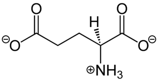
In neuroscience, glutamate is the anion of glutamic acid in its role as a neurotransmitter. It is by a wide margin the most abundant excitatory neurotransmitter in the vertebrate nervous system. It is used by every major excitatory function in the vertebrate brain, accounting in total for well over 90% of the synaptic connections in the human brain. It also serves as the primary neurotransmitter for some localized brain regions, such as cerebellum granule cells.

<span class="mw-page-title-main">Willardiine</span> Chemical compound

Willardiine (correctly spelled with two successive i's) or (S)-1-(2-amino-2-carboxyethyl)pyrimidine-2,4-dione is a chemical compound that occurs naturally in the seeds of Mariosousa willardiana and Acacia sensu lato. The seedlings of these plants contain enzymes capable of complex chemical substitutions that result in the formation of free amino acids (See:#Synthesis). Willardiine is frequently studied for its function in higher level plants. Additionally, many derivates of willardiine are researched for their potential in pharmaceutical development. Willardiine was first discovered in 1959 by R. Gmelin, when he isolated several free, non-protein amino acids from Acacia willardiana (another name for Mariosousa willardiana) when he was studying how these families of plants synthesize uracilyalanines. A related compound, Isowillardiine, was concurrently isolated by a different group, and it was discovered that the two compounds had different structural and functional properties. Subsequent research on willardiine has focused on the functional significance of different substitutions at the nitrogen group and the development of analogs of willardiine with different pharmacokinetic properties. In general, Willardiine is the one of the first compounds studied in which slight changes to molecular structure result in compounds with significantly different pharmacokinetic properties.

GRIN disorders are a group of neurodevelopmental disorders that result from mutations in genes coding for subunits of an N-methyl-D-aspartate (NMDA) receptor, which leads to dysfunction of glutamate signaling. GRIN disorders are universally characterized by a varying degree of developmental delay and intellectual disability, as well as epileptic seizures. Other clinical features vary depending on the affected gene and may include muscular hypotonia, spasticity, and movement disorders. GRIN disorders are confirmed with genetic testing and managed symptomatically since there is currently no cure for the disorder.

References

  1. Brassai A, Suvanjeiev RG, Bán EG, Lakatos M (March 2015). "Role of synaptic and nonsynaptic glutamate receptors in ischaemia induced neurotoxicity". Review. Brain Research Bulletin. 112 (March 2015): 1–6. doi:10.1016/j.brainresbull.2014.12.007. PMID   25540918. S2CID   11292811.
  2. 1 2 3 Petroff OA (December 2002). "GABA and glutamate in the human brain". Neuroscientist. 8 (6): 562–573. doi:10.1177/1073858402238515. PMID   12467378. S2CID   84891972.Glutamate is the main excitatory and GABA the main inhibitory neurotransmitter in the mammalian cortex
  3. "Glutamate Receptors - Structures and Functions". Centre of Synaptic Plasticity. University of Bristol. 2007-01-04. Archived from the original on 2007-09-15. Retrieved 2009-12-07.
  4. Watanabe M, Maemura K, Kanbara K, Tamayama T, Hayasaki H (2002). "GABA and GABA receptors in the central nervous system and other organs". A Survey of Cell Biology. International Review of Cytology. Vol. 213. pp. 1–47. doi:10.1016/S0074-7696(02)13011-7. ISBN   978-0-12-364617-0. PMID   11837891.{{cite book}}: |journal= ignored (help)
  5. Reiner A, Levitz J (June 2018). "Glutamatergic Signaling in the Central Nervous System: Ionotropic and Metabotropic Receptors in Concert". Neuron. 98 (6): 1080–1098. doi:10.1016/j.neuron.2018.05.018. PMC   6484838 . PMID   29953871.
  6. Debanne D, Daoudal G, Sourdet V, Russier M (2003). "Brain plasticity and ion channels". Journal of Physiology, Paris. 97 (4–6): 403–14. doi:10.1016/j.jphysparis.2004.01.004. PMID   15242652. S2CID   19116187.
  7. Maren S, Tocco G, Standley S, Baudry M, Thompson RF (October 1993). "Postsynaptic factors in the expression of long-term potentiation (LTP): increased glutamate receptor binding following LTP induction in vivo". Proceedings of the National Academy of Sciences of the United States of America. 90 (20): 9654–8. Bibcode:1993PNAS...90.9654M. doi: 10.1073/pnas.90.20.9654 . PMC   47628 . PMID   8415757.
  8. Pérez-Otaño I, Ehlers MD (May 2005). "Homeostatic plasticity and NMDA receptor trafficking". Trends in Neurosciences. 28 (5): 229–38. doi:10.1016/j.tins.2005.03.004. PMID   15866197. S2CID   22901201.
  9. Asztély F, Gustafsson B (February 1996). "Ionotropic glutamate receptors. Their possible role in the expression of hippocampal synaptic plasticity". Molecular Neurobiology. 12 (1): 1–11. doi:10.1007/BF02740744. PMID   8732537. S2CID   42232786.
  10. Weiler IJ, Greenough WT (August 1993). "Metabotropic glutamate receptors trigger postsynaptic protein synthesis". Proceedings of the National Academy of Sciences of the United States of America. 90 (15): 7168–71. Bibcode:1993PNAS...90.7168W. doi: 10.1073/pnas.90.15.7168 . PMC   47097 . PMID   8102206.
  11. 1 2 Teichberg VI (December 1991). "Glial glutamate receptors: likely actors in brain signaling". FASEB Journal. 5 (15): 3086–91. doi: 10.1096/fasebj.5.15.1660422 . PMID   1660422. S2CID   15094280.
  12. 1 2 3 Steinhäuser C, Gallo V (August 1996). "News on glutamate receptors in glial cells". Trends in Neurosciences. 19 (8): 339–45. doi:10.1016/0166-2236(96)10043-6. PMID   8843603. S2CID   31596399.
  13. 1 2 Hansen, Kasper B.; Wollmuth, Lonnie P.; Bowie, Derek; Furukawa, Hiro; Menniti, Frank S.; Sobolevsky, Alexander I.; Swanson, Geoffrey T.; Swanger, Sharon A.; Greger, Ingo H.; Nakagawa, Terunaga; McBain, Chris J.; Jayaraman, Vasanthi; Low, Chian-Ming; Dell'Acqua, Mark L.; Diamond, Jeffrey S. (October 2021). "Structure, Function, and Pharmacology of Glutamate Receptor Ion Channels". Pharmacological Reviews. 73 (4): 298–487. doi:10.1124/pharmrev.120.000131. ISSN   1521-0081. PMC   8626789 . PMID   34753794.
  14. 1 2 3 Hansen, Kasper B.; Yi, Feng; Perszyk, Riley E.; Furukawa, Hiro; Wollmuth, Lonnie P.; Gibb, Alasdair J.; Traynelis, Stephen F. (2018-08-06). "Structure, function, and allosteric modulation of NMDA receptors". The Journal of General Physiology. 150 (8): 1081–1105. doi:10.1085/jgp.201812032. ISSN   1540-7748. PMC   6080888 . PMID   30037851.
  15. 1 2 Kandel ER (2013). Principles of neural science. McGraw Hill Professional. ISBN   9780071390118. OCLC   970875967.
  16. Suchanek B, Seeburg PH, Sprengel R (January 1995). "Gene structure of the murine N-methyl D-aspartate receptor subunit NR2C". J. Biol. Chem. 270 (1): 41–4. doi: 10.1074/jbc.270.1.41 . PMID   7814402. S2CID   6758394.
  17. Goto H, Watanabe K, Araragi N, Kageyama R, Tanaka K, Kuroki Y, Toyoda A, Hattori M, Sakaki Y, Fujiyama A, Fukumaki Y, Shibata H (2009). "The identification and functional implications of human-specific "fixed" amino acid substitutions in the glutamate receptor family". BMC Evol. Biol. 9 (1): 224. Bibcode:2009BMCEE...9..224G. doi: 10.1186/1471-2148-9-224 . PMC   2753569 . PMID   19737383.
  18. Dingledine R, Borges K, Bowie D, Traynelis SF (March 1999). "The glutamate receptor ion channels". Pharmacol. Rev. 51 (1): 7–61. PMID   10049997.
  19. Andersson O, Stenqvist A, Attersand A, von Euler G (December 2001). "Nucleotide sequence, genomic organization, and chromosomal localization of genes encoding the human NMDA receptor subunits NR3A and NR3B". Genomics. 78 (3): 178–84. doi:10.1006/geno.2001.6666. PMID   11735224.
  20. Conn PJ, et al. (2005). "Metabotropic glutamate receptors in the basal ganglia motor circuit". Nature Reviews Neuroscience. 6 (10): 787–98. doi:10.1038/nrn1763. PMID   16276355. S2CID   9473598.
  21. Johnson JW, Ascher P (May 1990). "Voltage-dependent block by intracellular Mg2+ of N-methyl-D-aspartate-activated channels". Biophys. J. 57 (5): 1085–90. Bibcode:1990BpJ....57.1085J. doi:10.1016/S0006-3495(90)82626-6. PMC   1280813 . PMID   1692749.
  22. Crowder JM, Croucher MJ, Bradford HF, Collins JF (June 1987). "Excitatory amino acid receptors and depolarization-induced Ca2+ influx into hippocampal slices". J. Neurochem. 48 (6): 1917–24. doi:10.1111/j.1471-4159.1987.tb05756.x. PMID   2437250. S2CID   12586076.
  23. Gover TD, Moreira TH, Weinreich D (2009). "Role of Calcium in Regulating Primary Sensory Neuronal Excitability". Sensory Nerves. Handbook of Experimental Pharmacology. Vol. 194. pp. 563–87. doi:10.1007/978-3-540-79090-7_16. ISBN   978-3-540-79089-1. PMID   19655118.
  24. Barbado M, Fablet K, Ronjat M, De Waard M (June 2009). "Gene regulation by voltage-dependent calcium channels" (PDF). Biochim. Biophys. Acta. 1793 (6): 1096–104. doi:10.1016/j.bbamcr.2009.02.004. PMID   19250948. S2CID   2807929.
  25. 1 2 Muto T, Tsuchiya D, Morikawa K, Jingami H (March 2007). "Structures of the extracellular regions of the group II/III metabotropic glutamate receptors". Proc. Natl. Acad. Sci. U.S.A. 104 (10): 3759–64. Bibcode:2007PNAS..104.3759M. doi: 10.1073/pnas.0611577104 . PMC   1820657 . PMID   17360426.
  26. 1 2 Pin JP, Acher F (June 2002). "The metabotropic glutamate receptors: structure, activation mechanism and pharmacology". Curr Drug Targets CNS Neurol Disord. 1 (3): 297–317. doi:10.2174/1568007023339328. PMID   12769621.
  27. Platt SR (2007). "The role of glutamate in central nervous system health and disease--a review". Vet. J. 173 (2): 278–86. doi:10.1016/j.tvjl.2005.11.007. PMID   16376594.
  28. Kinnamon SC, Vandenbeuch A (July 2009). "Receptors and transduction of umami taste stimuli". Ann. N. Y. Acad. Sci. 1170 (1): 55–9. Bibcode:2009NYASA1170...55K. doi:10.1111/j.1749-6632.2009.04106.x. PMID   19686108. S2CID   20962248.
  29. Gill SS, Pulido OM, Mueller RW, McGuire PF (July 1998). "Molecular and immunochemical characterization of the ionotropic glutamate receptors in the rat heart". Brain Res. Bull. 46 (5): 429–34. doi:10.1016/S0361-9230(98)00012-4. PMID   9739005. S2CID   36044371.
  30. 1 2 Weaver CD, Yao TL, Powers AC, Verdoorn TA (May 1996). "Differential expression of glutamate receptor subtypes in rat pancreatic islets". J. Biol. Chem. 271 (22): 12977–84. doi: 10.1074/jbc.271.22.12977 . PMID   8662728. S2CID   36816887.
  31. Bertrand G, Gross R, Puech R, Loubatières-Mariani MM, Bockaert J (June 1993). "Glutamate stimulates glucagon secretion via an excitatory amino acid receptor of the AMPA subtype in rat pancreas". Eur. J. Pharmacol. 237 (1): 45–50. doi:10.1016/0014-2999(93)90091-U. PMID   7689469.
  32. Weaver CD, Gundersen V, Verdoorn TA (January 1998). "A high affinity glutamate/aspartate transport system in pancreatic islets of Langerhans modulates glucose-stimulated insulin secretion". J. Biol. Chem. 273 (3): 1647–53. doi: 10.1074/jbc.273.3.1647 . PMID   9430708. S2CID   27129532.
  33. Carlton SM, Hargett GL, Coggeshall RE (September 1995). "Localization and activation of glutamate receptors in unmyelinated axons of rat glabrous skin". Neurosci. Lett. 197 (1): 25–8. doi:10.1016/0304-3940(95)11889-5. PMID   8545047. S2CID   44486341.
  34. Carlson NG, Gahring LC, Twyman RE, Rogers SW (April 1997). "Identification of amino acids in the glutamate receptor, GluR3, important for antibody-binding and receptor-specific activation". J. Biol. Chem. 272 (17): 11295–301. doi: 10.1074/jbc.272.17.11295 . PMID   9111034. S2CID   7239019.
  35. 1 2 Gahring LC, Rogers SW, Twyman RE (February 1997). "Autoantibodies to glutamate receptor subunit GluR2 in nonfamilial olivopontocerebellar degeneration". Neurology. 48 (2): 494–500. doi:10.1212/wnl.48.2.494. PMID   9040745. S2CID   20274921.
  36. 1 2 Autoantibodies to glutamate receptor GluR3 in Rasmussen's encephalitis - 1994
  37. He XP, Patel M, Whitney KD, Janumpalli S, Tenner A, McNamara JO (January 1998). "Glutamate receptor GluR3 antibodies and death of cortical cells". Neuron. 20 (1): 153–63. doi: 10.1016/S0896-6273(00)80443-2 . PMID   9459451. S2CID   1742781.
  38. 1 2 Dubinsky JM (February 1993). "Intracellular calcium levels during the period of delayed excitotoxicity". J. Neurosci. 13 (2): 623–31. doi:10.1523/JNEUROSCI.13-02-00623.1993. PMC   6576661 . PMID   8093901.
  39. Manev H, Favaron M, Guidotti A, Costa E (July 1989). "Delayed increase of Ca2+ influx elicited by glutamate: role in neuronal death". Mol. Pharmacol. 36 (1): 106–12. PMID   2568579.
  40. 1 2 3 4 5 Meldrum B (1993). "Amino acids as dietary excitotoxins: a contribution to understanding neurodegenerative disorders". Brain Res. Brain Res. Rev. 18 (3): 293–314. doi:10.1016/0165-0173(93)90014-Q. PMID   8401596. S2CID   11003515.
  41. Aoyama K, Watabe M, Nakaki T (November 2008). "Regulation of neuronal glutathione synthesis". J. Pharmacol. Sci. 108 (3): 227–38. doi: 10.1254/jphs.08R01CR . PMID   19008644.
  42. 1 2 Markowitz AJ, White MG, Kolson DL, Jordan-Sciutto KL (July 2007). "Cellular interplay between neurons and glia: toward a comprehensive mechanism for excitotoxic neuronal loss in neurodegeneration". Cellscience. 4 (1): 111–146. PMC   2613343 . PMID   19122795.
  43. Nicholls DG (December 2009). "Spare respiratory capacity, oxidative stress and excitotoxicity". Biochem. Soc. Trans. 37 (Pt 6): 1385–8. doi:10.1042/BST0371385. PMID   19909281. S2CID   14826555.
  44. Hirsch JA, Gibson GE (August 1984). "Selective alteration of neurotransmitter release by low oxygen in vitro". Neurochem. Res. 9 (8): 1039–49. doi:10.1007/BF00964800. PMID   6149480. S2CID   22594469.
  45. Obrenovitch TP, Richards DA (1995). "Extracellular neurotransmitter changes in cerebral ischaemia". Cerebrovasc Brain Metab Rev. 7 (1): 1–54. PMID   7742171.
  46. 1 2 Beal MF (December 1992). "Mechanisms of excitotoxicity in neurologic diseases". FASEB J. 6 (15): 3338–44. doi: 10.1096/fasebj.6.15.1464368 . PMID   1464368. S2CID   22958157.
  47. Nechifor M (March 2008). "Magnesium in drug dependences". Magnesium Research. 21 (1): 5–15. doi:10.1684/mrh.2008.0124 (inactive 31 January 2024). PMID   18557129.{{cite journal}}: CS1 maint: DOI inactive as of January 2024 (link)
  48. "Glutamate Receptor Summary Report". CureHunter. Retrieved 2009-12-07.
  49. Vadasz C, Saito M, Gyetvai BM, Oros M, Szakall I, Kovacs KM, Prasad VV, Toth R (2007). "Glutamate receptor metabotropic 7 is cis-regulated in the mouse brain and modulates alcohol drinking". Genomics. 90 (6): 690–702. doi: 10.1016/j.ygeno.2007.08.006 . PMID   17936574.
  50. 1 2 Elia J, Glessner JT, Wang K, Takahashi N, Shtir CJ, Hadley D, Sleiman PM, Zhang H, Kim CE, Robison R, et al. (January 2012). "Genome-wide copy number variation study associates metabotropic glutamate receptor gene networks with attention deficit hyperactivity disorder". Nat. Genet. 44 (1): 78–84. doi:10.1038/ng.1013. PMC   4310555 . PMID   22138692.
  51. 1 2 Schmeisser MJ, Ey E, Wegener S, Bockmann J, Stempel AV, Kuebler A, Janssen AL, Udvardi PT, Shiban E, Spilker C, et al. (June 2012). "Autistic-like behaviours and hyperactivity in mice lacking ProSAP1/Shank2". Nature. 486 (7402): 256–60. Bibcode:2012Natur.486..256S. doi:10.1038/nature11015. PMID   22699619. S2CID   4412353.
  52. Plumb B. "Glutamate - A Pain Transmitter Your Subconscious May Be Able To Limit". EzineArticles.com.{{cite web}}: Missing or empty |url= (help)[ self-published source? ]
  53. Dorval KM, Wigg KG, Crosbie J, Tannock R, Kennedy JL, Ickowicz A, Pathare T, Malone M, Schachar R, Barr CL (July 2007). "Association of the glutamate receptor subunit gene GRIN2B with attention-deficit/hyperactivity disorder". Genes Brain Behav. 6 (5): 444–52. doi:10.1111/j.1601-183X.2006.00273.x. PMC   4833492 . PMID   17010153.
  54. Gainetdinov RR, Mohn AR, Bohn LM, Caron MG (September 2001). "Glutamatergic modulation of hyperactivity in mice lacking the dopamine transporter". Proc. Natl. Acad. Sci. U.S.A. 98 (20): 11047–54. Bibcode:2001PNAS...9811047G. doi: 10.1073/pnas.191353298 . PMC   58681 . PMID   11572967.
  55. Turic D, Langley K, Williams H, Norton N, Williams NM, Moskvina V, Van den Bree MB, Owen MJ, Thapar A, O'Donovan MC (June 2005). "A family based study implicates solute carrier family 1-member 3 (SLC1A3) gene in attention-deficit/hyperactivity disorder". Biol. Psychiatry. 57 (11): 1461–6. doi:10.1016/j.biopsych.2005.03.025. PMID   15950021. S2CID   26867098.
  56. Osherovich L (2012). "Fine-tuning mGluRs". Science-Business EXchange. 5 (1): 3. doi: 10.1038/scibx.2012.3 .
  57. Chez MG, Burton Q, Dowling T, Chang M, Khanna P, Kramer C (May 2007). "Memantine as adjunctive therapy in children diagnosed with autistic spectrum disorders: an observation of initial clinical response and maintenance tolerability". Journal of Child Neurology. 22 (5): 574–9. doi:10.1177/0883073807302611. PMID   17690064. S2CID   41428656.
  58. Erickson CA, Posey DJ, Stigler KA, Mullett J, Katschke AR, McDougle CJ (March 2007). "A retrospective study of memantine in children and adolescents with pervasive developmental disorders". Psychopharmacology. 191 (1): 141–7. doi:10.1007/s00213-006-0518-9. PMID   17016714. S2CID   13128356.
  59. Clinical trial number NCT01333865 for "A Study of Memantine Hydrochloride (Namenda®) for Cognitive and Behavioral Impairment in Adults With Autism Spectrum Disorders" at ClinicalTrials.gov
  60. Trudeau F, Gagnon S, Massicotte G (April 2004). "Hippocampal synaptic plasticity and glutamate receptor regulation: influences of diabetes mellitus". Eur. J. Pharmacol. 490 (1–3): 177–86. doi:10.1016/j.ejphar.2004.02.055. PMID   15094084.
  61. Diguet E, Fernagut PO, Normand E, Centelles L, Mulle C, Tison F (April 2004). "Experimental basis for the putative role of GluR6/kainate glutamate receptor subunit in Huntington's disease natural history". Neurobiol. Dis. 15 (3): 667–75. doi:10.1016/j.nbd.2003.12.010. PMID   15056475. S2CID   41752581.
  62. Wu J, Tang T, Bezprozvanny I (October 2006). "Evaluation of clinically relevant glutamate pathway inhibitors in in vitro model of Huntington's disease". Neurosci. Lett. 407 (3): 219–23. doi:10.1016/j.neulet.2006.08.036. PMID   16959411. S2CID   18046017.
  63. Nishizawa Y. (2001). "Glutamate release and neuronal damage in ischemia". Life Sci. 69 (4): 369–81. doi:10.1016/S0024-3205(01)01142-0. PMID   11459428.
  64. 1 2 Ottersen OP. "Sources of Glutamate in Ischemia". Neurodegeneration Research Group. University of Oslo. Archived from the original on 1998-12-02. Retrieved 2009-12-07.
  65. Meldrum BS (2000). "Glutamate as a neurotransmitter in the brain: review of physiology and pathology". J. Nutr. 130 (4S Suppl): 1007S–15S. doi: 10.1093/jn/130.4.1007s . PMID   10736372.
  66. 1 2 Bolton C, Paul C (2006). "Glutamate receptors in neuroinflammatory demyelinating disease". Mediators of Inflammation. 2006 (2): 93684. doi: 10.1155/MI/2006/93684 . PMC   1592583 . PMID   16883070.
  67. Cuomo D, et al. (2009). "Metabotropic glutamate receptor subtype 4 selectively modulates both glutamate and GABA transmission in the striatum: implications for Parkinson's disease treatment". J Neurochem. 109 (4): 1096–105. doi: 10.1111/j.1471-4159.2009.06036.x . PMID   19519781. S2CID   23414211.
  68. 1 2 3 Bitanihirwe BK, Lim MP, Kelley JF, Kaneko T, Woo TU (November 2009). "Glutamatergic deficits and parvalbumin-containing inhibitory neurons in the prefrontal cortex in schizophrenia". BMC Psychiatry. 9: 71. doi: 10.1186/1471-244X-9-71 . PMC   2784456 . PMID   19917116.
  69. Woo TU, Shrestha K, Lamb D, Minns MM, Benes FM (April 2008). "N-Methyl-D-Aspartate Receptor and Calbindin-Containing Neurons in the Anterior Cingulate Cortex in Schizophrenia and Bipolar Disorder". Biol Psychiatry. 64 (9): 803–809. doi:10.1016/j.biopsych.2008.04.034. PMC   3877780 . PMID   18585682.
  70. Woo TU, Shrestha K, Armstrong C, Minns MM, Walsh JP, Benes FM (August 2007). "Differential alterations of kainate receptor subunits in inhibitory interneurons in the anterior cingulate cortex in schizophrenia and bipolar disorder". Schizophrenia Research. 96 (1–3): 46–61. doi:10.1016/j.schres.2007.06.023. PMC   2712609 . PMID   17698324.
  71. de Lucena D, Fernandes BS, Berk M, et al. (October 2009). "Improvement of negative and positive symptoms in treatment-refractory schizophrenia: a double-blind, randomized, placebo-controlled trial with memantine as add-on therapy to clozapine". J Clin Psychiatry. 70 (10): 1416–23. doi:10.4088/JCP.08m04935gry. PMID   19906345. S2CID   26374475.
  72. López-Gil X, Artigas F, Adell A (November 2009). "Unraveling Monoamine Receptors Involved in the Action of Typical and Atypical Antipsychotics on Glutamatergic and Serotonergic Transmission in Prefrontal Cortex". Curr. Pharm. Des. 16 (5): 502–15. doi:10.2174/138161210790361416. hdl: 10261/77721 . PMID   19909228.
  73. Chapman AG (April 2000). "Glutamate and epilepsy". J. Nutr. 130 (4S Suppl): 1043S–5S. doi: 10.1093/jn/130.4.1043S . PMID   10736378.
  74. Moldrich RX, Chapman AG, De Sarro G, Meldrum BS (August 2003). "Glutamate metabotropic receptors as targets for drug therapy in epilepsy". Eur. J. Pharmacol. 476 (1–2): 3–16. doi:10.1016/S0014-2999(03)02149-6. PMID   12969743.
  75. Gillessen T, Budd SL, Lipton SA (2003). "Excitatory Amino Acid Neurotoxicity". Molecular and Cellular Biology of Neuroprotection in the CNS. Advances in Experimental Medicine and Biology. Vol. 513. pp. 3–40. doi:10.1007/978-1-4615-0123-7_1. ISBN   978-1-4613-4934-1. PMID   12575816.