Opioid receptor

Last updated
An animated view of the human k-opioid receptor in complex with the antagonist JDTic. 4DJH anim.2.gif
An animated view of the human κ-opioid receptor in complex with the antagonist JDTic.

Opioid receptors are a group of inhibitory G protein-coupled receptors with opioids as ligands. [1] [2] [3] The endogenous opioids are dynorphins, enkephalins, endorphins, endomorphins and nociceptin. The opioid receptors are ~40% identical to somatostatin receptors (SSTRs). Opioid receptors are distributed widely in the brain, in the spinal cord, on peripheral neurons, and digestive tract.

Contents

Discovery

By the mid-1960s, it had become apparent from pharmacologic studies that opioids were likely to exert their actions at specific receptor sites, and that there were likely to be multiple such sites. [4] Early studies had indicated that opiates appeared to accumulate in the brain. [5] The receptors were first identified as specific molecules through the use of binding studies, in which opiates that had been labeled with radioisotopes were found to bind to brain membrane homogenates. The first such study was published in 1971, using 3H-levorphanol. [6] In 1973, Candace Pert and Solomon H. Snyder published the first detailed binding study of what would turn out to be the μ opioid receptor, using 3H-naloxone. [7] That study has been widely credited as the first definitive finding of an opioid receptor, although two other studies followed shortly after. [8] [9]

Purification

Purification of the receptor further verified its existence. The first attempt to purify the receptor involved the use of a novel opioid antagonist called chlornaltrexamine that was demonstrated to bind to the opioid receptor. [10] Caruso later purified the detergent-extracted component of rat brain membrane that eluted with the specifically bound 3H-chlornaltrexamine. [11]

Major subtypes

There are four major subtypes of opioid receptors. [12] OGFr was originally discovered and named as a new opioid receptor zeta (ζ). However it was subsequently found that it shares little sequence similarity with the other opioid receptors, and has quite different function.

ReceptorSubtypesLocation [13] [14] Function [13] [14] G protein subunit
delta (δ)
DOR
OP1(I)
δ1, [15] δ2Gi
kappa (κ)
KOR
OP2(I)
κ1, κ2, κ3Gi
mu (μ)
MOR
OP3(I)
μ1, μ2, μ3μ1:

μ2:

μ3:

Gi
Nociceptin receptor
NOR
OP4(I)
ORL1
zeta (ζ)
ZOR

(I). Name based on order of discovery

Evolution

The opioid receptor (OR) family originated from two duplication events of a single ancestral opioid receptor early in vertebrate evolution. Phylogenetic analysis demonstrates that the family of opioid receptors was already present at the origin of jawed vertebrates over 450 million years ago. In humans, this paralogon resulting from a double tetraploidization event resulted in the receptor genes being located on chromosomes 1, 6, 8, and 20. Tetraploidization events often result in the loss of one or more of the duplicated genes, but in this case, nearly all species retain all four opioid receptors, indicating biological significance of these systems. Stefano traced the co-evolution of OR and the immune system underlying the fact that these receptors helped earlier animals to survive pain and inflammation shock in aggressive environments. [16]

The receptor families delta, kappa, and mu demonstrate 55–58% identity to one another, and a 48–49% homology to the nociceptin receptor. Taken together, this indicates that the NOP receptor gene, OPRL1, has equal evolutionary origin, but a higher mutation rate, than the other receptor genes. [17]

Although opioid receptor families share many similarities, their structural differences lead to functional difference. Thus, mu-opioid receptors induce relaxation, trust, satisfaction, and analgesia. [18] [19] This system may also help mediate stable, emotionally committed relationships. Experiments with juvenile guinea pigs showed that social attachment is mediated by the opioid system. The evolutionary role of opioid signaling in these behaviors was confirmed in dogs, chicks, and rats. [18] Opioid receptors also have a role in mating behaviors. [20] However, mu-opioid receptors do not just control social behavior because they also make individuals feel relaxed in a wide range of other situations.[ citation needed ]

Kappa- and delta-opioid receptors may be less associated with relaxation and analgesia because kappa-opioid receptor suppresses mu-opioid receptor activation, and delta-opioid receptor interacts differently with agonists and antagonists. Kappa-opioid receptors are involved in chronic anxiety's perceptual mobilization, whereas delta-opioid receptors induce action initiation, impulsivity, and behavioural mobilization. [19] [21] These differences led some researches to suggest that up- or down-regulations within three opioid receptors families are the basis of different dispositional emotionality seen in psychiatric disorders. [22] [23] [24]

Human-specific opioid-modulated cognitive features are not attributable to coding differences for receptors or ligands, which share 99% similarity with primates, but to regulatory changes in expression levels. [25] [26]

Nomenclature

The receptors were named using the first letter of the first ligand that was found to bind to them. Morphine was the first chemical shown to bind to "mu" receptors. The first letter of the drug morphine is m, rendered as the corresponding Greek letter μ. In similar manner, a drug known as ketocyclazocine was first shown to attach itself to "κ" (kappa) receptors, [27] while the "δ" (delta) receptor was named after the mouse vas deferens tissue in which the receptor was first characterised. [28] An additional opioid receptor was later identified and cloned based on homology with the cDNA. This receptor is known as the nociceptin receptor or ORL1 (opiate receptor-like 1).

The opioid receptor types are nearly 70% identical, with the differences located at the N and C termini. The μ receptor is perhaps the most important. It is thought that the G protein binds to the third intracellular loop of all opioid receptors. Both in mice and humans, the genes for the various receptor subtypes are located on separate chromosomes.

Separate opioid receptor subtypes have been identified in human tissue. Research has so far failed to identify the genetic evidence of the subtypes, and it is thought that they arise from post-translational modification of cloned receptor types. [29]

An IUPHAR subcommittee [30] [31] has recommended that appropriate terminology for the 3 classical (μ, δ, κ) receptors, and the non-classical (nociceptin) receptor, should be MOP ("Mu OPiate receptor"), DOP, KOP and NOP respectively.

Additional receptors

Sigma (σ) receptors were once considered to be opioid receptors due to the antitussive actions of many opioid drugs' being mediated via σ receptors, and the first selective σ agonists being derivatives of opioid drugs (e.g., allylnormetazocine). However, σ receptors were found to not be activated by endogenous opioid peptides, and are quite different from the other opioid receptors in both function and gene sequence, so they are now not usually classified with the opioid receptors.

The existence of further opioid receptors (or receptor subtypes) has also been suggested because of pharmacological evidence of actions produced by endogenous opioid peptides, but shown not to be mediated through any of the four known opioid receptor subtypes. The existence of receptor subtypes or additional receptors other than the classical opioid receptors (μ, δ, κ) has been based on limited evidence, since only three genes for the three main receptors have been identified. [32] [33] [34] The only one of these additional receptors to have been definitively identified is the zeta (ζ) opioid receptor, which has been shown to be a cellular growth factor modulator with met-enkephalin being the endogenous ligand. This receptor is now most commonly referred to as the opioid growth factor receptor (OGFr). [35] [36]

Epsilon (ε) opioid receptor

Another postulated opioid receptor is the ε opioid receptor. The existence of this receptor was suspected after the endogenous opioid peptide beta-endorphin was shown to produce additional actions that did not seem to be mediated through any of the known opioid receptors. [37] [38] Activation of this receptor produces strong analgesia and release of met-enkephalin; a number of widely used opioid agonists, such as the μ agonist etorphine and the κ agonist bremazocine, have been shown to act as agonists for this effect (even in the presence of antagonists to their more well known targets), [39] while buprenorphine has been shown to act as an epsilon antagonist. Several selective agonists and antagonists are now available for the putative epsilon receptor; [40] [41] however, efforts to locate a gene for this receptor have been unsuccessful, and epsilon-mediated effects were absent in μ/δ/κ "triple knockout" mice, [42] suggesting the epsilon receptor is likely to be either a splice variant derived from alternate post-translational modification, or a heteromer derived from hybridization of two or more of the known opioid receptors.

Mechanism of activation

Opioid receptors are a type of G protein–coupled receptor (GPCR). These receptors are distributed throughout the central nervous system and within the peripheral tissue of neural and non-neural origin. They are also located in high concentrations in the Periaqueductal gray, Locus coeruleus, and the Rostral ventromedial medulla. [43] The receptors consist of an extracellular amino acid N-terminus, seven trans-membrane helical loops, three extracellular loops, three intracellular loops, and an intracellular carboxyl C-terminus. Three GPCR extracellular loops provide a compartment where signaling molecules can attach to generate a response. Heterotrimeric G protein contain three different sub-units, which include an alpha (α) subunit, a beta (β) subunit, and a gamma (γ) sub-unit. [44] The gamma and beta sub-units are permanently bound together, producing a single Gβγ sub-unit. Heterotrimeric G proteins act as ‘molecular switches’, which play a key role in signal transduction, because they relay information from activated receptors to appropriate effector proteins. All G protein α sub-units contain palmitate, which is a 16-carbon saturated fatty acid, that is attached near the N-terminus through a labile, reversible thioester linkage to a cysteine amino acid. It is this palmitoylation that allows the G protein to interact with membrane phospholipids due to the hydrophobic nature of the alpha sub-units. The gamma sub-unit is also lipid modified and can attach to the plasma membrane as well. These properties of the two sub-units, allow the opioid receptor's G protein to permanently interact with the membrane via lipid anchors. [45]

When an agonistic ligand binds to the opioid receptor, a conformational change occurs, and the GDP molecule is released from the Gα sub-unit. This mechanism is complex, and is a major stage of the signal transduction pathway. When the GDP molecule is attached, the Gα sub-unit is in its inactive state, and the nucleotide-binding pocket is closed off inside the protein complex. However, upon ligand binding, the receptor switches to an active conformation, and this is driven by intermolecular rearrangement between the trans-membrane helices. The receptor activation releases an ‘ionic lock’ which holds together the cytoplasmic sides of transmembrane helices three and six, causing them to rotate. This conformational change exposes the intracellular receptor domains at the cytosolic side, which further leads to the activation of the G protein. When the GDP molecule dissociates from the Gα sub-unit, a GTP molecule binds to the free nucleotide-binding pocket, and the G protein becomes active. A Gα(GTP) complex is formed, which has a weaker affinity for the Gβγ sub-unit than the Gα(GDP) complex, causing the Gα sub-unit to separate from the Gβγ sub-unit, forming two sections of the G protein. The sub-units are now free to interact with effector proteins; however, they are still attached to the plasma membrane by lipid anchors. [46] After binding, the active G protein sub-units diffuses within the membrane and acts on various intracellular effector pathways. This includes inhibiting neuronal adenylate cyclase activity, as well as increasing membrane hyper-polarisation. When the adenylyl cyclase enzyme complex is stimulated, it results in the formation of Cyclic Adenosine 3', 5'-Monophosphate (cAMP), from Adenosine 5' Triphosphate (ATP). cAMP acts as a secondary messenger, as it moves from the plasma membrane into the cell and relays the signal. [47]

cAMP binds to, and activates cAMP-dependent protein kinase A (PKA), which is located intracellularly in the neuron. The PKA consists of a holoenzyme - it is a compound which becomes active due to the combination of an enzyme with a coenzyme. The PKA enzyme also contains two catalytic PKS-Cα subunits, and a regulator PKA-R subunit dimer. The PKA holoenzyme is inactive under normal conditions, however, when cAMP molecules that are produced earlier in the signal transduction mechanism combine with the enzyme, PKA undergoes a conformational change. This activates it, giving it the ability to catalyse substrate phosphorylation. [48] CREB (cAMP response element binding protein) belongs to a family of transcription factors and is positioned in the nucleus of the neuron. When the PKA is activated, it phosphorylates the CREB protein (adds a high energy phosphate group) and activates it. The CREB protein binds to cAMP response elements CRE, and can either increase or decrease the transcription of certain genes. The cAMP/PKA/CREB signalling pathway described above is crucial in memory formation and pain modulation. [49] It is also significant in the induction and maintenance of long-term potentiation, which is a phenomenon that underlies synaptic plasticity - the ability of synapses to strengthen or weaken over time.

Voltage-gated dependent calcium channel, (VDCCs), are key in the depolarisation of neurons, and play a major role in promoting the release of neurotransmitters. When agonists bind to opioid receptors, G proteins activate and dissociate into their constituent Gα and Gβγ sub-units. The Gβγ sub-unit binds to the intracellular loop between the two trans-membrane helices of the VDCC. When the sub-unit binds to the voltage-dependent calcium channel, it produces a voltage-dependent block, which inhibits the channel, preventing the flow of calcium ions into the neuron. Embedded in the cell membrane is also the G protein-coupled inwardly-rectifying potassium channel. When a Gβγ or Gα(GTP) molecule binds to the C-terminus of the potassium channel, it becomes active, and potassium ions are pumped out of the neuron. [50] The activation of the potassium channel and subsequent deactivation of the calcium channel causes membrane hyperpolarization. This is when there is a change in the membrane's potential, so that it becomes more negative. The reduction in calcium ions causes a reduction neurotransmitter release because calcium is essential for this event to occur. [51] This means that neurotransmitters such as glutamate and substance P cannot be released from the presynaptic terminal of the neurons. These neurotransmitters are vital in the transmission of pain, so opioid receptor activation reduces the release of these substances, thus creating a strong analgesic effect.

Pathology

Some forms of mutations in δ-opioid receptors have resulted in constant receptor activation. [52]

Protein–protein interactions

Receptor heteromers

See also

Related Research Articles

<span class="mw-page-title-main">G protein-coupled receptor</span> Class of cell surface receptors coupled to G-protein-associated intracellular signaling

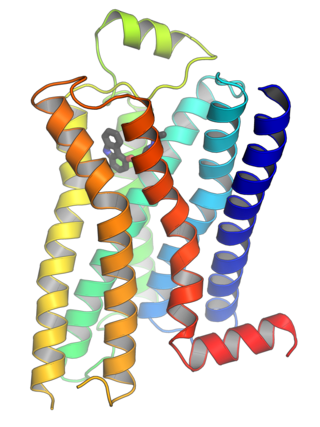
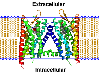
G protein-coupled receptors (GPCRs), also known as seven-(pass)-transmembrane domain receptors, 7TM receptors, heptahelical receptors, serpentine receptors, and G protein-linked receptors (GPLR), form a large group of evolutionarily related proteins that are cell surface receptors that detect molecules outside the cell and activate cellular responses. They are coupled with G proteins. They pass through the cell membrane seven times in the form of six loops of amino acid residues, which is why they are sometimes referred to as seven-transmembrane receptors. Ligands can bind either to the extracellular N-terminus and loops or to the binding site within transmembrane helices. They are all activated by agonists, although a spontaneous auto-activation of an empty receptor has also been observed.

<span class="mw-page-title-main">G protein</span> Type of proteins

G proteins, also known as guanine nucleotide-binding proteins, are a family of proteins that act as molecular switches inside cells, and are involved in transmitting signals from a variety of stimuli outside a cell to its interior. Their activity is regulated by factors that control their ability to bind to and hydrolyze guanosine triphosphate (GTP) to guanosine diphosphate (GDP). When they are bound to GTP, they are 'on', and, when they are bound to GDP, they are 'off'. G proteins belong to the larger group of enzymes called GTPases.

<span class="mw-page-title-main">Agonist</span> Chemical which binds to and activates a biochemical receptor

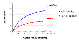
An agonist is a chemical that activates a receptor to produce a biological response. Receptors are cellular proteins whose activation causes the cell to modify what it is currently doing. In contrast, an antagonist blocks the action of the agonist, while an inverse agonist causes an action opposite to that of the agonist.

β-Endorphin Peptide hormone in humans

β-Endorphin (beta-endorphin) is an endogenous opioid neuropeptide and peptide hormone that is produced in certain neurons within the central nervous system and peripheral nervous system. It is one of three endorphins that are produced in humans, the others of which include α-endorphin and γ-endorphin.

Functional selectivity is the ligand-dependent selectivity for certain signal transduction pathways relative to a reference ligand at the same receptor. Functional selectivity can be present when a receptor has several possible signal transduction pathways. To which degree each pathway is activated thus depends on which ligand binds to the receptor. Functional selectivity, or biased signaling, is most extensively characterized at G protein coupled receptors (GPCRs). A number of biased agonists, such as those at muscarinic M2 receptors tested as analgesics or antiproliferative drugs, or those at opioid receptors that mediate pain, show potential at various receptor families to increase beneficial properties while reducing side effects. For example, pre-clinical studies with G protein biased agonists at the μ-opioid receptor show equivalent efficacy for treating pain with reduced risk for addictive potential and respiratory depression. Studies within the chemokine receptor system also suggest that GPCR biased agonism is physiologically relevant. For example, a beta-arrestin biased agonist of the chemokine receptor CXCR3 induced greater chemotaxis of T cells relative to a G protein biased agonist.

<span class="mw-page-title-main">Opioid peptide</span> Class of peptides that bind to opioid receptors

Opioid peptides or opiate peptides are peptides that bind to opioid receptors in the brain; opiates and opioids mimic the effect of these peptides. Such peptides may be produced by the body itself, for example endorphins. The effects of these peptides vary, but they all resemble those of opiates. Brain opioid peptide systems are known to play an important role in motivation, emotion, attachment behaviour, the response to stress and pain, control of food intake, and the rewarding effects of alcohol and nicotine.

κ-opioid receptor Protein-coding gene in the species Homo sapiens, named for ketazocine

The κ-opioid receptor or kappa opioid receptor, abbreviated KOR or KOP for its ligand ketazocine, is a G protein-coupled receptor that in humans is encoded by the OPRK1 gene. The KOR is coupled to the G protein Gi/G0 and is one of four related receptors that bind opioid-like compounds in the brain and are responsible for mediating the effects of these compounds. These effects include altering nociception, consciousness, motor control, and mood. Dysregulation of this receptor system has been implicated in alcohol and drug addiction.

<span class="mw-page-title-main">Endomorphin</span> Chemical compound

Endomorphins are considered to be natural opioid neurotransmitters central to pain relief. The two known endomorphins, endomorphin-1 and endomorphin-2, are tetrapeptides, consisting of Tyr-Pro-Trp-Phe and Tyr-Pro-Phe-Phe amino acid sequences respectively. These sequences fold into tertiary structures with high specificity and affinity for the μ-opioid receptor, binding it exclusively and strongly. Bound μ-opioid receptors typically induce inhibitory effects on neuronal activity. Endomorphin-like immunoreactivity exists within the central and peripheral nervous systems, where endomorphin-1 appears to be concentrated in the brain and upper brainstem, and endomorphin-2 in the spinal cord and lower brainstem. Because endomorphins activate the μ-opioid receptor, which is the target receptor of morphine and its derivatives, endomorphins possess significant potential as analgesics with reduced side effects and risk of addiction.

<span class="mw-page-title-main">Heterotrimeric G protein</span> Class of enzymes

Heterotrimeric G protein, also sometimes referred to as the "large" G proteins are membrane-associated G proteins that form a heterotrimeric complex. The biggest non-structural difference between heterotrimeric and monomeric G protein is that heterotrimeric proteins bind to their cell-surface receptors, called G protein-coupled receptors, directly. These G proteins are made up of alpha (α), beta (β) and gamma (γ) subunits. The alpha subunit is attached to either a GTP or GDP, which serves as an on-off switch for the activation of G-protein.

μ-opioid receptor Protein-coding gene in the species Homo sapiens, named for its ligand morphine

The μ-opioid receptors (MOR) are a class of opioid receptors with a high affinity for enkephalins and beta-endorphin, but a low affinity for dynorphins. They are also referred to as μ(mu)-opioid peptide (MOP) receptors. The prototypical μ-opioid receptor agonist is morphine, the primary psychoactive alkaloid in opium and for which the receptor was named, with mu being the first letter of Morpheus, the compound's namesake in the original Greek. It is an inhibitory G-protein coupled receptor that activates the Gi alpha subunit, inhibiting adenylate cyclase activity, lowering cAMP levels.

<span class="mw-page-title-main">Nociceptin receptor</span> Protein-coding gene in the species Homo sapiens

The nociceptin opioid peptide receptor (NOP), also known as the nociceptin/orphanin FQ (N/OFQ) receptor or kappa-type 3 opioid receptor, is a protein that in humans is encoded by the OPRL1 gene. The nociceptin receptor is a member of the opioid subfamily of G protein-coupled receptors whose natural ligand is the 17 amino acid neuropeptide known as nociceptin (N/OFQ). This receptor is involved in the regulation of numerous brain activities, particularly instinctive and emotional behaviors. Antagonists targeting NOP are under investigation for their role as treatments for depression and Parkinson's disease, whereas NOP agonists have been shown to act as powerful, non-addictive painkillers in non-human primates.

δ-opioid receptor Opioid receptor named for the mouse vas deferens, where it was first characterized

The δ-opioid receptor, also known as delta opioid receptor or simply delta receptor, abbreviated DOR or DOP, is an inhibitory 7-transmembrane G-protein coupled receptor coupled to the G protein Gi/G0 and has enkephalins as its endogenous ligands. The regions of the brain where the δ-opioid receptor is largely expressed vary from species model to species model. In humans, the δ-opioid receptor is most heavily expressed in the basal ganglia and neocortical regions of the brain.

G<sub>s</sub> alpha subunit Mammalian protein found in Homo sapiens

The Gs alpha subunit is a subunit of the heterotrimeric G protein Gs that stimulates the cAMP-dependent pathway by activating adenylyl cyclase. Gsα is a GTPase that functions as a cellular signaling protein. Gsα is the founding member of one of the four families of heterotrimeric G proteins, defined by the alpha subunits they contain: the Gαs family, Gαi/Gαo family, Gαq family, and Gα12/Gα13 family. The Gs-family has only two members: the other member is Golf, named for its predominant expression in the olfactory system. In humans, Gsα is encoded by the GNAS complex locus, while Golfα is encoded by the GNAL gene.

Gi protein alpha subunit is a family of heterotrimeric G protein alpha subunits. This family is also commonly called the Gi/o family or Gi/o/z/t family to include closely related family members. G alpha subunits may be referred to as Gi alpha, Gαi, or Giα.

<span class="mw-page-title-main">Alazocine</span> Synthetic opioid analgesic

Alazocine, also known more commonly as N-allylnormetazocine (NANM), is a synthetic opioid analgesic of the benzomorphan family related to metazocine, which was never marketed. In addition to its opioid activity, the drug is a sigma receptor agonist, and has been used widely in scientific research in studies of this receptor. Alazocine is described as a potent analgesic, psychotomimetic or hallucinogen, and opioid antagonist. Moreover, one of its enantiomers was the first compound that was found to selectively label the σ1 receptor, and led to the discovery and characterization of the receptor.

<span class="mw-page-title-main">G beta-gamma complex</span>

The G beta-gamma complex (Gβγ) is a tightly bound dimeric protein complex, composed of one Gβ and one Gγ subunit, and is a component of heterotrimeric G proteins. Heterotrimeric G proteins, also called guanosine nucleotide-binding proteins, consist of three subunits, called alpha, beta, and gamma subunits, or Gα, Gβ, and Gγ. When a G protein-coupled receptor (GPCR) is activated, Gα dissociates from Gβγ, allowing both subunits to perform their respective downstream signaling effects. One of the major functions of Gβγ is the inhibition of the Gα subunit.

<span class="mw-page-title-main">ICI-199,441</span> Chemical compound

ICI-199,441 is a drug which acts as a potent and selective κ-opioid agonist, and has analgesic effects. It is a biased agonist of the KOR, and is one of relatively few KOR ligands that is G protein-biased rather than β-arrestin-biased.

<span class="mw-page-title-main">Hemorphin-4</span> Endogenous opioid peptide

Hemorphin-4 is an endogenous opioid peptide of the hemorphin family which possesses antinociceptive properties and is derived from the β-chain of hemoglobin in the bloodstream. It is a tetrapeptide with the amino acid sequence Tyr-Pro-Trp-Thr. Hemorphin-4 has affinities for the μ-, δ-, and κ-opioid receptors that are in the same range as the structurally related β-casomorphins, although affinity to the κ-opioid receptor is markedly higher in comparison. It acts as an agonist at these sites. Hemorphin-4 also has inhibitory effects on angiotensin-converting enzyme (ACE), and as a result, may play a role in the regulation of blood pressure. Notably, inhibition of ACE also reduces enkephalin catabolism.

<span class="mw-page-title-main">Buprenorphine/samidorphan</span> Combination drug formulation

Buprenorphine/samidorphan is a combination formulation of buprenorphine and samidorphan which is under development as an add on to antidepressants in treatment-resistant depression (TRD).

<span class="mw-page-title-main">Axelopran</span> Chemical compound

Axelopran is a drug which is under development by Theravance Biopharma and licensed to Glycyx for all indications. It acts as a peripherally acting μ-opioid receptor antagonist and also acts on κ-, and δ-opioid receptors, with similar affinity for the μ- and κ-opioid receptors and about an order of magnitude lower affinity for the δ-opioid receptor. Recent data suggests that μ-opioid antagonists have a direct effect on overall survival in patients with advanced cancer.

References

  1. Dhawan BN, Cesselin F, Raghubir R, Reisine T, Bradley PB, Portoghese PS, Hamon M (December 1996). "International Union of Pharmacology. XII. Classification of opioid receptors". Pharmacological Reviews. 48 (4): 567–92. PMID   8981566. Open Access logo PLoS transparent.svg
  2. Janecka A, Fichna J, Janecki T (2004). "Opioid receptors and their ligands". Current Topics in Medicinal Chemistry. 4 (1): 1–17. doi:10.2174/1568026043451618. PMID   14754373. Open Access logo PLoS transparent.svg
  3. Waldhoer M, Bartlett SE, Whistler JL (2004). "Opioid receptors". Annual Review of Biochemistry. 73: 953–90. doi:10.1146/annurev.biochem.73.011303.073940. PMID   15189164. Closed Access logo transparent.svg
  4. Martin WR (December 1967). "Opioid antagonists". Pharmacological Reviews. 19 (4): 463–521. PMID   4867058. Closed Access logo transparent.svg
  5. Ingoglia NA, Dole VP (October 1970). "Localization of d- and l-methadone after intraventricular injection into rat brains". The Journal of Pharmacology and Experimental Therapeutics. 175 (1): 84–7. PMID   5471456. Closed Access logo transparent.svg
  6. Goldstein A, Lowney LI, Pal BK (August 1971). "Stereospecific and nonspecific interactions of the morphine congener levorphanol in subcellular fractions of mouse brain". Proceedings of the National Academy of Sciences of the United States of America. 68 (8): 1742–7. Bibcode:1971PNAS...68.1742G. doi: 10.1073/pnas.68.8.1742 . PMC   389284 . PMID   5288759. Open Access logo PLoS transparent.svg
  7. Pert CB, Snyder SH (March 1973). "Opiate receptor: demonstration in nervous tissue". Science. 179 (4077): 1011–4. Bibcode:1973Sci...179.1011P. doi:10.1126/science.179.4077.1011. PMID   4687585. S2CID   21783674. Closed Access logo transparent.svg
  8. Terenius L (1973). "Stereospecific interaction between narcotic analgesics and a synaptic plasm a membrane fraction of rat cerebral cortex". Acta Pharmacologica et Toxicologica. 32 (3): 317–20. doi:10.1111/j.1600-0773.1973.tb01477.x. PMID   4801733.
  9. Simon EJ, Hiller JM, Edelman I (July 1973). "Stereospecific binding of the potent narcotic analgesic (3H) Etorphine to rat-brain homogenate". Proceedings of the National Academy of Sciences of the United States of America. 70 (7): 1947–9. Bibcode:1973PNAS...70.1947S. doi: 10.1073/pnas.70.7.1947 . PMC   433639 . PMID   4516196.
  10. Caruso TP, Takemori AE, Larson DL, Portoghese PS (April 1979). "Chloroxymorphamine, and opioid receptor site-directed alkylating agent having narcotic agonist activity". Science. 204 (4390): 316–8. Bibcode:1979Sci...204..316C. doi:10.1126/science.86208. PMID   86208.
  11. Caruso TP, Larson DL, Portoghese PS, Takemori AE (December 1980). "Isolation of selective 3H-chlornaltrexamine-bound complexes, possible opioid receptor components in brains of mice". Life Sciences. 27 (22): 2063–9. doi:10.1016/0024-3205(80)90485-3. PMID   6259471.
  12. Corbett AD, Henderson G, McKnight AT, Paterson SJ (January 2006). "75 years of opioid research: the exciting but vain quest for the Holy Grail". British Journal of Pharmacology. 147 (Suppl 1): S153-62. doi:10.1038/sj.bjp.0706435. PMC   1760732 . PMID   16402099.
  13. 1 2 Stein C, Schäfer M, Machelska H (August 2003). "Attacking pain at its source: new perspectives on opioids". Nature Medicine. 9 (8): 1003–8. doi:10.1038/nm908. PMID   12894165. S2CID   25453057.
  14. 1 2 Fine PG, Portenoy RK (2004). "Chapter 2: The Endogenous Opioid System" (PDF). A Clinical Guide to Opioid Analgesia. McGraw Hill. Archived from the original (PDF) on 2011-07-19.
  15. Portoghese PS, Lunzer MM (April 2003). "Identity of the putative delta1-opioid receptor as a delta-kappa heteromer in the mouse spinal cord". European Journal of Pharmacology. 467 (1–3): 233–4. doi:10.1016/s0014-2999(03)01599-1. PMID   12706480.
  16. Stefano GB, Kream RM (June 2010). "Opioid peptides and opiate alkaloids in immunoregulatory processes". Archives of Medical Science. 6 (3): 456–60. doi:10.5114/aoms.2010.14271. PMC   3282526 . PMID   22371785.
  17. Stevens CW (January 2009). "The evolution of vertebrate opioid receptors". Frontiers in Bioscience. 14 (14): 1247–69. doi:10.2741/3306. PMC   3070387 . PMID   19273128.
  18. 1 2 Furay AR, Neumaier JF (October 2011). "Opioid receptors: binding that ties". Neuropsychopharmacology. 36 (11): 2157–8. doi:10.1038/npp.2011.147. PMC   3176578 . PMID   21918519.
  19. 1 2 Bodnar RJ (January 2016). "Endogenous opiates and behavior: 2014". Peptides. 75: 18–70. doi:10.1016/j.peptides.2015.10.009. PMID   26551874. S2CID   34578840.
  20. Burkett JP, Spiegel LL, Inoue K, Murphy AZ, Young LJ (October 2011). "Activation of μ-opioid receptors in the dorsal striatum is necessary for adult social attachment in monogamous prairie voles". Neuropsychopharmacology. 36 (11): 2200–10. doi:10.1038/npp.2011.117. PMC   3176565 . PMID   21734650.
  21. Olmstead MC, Ouagazzal AM, Kieffer BL (2009). "Mu and delta opioid receptors oppositely regulate motor impulsivity in the signaled nose poke task". PLOS ONE. 4 (2): e4410. Bibcode:2009PLoSO...4.4410O. doi: 10.1371/journal.pone.0004410 . PMC   2635474 . PMID   19198656.
  22. Akil H, Owens C, Gutstein H, Taylor L, Curran E, Watson S (1999). "Endogenous opioids: overview and current issues". Drug and Alcohol Dependence. 51 (1–2): 127–40. doi:10.1016/s0376-8716(98)00071-4. PMID   9716935.
  23. Trofimova I (April 2018). "Functionality versus dimensionality in psychological taxonomies, and a puzzle of emotional valence". Philosophical Transactions of the Royal Society of London. Series B, Biological Sciences. 373 (1744): 20170167. doi:10.1098/rstb.2017.0167. PMC   5832691 . PMID   29483351.
  24. Tuominen L, Salo J, Hirvonen J, Någren K, Laine P, Melartin T, et al. (July 2012). "Temperament trait Harm Avoidance associates with μ-opioid receptor availability in frontal cortex: a PET study using [(11)C]carfentanil". NeuroImage. 61 (3): 670–6. doi:10.1016/j.neuroimage.2012.03.063. PMID   22484309. S2CID   26046363.
  25. Cruz-Gordillo P, Fedrigo O, Wray GA, Babbitt CC (2010). "Extensive changes in the expression of the opioid genes between humans and chimpanzees". Brain, Behavior and Evolution. 76 (2): 154–62. doi:10.1159/000320968. PMID   21079395. S2CID   252466.
  26. Rockman MV, Hahn MW, Soranzo N, Zimprich F, Goldstein DB, Wray GA (December 2005). "Ancient and recent positive selection transformed opioid cis-regulation in humans". PLOS Biology. 3 (12): e387. doi: 10.1371/journal.pbio.0030387 . PMC   1283535 . PMID   16274263. Open Access logo PLoS transparent.svg
  27. Aggrawal A (May 1, 1995). "Opium: the king of narcotics". Opioids: past, present and future. BLTC Research. Archived from the original on May 26, 2012. Retrieved December 29, 2013.
  28. Lord JA, Waterfield AA, Hughes J, Kosterlitz HW (June 1977). "Endogenous opioid peptides: multiple agonists and receptors". Nature. 267 (5611): 495–9. Bibcode:1977Natur.267..495L. doi:10.1038/267495a0. PMID   195217. S2CID   4160761.
  29. Lemke, Thomas L.; Williams, David H.; Foye, William O. (2002). "Opioid Analgesics; Fries, DS". Foye's principles of medicinal chemistry. Hagerstown, MD: Lippincott Williams & Wilkins. ISBN   978-0-683-30737-5.
  30. Girdlestone D (October 2000). "Opioid receptors; Cox BM, Chavkin C, Christie MJ, Civelli O, Evans C, Hamon MD, et al.". The IUPHAR Compendium of Receptor Characterization and Classification (2nd ed.). London: IUPHAR Media. pp. 321–333.
  31. "Opioid receptors". IUPHAR Database. International Union of Pharmacology (2008-08-01).
  32. Grevel J, Yu V, Sadée W (May 1985). "Characterization of a labile naloxone binding site (lambda site) in rat brain". Journal of Neurochemistry. 44 (5): 1647–56. doi:10.1111/j.1471-4159.1985.tb08808.x. PMID   2985759. S2CID   84330114.
  33. Mizoguchi H, Narita M, Nagase H, Tseng LF (October 2000). "Activation of G-proteins in the mouse pons/medulla by beta-endorphin is mediated by the stimulation of mu- and putative epsilon-receptors". Life Sciences. 67 (22): 2733–43. doi:10.1016/S0024-3205(00)00852-3. PMID   11105989.
  34. Wollemann M, Benyhe S (June 2004). "Non-opioid actions of opioid peptides". Life Sciences. 75 (3): 257–70. doi:10.1016/j.lfs.2003.12.005. PMID   15135648.
  35. Zagon IS, Verderame MF, Allen SS, McLaughlin PJ (February 2000). "Cloning, sequencing, chromosomal location, and function of cDNAs encoding an opioid growth factor receptor (OGFr) in humans". Brain Research. 856 (1–2): 75–83. doi:10.1016/S0006-8993(99)02330-6. PMID   10677613. S2CID   37516655.
  36. Zagon IS, Verderame MF, McLaughlin PJ (February 2002). "The biology of the opioid growth factor receptor (OGFr)". Brain Research. Brain Research Reviews. 38 (3): 351–76. doi:10.1016/S0165-0173(01)00160-6. PMID   11890982. S2CID   37812525.
  37. Wüster M, Schulz R, Herz A (December 1979). "Specificity of opioids towards the mu-, delta- and epsilon-opiate receptors". Neuroscience Letters. 15 (2–3): 193–8. doi:10.1016/0304-3940(79)96112-3. PMID   231238. S2CID   53251283.
  38. Schulz R, Wüster M, Herz A (March 1981). "Pharmacological characterization of the epsilon-opiate receptor". The Journal of Pharmacology and Experimental Therapeutics. 216 (3): 604–6. PMID   6259326.
  39. Narita M, Tseng LF (March 1998). "Evidence for the existence of the beta-endorphin-sensitive "epsilon-opioid receptor" in the brain: the mechanisms of epsilon-mediated antinociception". Japanese Journal of Pharmacology. 76 (3): 233–53. doi: 10.1254/jjp.76.233 . PMID   9593217.
  40. Fujii H, Narita M, Mizoguchi H, Murachi M, Tanaka T, Kawai K, et al. (August 2004). "Drug design and synthesis of epsilon opioid receptor agonist: 17-(cyclopropylmethyl)-4,5alpha-epoxy-3,6beta-dihydroxy-6,14-endoethenomorphinan-7alpha-(N-methyl-N-phenethyl)carboxamide (TAN-821) inducing antinociception mediated by putative epsilon opioid receptor". Bioorganic & Medicinal Chemistry. 12 (15): 4133–45. doi:10.1016/j.bmc.2004.05.024. PMID   15246090.
  41. Fujii H, Nagase H (2006). "Rational drug design of selective epsilon opioid receptor agonist TAN-821 and antagonist TAN-1014". Current Medicinal Chemistry. 13 (10): 1109–18. doi:10.2174/092986706776360851. PMID   16719773.
  42. Contet C, Matifas A, Kieffer BL (May 2004). "No evidence for G-protein-coupled epsilon receptor in the brain of triple opioid receptor knockout mouse". European Journal of Pharmacology. 492 (2–3): 131–6. doi:10.1016/j.ejphar.2004.03.056. PMID   15178356.
  43. Al-Hasani R, Bruchas MR (December 2011). "Molecular mechanisms of opioid receptor-dependent signaling and behavior". Anesthesiology. 115 (6): 1363–81. doi:10.1097/ALN.0b013e318238bba6. PMC   3698859 . PMID   22020140.
  44. "Introduction to Essentials of Cell Biology | Learn Science at Scitable". www.nature.com. Retrieved 2017-11-08.
  45. Wedegaertner PB, Wilson PT, Bourne HR (January 1995). "Lipid modifications of trimeric G proteins". The Journal of Biological Chemistry. 270 (2): 503–6. doi: 10.1074/jbc.270.2.503 . PMID   7822269.
  46. Philip F, Sengupta P, Scarlata S (June 2007). "Signaling through a G Protein-coupled receptor and its corresponding G protein follows a stoichiometrically limited model". The Journal of Biological Chemistry. 282 (26): 19203–16. doi: 10.1074/jbc.M701558200 . PMID   17420253.
  47. Steer ML (November 1975). "Adenyl cyclase". Annals of Surgery. 182 (5): 603–9. doi:10.1097/00000658-197511000-00012. PMC   1344045 . PMID   172034.
  48. Keshwani MM, Kanter JR, Ma Y, Wilderman A, Darshi M, Insel PA, Taylor SS (October 2015). "Mechanisms of cyclic AMP/protein kinase A- and glucocorticoid-mediated apoptosis using S49 lymphoma cells as a model system". Proceedings of the National Academy of Sciences of the United States of America. 112 (41): 12681–6. Bibcode:2015PNAS..11212681K. doi: 10.1073/pnas.1516057112 . PMC   4611605 . PMID   26417071.
  49. Shao XM, Sun J, Jiang YL, Liu BY, Shen Z, Fang F, et al. (2016). "Inhibition of the cAMP/PKA/CREB Pathway Contributes to the Analgesic Effects of Electroacupuncture in the Anterior Cingulate Cortex in a Rat Pain Memory Model". Neural Plasticity. 2016: 5320641. doi: 10.1155/2016/5320641 . PMC   5206448 . PMID   28090359.
  50. Yamada M, Inanobe A, Kurachi Y (December 1998). "G protein regulation of potassium ion channels". Pharmacological Reviews. 50 (4): 723–60. PMID   9860808.
  51. Kosten TR, George TP (July 2002). "The neurobiology of opioid dependence: implications for treatment". Science & Practice Perspectives. 1 (1): 13–20. doi:10.1151/spp021113. PMC   2851054 . PMID   18567959.
  52. Befort K, Zilliox C, Filliol D, Yue S, Kieffer BL (June 1999). "Constitutive activation of the delta opioid receptor by mutations in transmembrane domains III and VII". The Journal of Biological Chemistry. 274 (26): 18574–81. doi: 10.1074/jbc.274.26.18574 . PMID   10373467.
  53. Fujita W, Gomes I, Devi LA (September 2014). "Revolution in GPCR signalling: opioid receptor heteromers as novel therapeutic targets: IUPHAR review 10". British Journal of Pharmacology. 171 (18): 4155–76. doi:10.1111/bph.12798. PMC   4241085 . PMID   24916280.

Further reading