Golfer's elbow

Last updated
Golfer's elbow
Other namesMedial epicondylitis
Gray329-Medial epicondyle of the humerus.png
Left elbow-joint, showing anterior and ulnar collateral ligaments. (Medial epicondyle labeled at center top.)
Specialty Orthopedics

Golfer's elbow, or medial epicondylitis, is tendinosis (or more precisely enthesopathy) of the medial common flexor tendon on the inside of the elbow. [1] It is similar to tennis elbow, which affects the outside of the elbow at the lateral epicondyle. The tendinopathy results from overload or repetitive use of the arm, causing an injury similar to ulnar collateral ligament injury of the elbow in "pitcher's elbow". [1]

Contents

Description

The anterior-medial forearm contains several muscles that flex the wrist and pronate the forearm. [1] These muscles have a common tendinous attachment at the medial epicondyle of the humerus at the elbow joint. [1]

The flexor and pronator muscles of the forearm include the pronator teres, flexor carpi radialis, palmaris longus, and flexor digitorum superficialis, all of which originate on the medial epicondyle and are innervated by the median nerve. [1] The flexor carpi ulnaris muscle also inserts on the medial epicondyle and is innervated by the ulnar nerve. [1] Together, these five muscles have a common attachment (an enthesis) on the medial epicondyle of the humerus. [1] The flexor tendon is approximately 3 centimetres (1.2 in) long, crosses the medial aspect of the elbow, and runs parallel to the ulnar collateral ligament. [1]

The injury is not acute inflammation, but rather is a chronic disorder resulting from overuse of a repetitive arm motion. [1] [2] Repetitive activity leads to recurrent microtears within the flexor tendon, with remodeling of the collagen fibers and an increase in the amount of mucoid ground substance. [1] As a result, scar tissue formation and thickening of the tendon lead to reduced collagen strength and pain with repetitive use. [1]

Causes

Still shot from a 3D medical animation illustrating golfer's elbow affecting the medial epicondyle on the lower inside of the joint. Golfers-Elbow SAG.jpg
Still shot from a 3D medical animation illustrating golfer's elbow affecting the medial epicondyle on the lower inside of the joint.

The condition is referred to as golfer's elbow when a full golf swing causes elbow pain. It may also be called pitcher's elbow due to the same tendon being stressed by repetitive throwing of objects, such as a baseball or football. [1] [3] [4]

Golfer's elbow appears to occur from repetitive full swings during the period from the top of the backswing to just before ball impact. [1] The full swing motion causes high energy valgus forces during the late cocking (backswing in golf) and acceleration phase (downswing and impact). [1]

It is unknown whether this condition is any more common in labor-related occupations with forceful repetitive activities (such as in construction or plumbing) than it is in the general public. [1]

In adults, the pathophysiology may involve mucoid degeneration (disorganized collagen, increased extra-cellular matrix, and chondroid metaplasia). [5]

Diagnosis

Medial epicondylitis is diagnosed based on characteristic pain with activities using strength in wrist flexion and confirmed on examination with discrete point tenderness over the common flexor origin at the medial epicondyle, and pain with resisted wrist flexion and passive wrist extension. [1] [2] [4] [6]

Imaging may be employed, although radiography might show calcifications in the muscle origin. Ultrasound and magnetic resonance imaging can identify the mucoid degeneration, but are not necessary for diagnosis. [1] [2] [4]

Occurrence

Medial injury of the flexor tendon is estimated to occur in 0.4% of the population. [1] It occurs most often in people ages 45 to 64, is more common in women than in men, and with 75% of cases in the dominant arm. [1]

Risk factors for developing golfer's elbow include improper technique or lack of strength, endurance, or flexibility. [1] Risk factors in people performing manual labor include heavy and excessive repetition, high body mass index, the presence of comorbidities, and high work demands. [1]

Treatment

Non-specific treatments to alleviate pain include: [1] [2] [4]

Before anesthetics and steroids are used, conservative treatment with an occupational therapist may be attempted. Before therapy can commence, treatment such as rest, ice, compression and elevation (R.I.C.E.) will typically be used. [1] [2] [4] This will help to decrease the pain and inflammation; rest will alleviate discomfort because golfer's elbow is an overuse injury. The subject can use a tennis elbow splint for compression. A pad can be placed anteromedially on the proximal forearm. [7] The splint is made in 30–45 degrees of elbow flexion. A daytime elbow pad also may be useful, by limiting additional trauma to the nerve. [8]

Simple analgesic medication has a place, as does more specific treatment with oral anti-inflammatory medications. [1] [2] [4] These will help control pain and any inflammation. [7] A more invasive treatment is the injection into and around the inflamed and tender area of a glucocorticoid (steroid) agent. [9]

Physical therapy

Therapy includes a variety of exercises for muscle and tendon reconditioning, starting with stretching and gradual strengthening of the flexor-pronator muscles. [2] [4] [6] Strengthening will slowly begin with isometrics and progresses to eccentric exercises helping to extend the range of motion back to where it once was. After the strengthening exercises, it is common for the subject to ice the area. [7]

Surgery

After 6 months if the symptoms do not improve, surgery may be recommended. [1] [2] [4] Surgical debridement or cleaning of the area is one of the most common treatments. [6] The ulnar nerve may also be decompressed surgically. [7] If the appropriate remediation steps are taken – rest, ice, and rehabilitative exercise and stretching – recovery may follow. Few subjects will need to progress to steroid injection, and less than 10% will require surgical intervention. [7] Arthroscopy is not an option for treating golfer's elbow. [6]

See also

Related Research Articles

<span class="mw-page-title-main">Humerus</span> Long bone of the upper arm

The humerus is a long bone in the arm that runs from the shoulder to the elbow. It connects the scapula and the two bones of the lower arm, the radius and ulna, and consists of three sections. The humeral upper extremity consists of a rounded head, a narrow neck, and two short processes. The body is cylindrical in its upper portion, and more prismatic below. The lower extremity consists of 2 epicondyles, 2 processes, and 3 fossae. As well as its true anatomical neck, the constriction below the greater and lesser tubercles of the humerus is referred to as its surgical neck due to its tendency to fracture, thus often becoming the focus of surgeons.

<span class="mw-page-title-main">Median nerve</span> Nerve of the upper limb

The median nerve is a nerve in humans and other animals in the upper limb. It is one of the five main nerves originating from the brachial plexus.

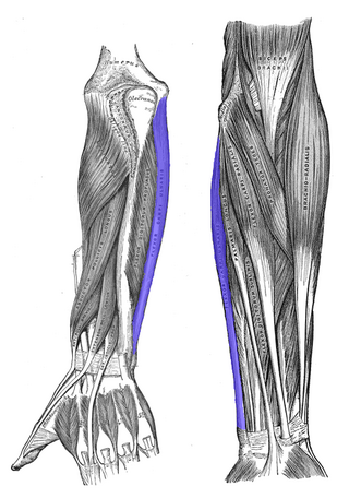
In human anatomy, the extensor carpi ulnaris is a skeletal muscle located on the ulnar side of the forearm. The extensor carpi ulnaris acts to extend and adduct at the carpus/wrist from anatomical position.

<span class="mw-page-title-main">Ulnar nerve</span> Nerve which runs near the ulna bone

In human anatomy, the ulnar nerve is a nerve that runs near the ulna bone. The ulnar collateral ligament of elbow joint is in relation with the ulnar nerve. The nerve is the largest in the human body unprotected by muscle or bone, so injury is common. This nerve is directly connected to the little finger, and the adjacent half of the ring finger, innervating the palmar aspect of these fingers, including both front and back of the tips, perhaps as far back as the fingernail beds.

<span class="mw-page-title-main">Upper limb</span> Consists of the arm, forearm, and hand

The upper limbs or upper extremities are the forelimbs of an upright-postured tetrapod vertebrate, extending from the scapulae and clavicles down to and including the digits, including all the musculatures and ligaments involved with the shoulder, elbow, wrist and knuckle joints. In humans, each upper limb is divided into the arm, forearm and hand, and is primarily used for climbing, lifting and manipulating objects.

<span class="mw-page-title-main">Tennis elbow</span> Condition in which the outer part of the elbow becomes sore and tender

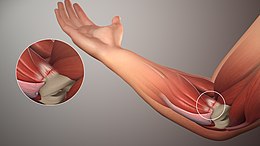
Tennis elbow, also known as lateral epicondylitis or enthesopathy of the extensor carpi radialis origin, is an enthesopathy of the origin of the extensor carpi radialis brevis on the lateral epicondyle. The outer part of the elbow becomes painful and tender. The pain may also extend into the back of the forearm. Onset of symptoms is generally gradual, although they can seem sudden and be misinterpreted as an injury. Golfer's elbow is a similar condition that affects the inside of the elbow.

Epicondylitis is the inflammation of an epicondyle or of adjacent tissues. Epicondyles are on the medial and lateral aspects of the elbow, consisting of the two bony prominences at the distal end of the humerus. These bony projections serve as the attachment point for the forearm musculature. Inflammation to the tendons and muscles at these attachment points can lead to medial and/or lateral epicondylitis. This can occur through a range of factors that overuse the muscles that attach to the epicondyles, such as sports or job-related duties that increase the workload of the forearm musculature and place stress on the elbow. Lateral epicondylitis is also known as “Tennis Elbow” due to its sports related association to tennis athletes, while medial epicondylitis is often referred to as “golfer's elbow.”

<span class="mw-page-title-main">Cubital fossa</span> The human elbow pit

The cubital fossa, chelidon or inside of elbow is the area on the anterior side of the upper part between the arm and forearm of a human or other hormid animals. It lies anteriorly to the elbow when in standard anatomical position.

<span class="mw-page-title-main">Flexor carpi ulnaris muscle</span> Muscle of the forearm

The flexor carpi ulnaris (FCU) is a muscle of the forearm that flexes and adducts at the wrist joint.

<span class="mw-page-title-main">Lateral epicondyle of the humerus</span> Structure of humerus

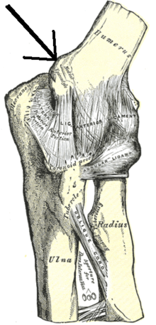
The lateral epicondyle of the humerus is a large, tuberculated eminence, curved a little forward, and giving attachment to the radial collateral ligament of the elbow joint, and to a tendon common to the origin of the supinator and some of the extensor muscles. Specifically, these extensor muscles include the anconeus muscle, the supinator, extensor carpi radialis brevis, extensor digitorum, extensor digiti minimi, and extensor carpi ulnaris. In birds, where the arm is somewhat rotated compared to other tetrapods, it is termed dorsal epicondyle of the humerus. In comparative anatomy, the term ectepicondyle is sometimes used.

The flexor pollicis longus is a muscle in the forearm and hand that flexes the thumb. It lies in the same plane as the flexor digitorum profundus. This muscle is unique to humans, being either rudimentary or absent in other primates. A meta-analysis indicated accessory flexor pollicis longus is present in around 48% of the population.

The pronator teres is a muscle that, along with the pronator quadratus, serves to pronate the forearm. It has two origins, at the medial humeral supracondylar ridge and the ulnar tuberosity, and inserts near the middle of the radius.

<span class="mw-page-title-main">Medial epicondyle of the humerus</span> Rounded eminence on the medial side of the humerus

The medial epicondyle of the humerus is an epicondyle of the humerus bone of the upper arm in humans. It is larger and more prominent than the lateral epicondyle and is directed slightly more posteriorly in the anatomical position. In birds, where the arm is somewhat rotated compared to other tetrapods, it is called the ventral epicondyle of the humerus. In comparative anatomy, the more neutral term entepicondyle is used.

<span class="mw-page-title-main">Anterior interosseous nerve</span>

The anterior interosseous nerve is a branch of the median nerve that supplies the deep muscles on the anterior of the forearm, except the ulnar (medial) half of the flexor digitorum profundus. Its nerve roots come from C8 and T1.

The posterior compartment of the forearm contains twelve muscles which primarily extend the wrist and digits. It is separated from the anterior compartment by the interosseous membrane between the radius and ulna.

<span class="mw-page-title-main">Idiopathic Ulnar neuropathy at the elbow</span> Medical condition

Idiopathic Ulnar neuropathy at the elbow is a condition where pressure on the ulnar nerve as it passes through the cubital tunnel causes nerve dysfunction (neuropathy). The symptoms of neuropathy are paresthesia (tingling) and numbness primarily affecting the little finger and ring finger of the hand. Ulnar neuropathy can progress to weakness and atrophy of the muscles in the hand. Symptoms can be alleviated by attempts to keep the elbow from flexing while sleeping, such as sticking one's arm in the pillow case, so the pillow restricts flexion.

<span class="mw-page-title-main">Ulnar neuropathy</span> Medical condition

Ulnar neuropathy is a disorder involving the ulnar nerve. Ulnar neuropathy may be caused by entrapment of the ulnar nerve with resultant numbness and tingling. It may also cause weakness or paralysis of the muscles supplied by the nerve.

<span class="mw-page-title-main">Elbow</span> Joint between the upper and lower parts of the arm

The elbow is the region between the upper arm and the forearm that surrounds the elbow joint. The elbow includes prominent landmarks such as the olecranon, the cubital fossa, and the lateral and the medial epicondyles of the humerus. The elbow joint is a hinge joint between the arm and the forearm; more specifically between the humerus in the upper arm and the radius and ulna in the forearm which allows the forearm and hand to be moved towards and away from the body. The term elbow is specifically used for humans and other primates, and in other vertebrates it is not used. In those cases, forelimb plus joint is used.

<span class="mw-page-title-main">Median nerve palsy</span> Medical condition

Injuries to the arm, forearm or wrist area can lead to various nerve disorders. One such disorder is median nerve palsy. The median nerve controls the majority of the muscles in the forearm. It controls abduction of the thumb, flexion of hand at wrist, flexion of digital phalanx of the fingers, is the sensory nerve for the first three fingers, etc. Because of this major role of the median nerve, it is also called the eye of the hand. If the median nerve is damaged, the ability to abduct and oppose the thumb may be lost due to paralysis of the thenar muscles. Various other symptoms can occur which may be repaired through surgery and tendon transfers. Tendon transfers have been very successful in restoring motor function and improving functional outcomes in patients with median nerve palsy.

<span class="mw-page-title-main">Ulnar collateral ligament injury of the elbow</span> Medical condition

Ulnar collateral ligament injuries can occur during certain activities such as overhead baseball pitching. Acute or chronic disruption of the ulnar collateral ligament result in medial elbow pain, valgus instability, and impaired throwing performance. There are both non-surgical and surgical treatment options.

References

  1. 1 2 3 4 5 6 7 8 9 10 11 12 13 14 15 16 17 18 19 20 21 22 23 24 25 Kiel, John; Kaiser, Kimberly (27 October 2018), Golfers elbow, StatPearls Publishing, PMID   30085542 , retrieved 2019-03-12
  2. 1 2 3 4 5 6 7 8 Childress MA, Beutler A (April 2013). "Management of chronic tendon injuries". American Family Physician. 87 (7): 486–90. PMID   23547590.
  3. "Pitcher's Elbow – Stanford Sports Medicine – Stanford Medical Outpatient Center". Stanford University Medical Center . Retrieved 9 September 2009.
  4. 1 2 3 4 5 6 7 8 Wilson, JJ; Best, TM (1 September 2005). "Common overuse tendon problems: A review and recommendations for treatment". American Family Physician. 72 (5): 811–818. PMID   16156339.
  5. Bruni, D.; Pierson, S.; Sarwar, F.; Ring, D.; Ramtin, S. (2023). "Are the Pathologic Features of Enthesopathy, Tendinopathy, and Labral and Articular Disc Disease Related to Mucoid Degeneration? A Systematic Review". Clinical Orthopaedics and Related Research. 481 (4): 641–650. doi:10.1097/CORR.0000000000002499. PMC   10013668 . PMID   36563131.
  6. 1 2 3 4 Amin, Nirav H.; Kumar, Neil S.; Schickendantz, Mark S. (June 2015). "Medial Epicondylitis: Evaluation and Management". Journal of the American Academy of Orthopaedic Surgeons. 23 (6): 348–355. doi: 10.5435/JAAOS-D-14-00145 . PMID   26001427. S2CID   31827631.
  7. 1 2 3 4 5 Medial Epicondylitis at eMedicine
  8. "Golfers Elbow | Orthopedic Solutions". orthopedicsolutionsokc.com.
  9. Jacobs, J.W.G.; Michels-van Amelsfort, J.M.R. (April 2013). "How to perform local soft-tissue glucocorticoid injections?". Best Practice & Research Clinical Rheumatology. 27 (2): 171–194. doi:10.1016/j.berh.2013.03.003. PMID   23731930.