Hemoprotein

Last updated
Binding of oxygen to a heme prosthetic group, which would be part of a hemoprotein. Mboxygenation.png
Binding of oxygen to a heme prosthetic group, which would be part of a hemoprotein.

A hemeprotein (or haemprotein; also hemoprotein or haemoprotein), or heme protein, is a protein that contains a heme prosthetic group. [1] They are a very large class of metalloproteins. The heme group confers functionality, which can include oxygen carrying, oxygen reduction, electron transfer, and other processes. Heme is bound to the protein either covalently or noncovalently or both. [2]

Contents

The heme consists of iron cation bound at the center of the conjugate base of the porphyrin, as well as other ligands attached to the "axial sites" of the iron. The porphyrin ring is a planar dianionic, tetradentate ligand. The iron is typically Fe2+ or Fe3+. One or two ligands are attached at the axial sites. The porphyrin ring has 4 nitrogen atoms that bind to the iron, leaving two other coordination positions of the iron available for bonding to the histidine of the protein and a divalent atom. [2]

Hemeproteins probably evolved to incorporate the iron atom contained within the protoporphyrin IX ring of heme into proteins. As it makes hemeproteins responsive to molecules that can bind divalent iron, this strategy has been maintained throughout evolution as it plays crucial physiological functions. The serum iron pool maintains iron in soluble form, making it more accessible for cells. [3] Oxygen (O2), nitric oxide (NO), carbon monoxide (CO) and hydrogen sulfide (H2S) bind to the iron atom in heme proteins. Once bound to the prosthetic heme groups, these molecules can modulate the activity/function of those hemeproteins, affording signal transduction. Therefore, when produced in biologic systems (cells), these gaseous molecules are referred to as gasotransmitters.

A model of the Fe-protoporphyrin IX subunit of the Heme B cofactor. Haem-B-3D-vdW.png
A model of the Fe-protoporphyrin IX subunit of the Heme B cofactor.

Because of their diverse biological functions and widespread abundance, hemeproteins are among the most studied biomolecules. [4] Data on heme protein structure and function has been aggregated into The Heme Protein Database (HPD), a secondary database to the Protein Data Bank. [5]

Roles

Hemeproteins have diverse biological functions including oxygen transport, which is completed via hemeproteins including hemoglobin, hemocyanin, [6] myoglobin, neuroglobin, cytoglobin, and leghemoglobin. [7]

Some hemeproteins - cytochrome P450s, cytochrome c oxidase, ligninases, catalase and peroxidases - are enzymes. They often activate O2 for oxidation or hydroxylation.

Hemeproteins also enable electron transfer as they form part of the electron transport chain. Cytochrome a, cytochrome b, and cytochrome c have such electron transfer functions. It is now known that cytochrome a and cytochrome a3 make up one protein and was deemed the name cytochrome aa3. [8] The sensory system also relies on some hemeproteins including FixL, an oxygen sensor, CooA, a carbon monoxide sensor, and soluble guanylyl cyclase.

Hemoglobin and myoglobin

Hemoglobin and myoglobin are examples of hemeproteins that respectively transport and store of oxygen in mammals and in some fish. [9] Hemoglobin is a quaternary protein that occurs in the red blood cell, whereas, myoglobin is a tertiary protein found in the muscle cells of mammals. Although they might differ in location and size, their function are similar. Being hemeproteins, they both contain a heme prosthetic group.

His-F8 of the myoglobin, also known as the proximal histidine, is covalently bonded to the 5th coordination position of the iron. Oxygen interacts with the distal His by way of a hydrogen bond, not a covalent one. It binds to the 6th coordination position of the iron, His-E7 of the myoglobin binds to the oxygen that is now covalently bonded to the iron. The same is true for hemoglobin; however, being a protein with four subunits, hemoglobin contains four heme units in total, allowing four oxygen molecules in total to bind to the protein.

Myoglobin and hemoglobin are globular proteins that serve to bind and deliver oxygen using a prosthetic group. These globins dramatically improve the concentration of molecular oxygen that can be carried in the biological fluids of vertebrates and some invertebrates.

Differences occur in ligand binding and allosteric regulation.

Myoglobin

Myoglobin is found in vertebrate muscle cells and is a water-soluble globular protein. [10] Muscle cells, when put into action, can quickly require a large amount of oxygen for respiration due to their energy requirements. Therefore, muscle cells use myoglobin to accelerate oxygen diffusion and act as localized oxygen reserves for times of intense respiration. Myoglobin also stores the required amount of oxygen and makes it available for the muscle cell mitochondria.

Hemoglobin

In vertebrates, hemoglobin is found in the cytosol of red blood cells. Hemoglobin is sometimes referred to as the oxygen transport protein, in order to contrast it with myoglobin, which is stationary.

In vertebrates, oxygen is taken into the body by the tissues of the lungs, and passed to the red blood cells in the bloodstream where it's used in aerobic metabolic pathways. [10] Oxygen is then distributed to all of the tissues in the body and offloaded from the red blood cells to respiring cells. The hemoglobin then picks up carbon dioxide to be returned to the lungs. Thus, hemoglobin binds and off-loads both oxygen and carbon dioxide at the appropriate tissues, serving to deliver the oxygen needed for cellular metabolism and removing the resulting waste product, CO2.

Neuroglobin

Found in neurons, neuroglobin is responsible for driving nitric oxide to promote neuron cell survival [11] Neuroglobin is believed to increase the oxygen supply for neurons, sustaining ATP production, but they also function as storage proteins. [12]

Peroxidases and catalases

Almost all human peroxidases are hemoproteins, except glutathione peroxidase. They use hydrogen peroxide as a substrate. Metalloenzymes catalyze reactions using peroxide as an oxidant. [13] Catalases are hemoproteins responsible for the catalysis of converting hydrogen peroxide into water and oxygen. [14] They are made up of 4 subunits, each subunit having a Fe3+ heme group. They have an average molecular weight of ~240,000 g/mol.

Electron transport chain and other redox catalysts

Cytochromes, cytochrome c oxidase, and coenzyme Q – cytochrome c reductase are heme-containing proteins or protein subunits embedded in the inner membrane of mitochondria which play an essential role in cellular respiration.

Sulfite oxidase, a molybdenum-dependent cytochrome, oxidizes sulfite to sulfate.

Nitric oxide synthase

Designed heme proteins

Pincer-1: A designed heme-binding peptide adopting an all-beta secondary structure. ABOVE: Topological representation of Pincer-1 showing the secondary structure and designed interacting residues. BELOW: All-atom 3-dimensional model of Pincer-1. This model was partially confirmed using NMR. Pincer-1 beta heme peptide.png
Pincer-1: A designed heme-binding peptide adopting an all-beta secondary structure. ABOVE: Topological representation of Pincer-1 showing the secondary structure and designed interacting residues. BELOW: All-atom 3-dimensional model of Pincer-1. This model was partially confirmed using NMR.

Due to the diverse functions of the heme molecule: as an electron transporter, an oxygen carrier, and as an enzyme cofactor, heme binding proteins have consistently attracted the attention of protein designers. Initial design attempts focused on α-helical heme binding proteins, in part, due to the relative simplicity of designing self-assembling helical bundles. Heme binding sites were designed inside the inter-helical hydrophobic grooves. Examples of such designs include:

Later design attempts focused on creating functional heme binding helical bundles, such as:

Design techniques have matured to such an extent that it is now possible to generate entire libraries of heme binding helical proteins. [31]

Recent design attempts have focused on creating all-beta heme binding proteins, whose novel topology is very rare in nature. Such designs include:

Some methodologies attempt to incorporate cofactors into the hemoproteins who typically endure harsh conditions. In order to incorporate a synthetic cofactor, what must first occur is the denaturing of the holoprotein to remove the heme. The apoprotein is then rebuilt with the cofactor. [35]

Related Research Articles

<span class="mw-page-title-main">Hemoglobin</span> Metalloprotein that binds with oxygen

Hemoglobin is a protein containing iron that facilitates the transport of oxygen in red blood cells. Almost all vertebrates contain hemoglobin, with the exception of the fish family Channichthyidae. Hemoglobin in the blood carries oxygen from the respiratory organs to the other tissues of the body, where it releases the oxygen to enable aerobic respiration which powers the animal's metabolism. A healthy human has 12 to 20 grams of hemoglobin in every 100 mL of blood. Hemoglobin is a metalloprotein, a chromoprotein, and globulin.

<span class="mw-page-title-main">Myoglobin</span> Iron and oxygen-binding protein

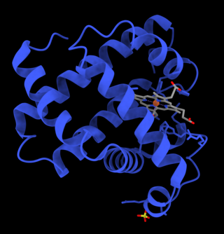
Myoglobin is an iron- and oxygen-binding protein found in the cardiac and skeletal muscle tissue of vertebrates in general and in almost all mammals. Myoglobin is distantly related to hemoglobin. Compared to hemoglobin, myoglobin has a higher affinity for oxygen and does not have cooperative binding with oxygen like hemoglobin does. Myoglobin consists of non-polar amino acids at the core of the globulin, where the heme group is non-covalently bounded with the surrounding polypeptide of myoglobin. In humans, myoglobin is found in the bloodstream only after muscle injury.

<span class="mw-page-title-main">Cytochrome c</span> Protein-coding gene in the species Homo sapiens

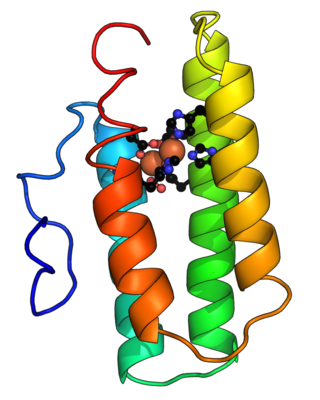
The cytochrome complex, or cyt c, is a small hemeprotein found loosely associated with the inner membrane of the mitochondrion where it plays a critical role in cellular respiration. It transfers electrons between Complexes III and IV. Cytochrome c is highly water-soluble, unlike other cytochromes. It is capable of undergoing oxidation and reduction as its iron atom converts between the ferrous and ferric forms, but does not bind oxygen. It also plays a major role in cell apoptosis. In humans, cytochrome c is encoded by the CYCS gene.

<span class="mw-page-title-main">Leghemoglobin</span> Phytoglobin

Leghemoglobin is an oxygen-carrying phytoglobin found in the nitrogen-fixing root nodules of leguminous plants. It is produced by these plants in response to the roots being colonized by nitrogen-fixing bacteria, termed rhizobia, as part of the symbiotic interaction between plant and bacterium: roots not colonized by Rhizobium do not synthesise leghemoglobin. Leghemoglobin has close chemical and structural similarities to hemoglobin, and, like hemoglobin, is red in colour. It was originally thought that the heme prosthetic group for plant leghemoglobin was provided by the bacterial symbiont within symbiotic root nodules. However, subsequent work shows that the plant host strongly expresses heme biosynthesis genes within nodules, and that activation of those genes correlates with leghemoglobin gene expression in developing nodules.

<span class="mw-page-title-main">Heme</span> Chemical coordination complex of an iron ion chelated to a porphyrin

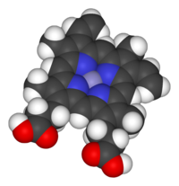
Heme, or haem, is a ring-shaped iron-containing molecular component of hemoglobin, which is necessary to bind oxygen in the bloodstream. It is composed of four pyrrole rings with 2 vinyl and 2 propionic acid side chains. Heme is biosynthesized in both the bone marrow and the liver.

<span class="mw-page-title-main">Hemerythrin</span> InterPro Family

Hemerythrin (also spelled haemerythrin; Ancient Greek: αἷμα, romanized: haîma, lit. 'blood', Ancient Greek: ἐρυθρός, romanized: erythrós, lit. 'red') is an oligomeric protein responsible for oxygen (O2) transport in the marine invertebrate phyla of sipunculids, priapulids, brachiopods, and in a single annelid worm genus, Magelona. Myohemerythrin is a monomeric O2-binding protein found in the muscles of marine invertebrates. Hemerythrin and myohemerythrin are essentially colorless when deoxygenated, but turn a violet-pink in the oxygenated state.

<span class="mw-page-title-main">Globin</span> Superfamily of oxygen-transporting globular proteins

The globins are a superfamily of heme-containing globular proteins, involved in binding and/or transporting oxygen. These proteins all incorporate the globin fold, a series of eight alpha helical segments. Two prominent members include myoglobin and hemoglobin. Both of these proteins reversibly bind oxygen via a heme prosthetic group. They are widely distributed in many organisms.

<span class="mw-page-title-main">Metalloprotein</span> Protein that contains a metal ion cofactor

Metalloprotein is a generic term for a protein that contains a metal ion cofactor. A large proportion of all proteins are part of this category. For instance, at least 1000 human proteins contain zinc-binding protein domains although there may be up to 3000 human zinc metalloproteins.

<span class="mw-page-title-main">Trace metal</span> Metals subset of trace elements

Trace metals are the metals subset of trace elements; that is, metals normally present in small but measurable amounts in animal and plant cells and tissues and that are a necessary part of nutrition and physiology. Some biometals are trace metals. Ingestion of, or exposure to, excessive quantities can be toxic. However, insufficient plasma or tissue levels of certain trace metals can cause pathology, as is the case with iron.

<span class="mw-page-title-main">Binding site</span> Molecule-specific coordinate bonding area in biological systems

In biochemistry and molecular biology, a binding site is a region on a macromolecule such as a protein that binds to another molecule with specificity. The binding partner of the macromolecule is often referred to as a ligand. Ligands may include other proteins, enzyme substrates, second messengers, hormones, or allosteric modulators. The binding event is often, but not always, accompanied by a conformational change that alters the protein's function. Binding to protein binding sites is most often reversible, but can also be covalent reversible or irreversible.

<span class="mw-page-title-main">Human iron metabolism</span> Iron metabolism in the body

Human iron metabolism is the set of chemical reactions that maintain human homeostasis of iron at the systemic and cellular level. Iron is both necessary to the body and potentially toxic. Controlling iron levels in the body is a critically important part of many aspects of human health and disease. Hematologists have been especially interested in systemic iron metabolism, because iron is essential for red blood cells, where most of the human body's iron is contained. Understanding iron metabolism is also important for understanding diseases of iron overload, such as hereditary hemochromatosis, and iron deficiency, such as iron-deficiency anemia.

<span class="mw-page-title-main">Heme oxygenase</span> Class of enzymes

Heme oxygenase, or haem oxygenase, is an enzyme that catalyzes the degradation of heme to produce biliverdin, ferrous ion, and carbon monoxide.

Iron-binding proteins are carrier proteins and metalloproteins that are important in iron metabolism and the immune response. Iron is required for life.

<span class="mw-page-title-main">Heme C</span> Chemical compound

Heme C is an important kind of heme.

<span class="mw-page-title-main">Heme B</span> Chemical compound

Heme B or haem B is the most abundant heme. Hemoglobin and myoglobin are examples of oxygen transport proteins that contain heme B. The peroxidase family of enzymes also contain heme B. The COX-1 and COX-2 enzymes (cyclooxygenase) of recent fame, also contain heme B at one of two active sites.

<span class="mw-page-title-main">Heme A</span> Chemical compound

Heme A is a heme, a coordination complex consisting of a macrocyclic ligand called a porphyrin, chelating an iron atom. Heme A is a biomolecule and is produced naturally by many organisms. Heme A, often appears a dichroic green/red when in solution, is a structural relative of heme B, a component of hemoglobin, the red pigment in blood.

<span class="mw-page-title-main">Iron in biology</span> Use of Iron by organisms

Iron is an important biological element. It is used in both the ubiquitous iron-sulfur proteins and in vertebrates it is used in hemoglobin which is essential for blood and oxygen transport.

<span class="mw-page-title-main">Nitric oxide dioxygenase</span>

Nitric oxide dioxygenase (EC 1.14.12.17) is an enzyme that catalyzes the conversion of nitric oxide (NO) to nitrate (NO
3
) . The net reaction for the reaction catalyzed by nitric oxide dioxygenase is shown below:

<span class="mw-page-title-main">Eosinophil peroxidase</span> Protein-coding gene in the species Homo sapiens

Eosinophil peroxidase is an enzyme found within the eosinophil granulocytes, innate immune cells of humans and mammals. This oxidoreductase protein is encoded by the gene EPX, expressed within these myeloid cells. EPO shares many similarities with its orthologous peroxidases, myeloperoxidase (MPO), lactoperoxidase (LPO), and thyroid peroxidase (TPO). The protein is concentrated in secretory granules within eosinophils. Eosinophil peroxidase is a heme peroxidase, its activities including the oxidation of halide ions to bacteriocidal reactive oxygen species, the cationic disruption of bacterial cell walls, and the post-translational modification of protein amino acid residues.

References

  1. "Heme Prosthetic Group Definition". earth.callutheran.edu. Retrieved 2023-04-27.
  2. 1 2 Nelson DL, Cox MN (2000). Lehninger, Principles of Biochemistry (3rd ed.). New Yorkm: Worth Publishing. ISBN   1-57259-153-6.
  3. Frazer, David M.; Anderson, Gregory J. (March 2014). "The regulation of iron transport: The Regulation of Iron Transport". BioFactors. 40 (2): 206–214. doi:10.1002/biof.1148. PMID   24132807. S2CID   2998785.
  4. Reedy CJ, Elvekrog MM, Gibney BR (January 2008). "Development of a heme protein structure-electrochemical function database". Nucleic Acids Research. 36 (Database issue): D307–D313. doi:10.1093/nar/gkm814. PMC   2238922 . PMID   17933771.
  5. Gibney BR. "Heme Protein Database". Brooklyn, NY: Brooklyn College.
  6. "hemoproteins - Humpath.com - Human pathology". www.humpath.com. Retrieved 2023-04-27.
  7. Lippard J, Berg JM (1994). Principles of Bioinorganic Chemistry. Mill Valley, CA: University Science Books. ISBN   0-935702-73-3.
  8. Mahinthichaichan, Paween; Gennis, Robert B.; Tajkhorshid, Emad (2018-04-10). "Cytochrome aa 3 Oxygen Reductase Utilizes the Tunnel Observed in the Crystal Structures To Deliver O 2 for Catalysis". Biochemistry. 57 (14): 2150–2161. doi:10.1021/acs.biochem.7b01194. ISSN   0006-2960. PMC   5936630 . PMID   29546752.
  9. Gerber, Lucie; Clow, Kathy A.; Driedzic, William R.; Gamperl, Anthony K. (July 2021). "The Relationship between Myoglobin, Aerobic Capacity, Nitric Oxide Synthase Activity and Mitochondrial Function in Fish Hearts". Antioxidants. 10 (7): 1072. doi: 10.3390/antiox10071072 . ISSN   2076-3921. PMC   8301165 . PMID   34356305.
  10. 1 2 "Hemoglobin and Myoglobin | Integrative Medical Biochemistry Examination and Board Review | AccessPharmacy | McGraw Hill Medical". accesspharmacy.mhmedical.com. Retrieved 2023-04-27.
  11. DellaValle, Brian; Hempel, Casper; Kurtzhals, Jørgen A. L.; Penkowa, Milena (2010-08-01). "In vivo expression of neuroglobin in reactive astrocytes during neuropathology in murine models of traumatic brain injury, cerebral malaria, and autoimmune encephalitis: Neuroglobin in Reactive Astrogliosis". Glia. 58 (10): 1220–1227. doi:10.1002/glia.21002. PMID   20544857. S2CID   8563830.
  12. Burmester, Thorsten; Hankeln, Thomas (June 2004). "Neuroglobin: A Respiratory Protein of the Nervous System". Physiology. 19 (3): 110–113. doi:10.1152/nips.01513.2003. ISSN   1548-9213. PMID   15143204.
  13. Winterbourn, Christine C. (2013-01-01), "Chapter One - The Biological Chemistry of Hydrogen Peroxide", in Cadenas, Enrique; Packer, Lester (eds.), Hydrogen Peroxide and Cell Signaling, Part C, Methods in Enzymology, vol. 528, Academic Press, pp. 3–25, doi:10.1016/B978-0-12-405881-1.00001-X, ISBN   978-0-12-405881-1, PMID   23849856 , retrieved 2023-04-27
  14. Brzozowska, Ewa; Bazan, Justyna; Gamian, Andrzej (2011-03-25). "Funkcje białek bakteriofagowych". Postępy Higieny i Medycyny Doświadczalnej. 65: 167–176. doi: 10.5604/17322693.936090 . ISSN   1732-2693. PMID   21502693.
  15. 1 2 Nagarajan D, Sukumaran S, Deka G, Krishnamurthy K, Atreya HS, Chandra N (June 2018). "Design of a heme-binding peptide motif adopting a β-hairpin conformation". The Journal of Biological Chemistry. 293 (24): 9412–9422. doi: 10.1074/jbc.RA118.001768 . PMC   6005436 . PMID   29695501.
  16. Sasaki T, Kaiser ET (1989-01-01). "Helichrome: synthesis and enzymic activity of a designed hemeprotein". Journal of the American Chemical Society. 111 (1): 380–381. doi:10.1021/ja00183a065. ISSN   0002-7863.
  17. Sasaki T, Kaiser ET (January 1990). "Synthesis and structural stability of helichrome as an artificial hemeproteins". Biopolymers. 29 (1): 79–88. doi:10.1002/bip.360290112. PMID   2328295. S2CID   35536899.
  18. Isogai Y, Ota M, Fujisawa T, Izuno H, Mukai M, Nakamura H, et al. (June 1999). "Design and synthesis of a globin fold". Biochemistry. 38 (23): 7431–7443. doi:10.1021/bi983006y. PMID   10360940.
  19. Rosenblatt MM, Wang J, Suslick KS (November 2003). "De novo designed cyclic-peptide heme complexes". Proceedings of the National Academy of Sciences of the United States of America. 100 (23): 13140–13145. Bibcode:2003PNAS..10013140R. doi: 10.1073/pnas.2231273100 . PMC   263730 . PMID   14595023.
  20. Robertson DE, Farid RS, Moser CC, Urbauer JL, Mulholland SE, Pidikiti R, et al. (March 1994). "Design and synthesis of multi-haem proteins". Nature. 368 (6470): 425–432. Bibcode:1994Natur.368..425R. doi:10.1038/368425a0. PMID   8133888. S2CID   4360174.
  21. Choma CT, Lear JD, Nelson MJ, Dutton PL, Robertson DE, DeGrado WF (1994-02-01). "Design of a heme-binding four-helix bundle". Journal of the American Chemical Society. 116 (3): 856–865. doi:10.1021/ja00082a005. ISSN   0002-7863.
  22. Discher BM, Noy D, Strzalka J, Ye S, Moser CC, Lear JD, et al. (September 2005). "Design of amphiphilic protein maquettes: controlling assembly, membrane insertion, and cofactor interactions". Biochemistry. 44 (37): 12329–12343. doi:10.1021/bi050695m. PMC   2574520 . PMID   16156646.
  23. Mahajan M, Bhattacharjya S (June 2014). "Designed di-heme binding helical transmembrane protein". ChemBioChem. 15 (9): 1257–1262. doi:10.1002/cbic.201402142. PMID   24829076. S2CID   20982919.
  24. Korendovych IV, Senes A, Kim YH, Lear JD, Fry HC, Therien MJ, et al. (November 2010). "De novo design and molecular assembly of a transmembrane diporphyrin-binding protein complex". Journal of the American Chemical Society. 132 (44): 15516–15518. doi:10.1021/ja107487b. PMC   3016712 . PMID   20945900.
  25. 1 2 Farid TA, Kodali G, Solomon LA, Lichtenstein BR, Sheehan MM, Fry BA, et al. (December 2013). "Elementary tetrahelical protein design for diverse oxidoreductase functions". Nature Chemical Biology. 9 (12): 826–833. doi:10.1038/nchembio.1362. PMC   4034760 . PMID   24121554.
  26. Huang SS, Koder RL, Lewis M, Wand AJ, Dutton PL (April 2004). "The HP-1 maquette: from an apoprotein structure to a structured hemoprotein designed to promote redox-coupled proton exchange". Proceedings of the National Academy of Sciences of the United States of America. 101 (15): 5536–5541. doi: 10.1073/pnas.0306676101 . PMC   397418 . PMID   15056758.
  27. Faiella M, Maglio O, Nastri F, Lombardi A, Lista L, Hagen WR, Pavone V (December 2012). "De novo design, synthesis and characterisation of MP3, a new catalytic four-helix bundle hemeprotein". Chemistry: A European Journal. 18 (50): 15960–15971. doi:10.1002/chem.201201404. PMID   23150230.
  28. Cherry JR, Lamsa MH, Schneider P, Vind J, Svendsen A, Jones A, Pedersen AH (April 1999). "Directed evolution of a fungal peroxidase". Nature Biotechnology. 17 (4): 379–384. doi:10.1038/7939. PMID   10207888. S2CID   41233353.
  29. Anderson JL, Armstrong CT, Kodali G, Lichtenstein BR, Watkins DW, Mancini JA, et al. (February 2014). "Constructing a man-made c-type cytochrome maquette in vivo: electron transfer, oxygen transport and conversion to a photoactive light harvesting maquette". Chemical Science. 5 (2): 507–514. doi:10.1039/C3SC52019F. PMC   3952003 . PMID   24634717.
  30. Koder RL, Anderson JL, Solomon LA, Reddy KS, Moser CC, Dutton PL (March 2009). "Design and engineering of an O(2) transport protein". Nature. 458 (7236): 305–309. Bibcode:2009Natur.458..305K. doi:10.1038/nature07841. PMC   3539743 . PMID   19295603.
  31. Moffet DA, Foley J, Hecht MH (September 2003). "Midpoint reduction potentials and heme binding stoichiometries of de novo proteins from designed combinatorial libraries". Biophysical Chemistry. Walter Kauzmann's 85th Birthday. 105 (2–3): 231–239. doi: 10.1016/S0301-4622(03)00072-3 . PMID   14499895.
  32. Mahajan M, Bhattacharjya S (June 2013). "β-Hairpin peptides: heme binding, catalysis, and structure in detergent micelles". Angewandte Chemie. 52 (25): 6430–6434. doi:10.1002/anie.201300241. PMID   23640811.
  33. D'Souza A, Wu X, Yeow EK, Bhattacharjya S (May 2017). "Designed Heme-Cage β-Sheet Miniproteins". Angewandte Chemie. 56 (21): 5904–5908. doi:10.1002/anie.201702472. PMID   28440962.
  34. D'Souza A, Mahajan M, Bhattacharjya S (April 2016). "Designed multi-stranded heme binding β-sheet peptides in membrane". Chemical Science. 7 (4): 2563–2571. doi:10.1039/C5SC04108B. PMC   5477022 . PMID   28660027.
  35. Lemon, Christopher M.; Marletta, Michael A. (2021-12-21). "Designer Heme Proteins: Achieving Novel Function with Abiological Heme Analogues". Accounts of Chemical Research. 54 (24): 4565–4575. doi:10.1021/acs.accounts.1c00588. ISSN   0001-4842. PMC   8754152 . PMID   34890183.