Hemoptysis

Last updated
Hemoptysis
Other namesHaemoptysis, coughing up of blood
Bronchitis Normal vs Affected Airway.jpg
Lower left: Inflammation of the bronchus can bring about bloody mucus.
Pronunciation
Specialty Pulmonology
Symptoms Spitting blood or bloody sputum
Complications Pulmonary aspiration, choking
Causes bronchitis, lung cancer, certain infections
Differential diagnosis epistaxis (combined with post-nasal drip)

Hemoptysis or haemoptysis is the discharge of blood or blood-stained mucus through the mouth coming from the bronchi, larynx, trachea, or lungs. It does not necessarily involve coughing. In other words, it is the airway bleeding. This can occur with lung cancer, infections such as tuberculosis, bronchitis, or pneumonia, and certain cardiovascular conditions. Hemoptysis is considered massive at 300 mL (11 imp fl oz; 10 US fl oz). In such cases, there are always severe injuries. The primary danger comes from choking, rather than blood loss. [1]

Contents

Diagnosis

Diagnostic approach to solving the puzzle of hemoptysis. Hemoptysis.svg
Diagnostic approach to solving the puzzle of hemoptysis.

Differential diagnosis

The most common causes for hemoptysis in adults are chest infections such as bronchitis or pneumonia. [1] In children, hemoptysis is commonly caused by the presence of a foreign body in the airway. Other common causes include lung cancers and tuberculosis. Less common causes include aspergilloma, bronchiectasis, coccidioidomycosis, pulmonary embolism, pneumonic plague, and cystic fibrosis. Rarer causes include hereditary hemorrhagic telangiectasia (HHT or Rendu-Osler-Weber syndrome), Goodpasture's syndrome, and granulomatosis with polyangiitis. A rare cause of hemoptysis in women is endometriosis, which leads to intermittent hemoptysis coinciding with menstrual periods in 7% of women with thoracic endometriosis syndrome. [4] Hemoptysis may be exacerbated or even caused by overtreatment with anticoagulant drugs such as warfarin.[ citation needed ]

Blood-laced mucus from the sinus or nose area can sometimes be misidentified as symptomatic of hemoptysis (such secretions can be a sign of nasal or sinus cancer, but also a sinus infection). Extensive non-respiratory injury can also cause one to cough up blood. Cardiac causes like congestive heart failure and mitral stenosis should be ruled out. The origin of blood can be identified by observing its color. Bright-red, foamy blood comes from the respiratory tract, whereas dark-red, coffee-colored blood comes from the gastrointestinal tract. Sometimes hemoptysis may be rust-colored.[ citation needed ]

Massive hemoptysis and mortality

Hemoptysis Raetchon a sonk.jpg
Hemoptysis

Although there are reports that the fatality rate is as high as 80%, the mortality rate for hospitalized hemoptysis patients is 2669/28539=9.4%, calculated from the data in the article by Kinoshita et al. [27] This is probably the most reasonable figure considering the overwhelming number of cases.[ citation needed ]

The general definition of massive hemoptysis is more than 200 ml within 24 hours, but there is a wide range in the literature (100-600 ml). Considering that the total volume of the tracheal and bronchial lumen is about 150 cc, [28] [29] it may be reasonable to define massive hemoptysis as 200 ml, which is a little more than 150 ml, in terms of setting the threshold for fatal hemoptysis. More than 400ml/day is not adequate for screening purposes.[ citation needed ]

Treatment

Treatment depends on the underlying cause. Treatments include iced saline, and topical vasoconstrictors such as adrenaline or vasopressin. Tranexamic acid was proved to improve in-hospital mortality. [27] Selective bronchial intubation can be used to collapse the lung that is bleeding. Also, endobronchial tamponade can be used. [30] Laser photocoagulation can be used to stop bleeding during bronchoscopy. Angiography of bronchial arteries can be performed to locate the bleeding, and it can often be embolized. [31] Bronchial artery embolization (BAE) is the first line treatment nowadays. [32] [33] [34] [35] [36] Surgical option is usually the last resort and can involve removal of a lung lobe or removal of the entire lung. Cough suppressants can increase the risk of choking. [1]

Related Research Articles

<span class="mw-page-title-main">Sputum</span> Mucus that is coughed up from the lower airways

Sputum is mucus that is coughed up from the lower airways. In medicine, sputum samples are usually used for a naked eye examination, microbiological investigation of respiratory infections and cytological investigations of respiratory systems. It is crucial that the specimen does not include any mucoid material from the nose or oral cavity.

<span class="mw-page-title-main">Acute bronchitis</span> Medical condition

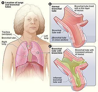
Acute bronchitis, also known as a chest cold, is short-term bronchitis – inflammation of the bronchi of the lungs. The most common symptom is a cough. Other symptoms include coughing up mucus, wheezing, shortness of breath, fever, and chest discomfort. The infection may last from a few to ten days. The cough may persist for several weeks afterward with the total duration of symptoms usually around three weeks. Some have symptoms for up to six weeks.

Salt water aspiration syndrome is a rare diving disorder suffered by scuba divers who inhale a mist of seawater, usually from a faulty demand valve, causing irritation of the lungs. It is not the same thing as aspiration of salt water as a bulk liquid, i.e. drowning. It can usually be treated by rest for several hours. If severe, medical assessment is required. First described by Carl Edmonds.

<span class="mw-page-title-main">Granulomatosis with polyangiitis</span> Medical condition

Granulomatosis with polyangiitis (GPA), also known as Wegener's granulomatosis (WG), after the German physician Friedrich Wegener, is a rare long-term systemic disorder that involves the formation of granulomas and inflammation of blood vessels (vasculitis). It is an autoimmune disease and a form of vasculitis that affects small- and medium-size vessels in many organs but most commonly affects the upper respiratory tract, lungs and kidneys. The signs and symptoms of GPA are highly varied and reflect which organs are supplied by the affected blood vessels. Typical signs and symptoms include nosebleeds, stuffy nose and crustiness of nasal secretions, and inflammation of the uveal layer of the eye. Damage to the heart, lungs and kidneys can be fatal.

<span class="mw-page-title-main">Hemothorax</span> Blood accumulation in the pleural cavity

A hemothorax is an accumulation of blood within the pleural cavity. The symptoms of a hemothorax may include chest pain and difficulty breathing, while the clinical signs may include reduced breath sounds on the affected side and a rapid heart rate. Hemothoraces are usually caused by an injury, but they may occur spontaneously due to cancer invading the pleural cavity, as a result of a blood clotting disorder, as an unusual manifestation of endometriosis, in response to Pneumothorax, or rarely in association with other conditions.

<span class="mw-page-title-main">Bronchial artery</span> Blood vessels supplying the lungs

In human anatomy, the bronchial arteries supply the lungs with oxygenated blood, and nutrition. Although there is much variation, there are usually two bronchial arteries that run to the left lung, and one to the right lung, and are a vital part of the respiratory system.

<span class="mw-page-title-main">Respiratory disease</span> Disease of the respiratory system

Respiratory diseases, or lung diseases, are pathological conditions affecting the organs and tissues that make gas exchange difficult in air-breathing animals. They include conditions of the respiratory tract including the trachea, bronchi, bronchioles, alveoli, pleurae, pleural cavity, the nerves and muscles of respiration. Respiratory diseases range from mild and self-limiting, such as the common cold, influenza, and pharyngitis to life-threatening diseases such as bacterial pneumonia, pulmonary embolism, tuberculosis, acute asthma, lung cancer, and severe acute respiratory syndromes, such as COVID-19. Respiratory diseases can be classified in many different ways, including by the organ or tissue involved, by the type and pattern of associated signs and symptoms, or by the cause of the disease.

<span class="mw-page-title-main">Aspergilloma</span> Medical condition

An aspergilloma is a clump of mold which exists in a body cavity such as a paranasal sinus or an organ such as the lung. By definition, it is caused by fungi of the genus Aspergillus.

Rasmussen aneurysm is a distinctive variant of pseudoaneurysm of a branch of the pulmonary artery, predominantly found adjacent to or within a lung cavity, both often arising as a complication of pulmonary tuberculosis. The condition was originally described by Fritz Valdemar Rasmussen in 1868. It is a relatively uncommon complication associated with cavitary tuberculosis of the lung, with its prevalence observed in approximately 5–8% of cases based on autopsy findings.

<span class="mw-page-title-main">Peribronchial cuffing</span> Localized patches of lung collapse caused by excess fluid or mucus buildup

Peribronchial cuffing, also referred to as peribronchial thickening or bronchial wall thickening, is a radiologic sign which occurs when excess fluid or mucus buildup in the small airway passages of the lung causes localized patches of atelectasis. This causes the area around the bronchus to appear more prominent on an X-ray. It has also been described as donut sign, considering the edge is thicker, and the center contains air.

Pulmonary-renal syndrome (PRS) is a rare medical syndrome in which respiratory failure involving bleeding in the lungs and kidney failure (glomerulonephritis) occur. PRS is associated with a high rate of morbidity and death. The term was first used by Goodpasture in 1919 to describe the association of respiratory and kidney failure.

<span class="mw-page-title-main">Hughes–Stovin syndrome</span> Autoimmune disorder

Hughes–Stovin syndrome (HSS) is a rare autoimmune disorder often described as inflammation in relation to blood vessels, a form of vasculitis. It is not associated with any known cause and is typically characterized by multiple aneurysms in pulmonary arteries and deep vein thromboses. It is named after the two British physicians, John Patterson Hughes and Peter George Ingle Stovin, who first described it in 1959. HSS is presumed to be a rare variant of Behçet's disease, which entails more general problems with the circulatory system. Due to its clinical similarity with Behçet's disease, it has also been referred to as 'Incomplete Behçet's disease.' Most patients are young adult males between the age of 20–40. Common clinical presentations include fever, cough, dyspnea and hemoptysis. Radiological features are similar to those of Behçet's disease.

<span class="mw-page-title-main">Eosinophilic bronchitis</span> Medical condition

Eosinophilic bronchitis (EB) is a type of airway inflammation due to excessive mast cell recruitment and activation in the superficial airways as opposed to the smooth muscles of the airways as seen in asthma. It often results in a chronic cough. Lung function tests are usually normal. Inhaled corticosteroids are often an effective treatment.

Swimming induced pulmonary edema (SIPE), also known as immersion pulmonary edema, is a life threatening condition that occurs when fluids from the blood leak abnormally from the small vessels of the lung (pulmonary capillaries) into the airspaces (alveoli).

<span class="mw-page-title-main">Chronic pulmonary aspergillosis</span> Fungal infection

Chronic pulmonary aspergillosis is a long-term fungal infection caused by members of the genus Aspergillus—most commonly Aspergillusfumigatus. The term describes several disease presentations with considerable overlap, ranging from an aspergilloma—a clump of Aspergillus mold in the lungs—through to a subacute, invasive form known as chronic necrotizing pulmonary aspergillosis which affects people whose immune system is weakened. Many people affected by chronic pulmonary aspergillosis have an underlying lung disease, most commonly tuberculosis, allergic bronchopulmonary aspergillosis, asthma, or lung cancer.

<span class="mw-page-title-main">Ground-glass opacity</span> Radiologic sign on radiographs and computed tomography scans

Ground-glass opacity (GGO) is a finding seen on chest x-ray (radiograph) or computed tomography (CT) imaging of the lungs. It is typically defined as an area of hazy opacification (x-ray) or increased attenuation (CT) due to air displacement by fluid, airway collapse, fibrosis, or a neoplastic process. When a substance other than air fills an area of the lung it increases that area's density. On both x-ray and CT, this appears more grey or hazy as opposed to the normally dark-appearing lungs. Although it can sometimes be seen in normal lungs, common pathologic causes include infections, interstitial lung disease, and pulmonary edema.

Thoracic endometriosis is a rare form of endometriosis where endometrial-like tissue is found in the lung parenchyma and/or the pleura. It can be classified as either pulmonary, or pleural, respectively. Endometriosis is characterized by the presence of tissue similar to the lining of the uterus forming abnormal growths elsewhere in the body. Usually these growths are found in the pelvis, between the rectum and the uterus, the ligaments of the pelvis, the bladder, the ovaries, and the sigmoid colon. The cause is not known. The most common symptom of thoracic endometriosis is chest pain occurring right before or during menstruation. Diagnosis is based on clinical history and examination, augmented with X-ray, CT scan, and magnetic resonance imaging of the chest. Treatment options include surgery and hormones.

Plastic bronchitis (PB) is a disorder in which branching casts of the airways are expectorated. PB is not a single disease with a defined mechanism that explains the cast formation in all conditions. Examples of diseases associated with expectoration of casts, and which sometimes are labeled PB include tuberculosis, atypical mycobacterial disease, allergic bronchopulmonary aspergillosis, and asthma.

<span class="mw-page-title-main">Lung cavity</span> Medical condition

A lung cavity or pulmonary cavity is an abnormal, thick-walled, air-filled space within the lung. Cavities in the lung can be caused by infections, cancer, autoimmune conditions, trauma, congenital defects, or pulmonary embolism. The most common cause of a single lung cavity is lung cancer. Bacterial, mycobacterial, and fungal infections are common causes of lung cavities. Globally, tuberculosis is likely the most common infectious cause of lung cavities. Less commonly, parasitic infections can cause cavities. Viral infections almost never cause cavities. The terms cavity and cyst are frequently used interchangeably; however, a cavity is thick walled, while a cyst is thin walled. The distinction is important because cystic lesions are unlikely to be cancer, while cavitary lesions are often caused by cancer.

Bronchial artery embolization is a treatment for hemoptysis, abbreviated as BAE. It is a kind of catheter intervention to control hemoptysis by embolizing the bronchial artery, which is a bleeding source. Embolic agents are particulate embolic material such as gelatin sponge or polyvinyl alcohol (PVA), and liquid embolic material such as NBCA, or metallic coils.

References

  1. 1 2 3 Sabatine MS (2014). Pocket medicine (Fifth ed.). [S.l.]: Aspen Publishers, Inc. ISBN   978-1451193787.
  2. "Hemoptysis". The Lecturio Medical Concept Library. Retrieved 24 July 2021.
  3. Richard F.LeBlond (2004). Diagnostics. US: McGraw-Hill Companies, Inc. ISBN   978-0-07-140923-0.
  4. McCann MR, Schenk WB, Nassar A, Maimone S (September 2020). "Thoracic endometriosis presenting as a catamenial hemothorax with discordant video-assisted thoracoscopic surgery". Radiology Case Reports. 15 (9): 1419–1422. doi:10.1016/j.radcr.2020.05.064. PMC   7334551 . PMID   32642009.
  5. Google Health – Google
  6. Google Health – Google
  7. "Sarcoidosis Signs & Symptoms – Sarcoidosis – HealthCommunities.com". Archived from the original on 2009-02-21. Retrieved 2010-02-05.
  8. MedlinePlus Encyclopedia : Pulmonary aspergilloma
  9. Google Health – Google
  10. "Histoplasmosis Symptoms – Diseases and Conditions – Mayo Clinic". Archived from the original on 2013-05-31. Retrieved 2010-02-05.
  11. "Pneumonia". The Lecturio Medical Concept Library. 27 August 2020. Retrieved 1 July 2021.
  12. Dorland's illustrated medical dictionary (32nd ed.). Saunders/Elsevier. 2 May 2011. p. 593. ISBN   9781416062578.
  13. Ware LB, Matthay MA (December 2005). "Clinical practice. Acute pulmonary edema". N. Engl. J. Med. 353 (26): 2788–96. doi:10.1056/NEJMcp052699. PMID   16382065.
  14. "Endometriosis". The Lecturio Medical Concept Library. Retrieved 25 July 2021.
  15. "Foreign Body Aspiration". The Lecturio Medical Concept Library. Retrieved 25 July 2021.
  16. Pediatric Goodpasture Syndrome at eMedicine
  17. Altaie R, Ditizio F, Fahy GT (March 2005). "Microscopic polyangitis presenting with sub-acute reversible optic neuropathy". Eye (Lond). 19 (3): 363–5. doi: 10.1038/sj.eye.6701479 . hdl: 10379/8830 . PMID   15272290.
  18. "Granulomatosis with Polyangiitis". www.mayoclinic.org. Mayo Foundation for Medical Education and Research. Archived from the original on 22 December 2017. Retrieved 3 March 2018.
  19. Adu, Emery & Madaio 2012, p. 125.
  20. "What Is Churg–Strauss Syndrome?". WebMD. 30 January 2019. Retrieved 8 March 2020.
  21. 1 2 "Hemoptysis Causes – Hemoptysis – HealthCommunities.com". Archived from the original on 2009-01-23. Retrieved 2010-02-05.
  22. 1 2 3 4 "Other Causes of Hemoptysis – Hemoptysis – HealthCommunities.com". Archived from the original on 2009-06-08. Retrieved 2010-02-05.
  23. "Mitral Stenosis". The Lecturio Medical Concept Library. Retrieved 24 July 2021.
  24. Jha, Suman K.; Karna, Bibek; Mahajan, Kunal (2020), "Tropical Pulmonary Eosinophilia", StatPearls, Treasure Island (FL): StatPearls Publishing, PMID   32491456 , retrieved 2020-12-01
  25. Hunt, Beverley J. (2014). "Bleeding and Coagulopathies in Critical Care". New England Journal of Medicine. 370 (9): 847–859. doi:10.1056/NEJMra1208626. ISSN   0028-4793. PMID   24571757.
  26. Hughes, JP; Stovin, PG (January 1959). "Segmental pulmonary artery aneurysms with peripheral venous thrombosis". British Journal of Diseases of the Chest. 53 (1): 19–27. doi:10.1016/S0007-0971(59)80106-6. PMID   13618502.
  27. 1 2 Kinoshita T, Ohbe H, Matsui H, Fushimi K, Ogura H, Yasunaga H (November 2019). "Effect of tranexamic acid on mortality in patients with haemoptysis: a nationwide study". Critical Care. 23 (1): 347. doi: 10.1186/s13054-019-2620-5 . PMC   6836388 . PMID   31694697.
  28. Patwa A, Shah A (September 2015). "Anatomy and physiology of respiratory system relevant to anaesthesia". Indian Journal of Anaesthesia. 59 (9): 533–41. doi: 10.4103/0019-5049.165849 . PMC   4613399 . PMID   26556911.
  29. Davidson K, Shojaee S (January 2020). "Managing Massive Hemoptysis". Chest. 157 (1): 77–88. doi:10.1016/j.chest.2019.07.012. PMID   31374211. S2CID   199388328.
  30. Valipour A, Kreuzer A, Koller H, Koessler W, Burghuber OC (June 2005). "Bronchoscopy-guided topical hemostatic tamponade therapy for the management of life-threatening hemoptysis". Chest. 127 (6): 2113–8. doi:10.1378/chest.127.6.2113. PMID   15947328.
  31. Hanson C, Karlsson CA, Kämpe M, Lamberg K, Lindberg E, Boman LM, Stålenheim G (August 2004). Guidelines for treatment of acute lung diseases (Report). Uppsala Academic Hospital.
  32. Woo S, Yoon CJ, Chung JW, Kang SG, Jae HJ, Kim HC, et al. (November 2013). "Bronchial artery embolization to control hemoptysis: comparison of N-butyl-2-cyanoacrylate and polyvinyl alcohol particles". Radiology. 269 (2): 594–602. doi:10.1148/radiol.13130046. PMID   23801773. S2CID   21597160.
  33. Ishikawa H, Hara M, Ryuge M, Takafuji J, Youmoto M, Akira M, et al. (February 2017). "Efficacy and safety of super selective bronchial artery coil embolisation for haemoptysis: a single-centre retrospective observational study". BMJ Open. 7 (2): e014805. doi:10.1136/bmjopen-2016-014805. PMC   5318547 . PMID   28213604.
  34. Ryuge M, Hara M, Hiroe T, Omachi N, Minomo S, Kitaguchi K, et al. (February 2019). "Mechanisms of recurrent haemoptysis after super-selective bronchial artery coil embolisation: a single-centre retrospective observational study". European Radiology. 29 (2): 707–715. doi:10.1007/s00330-018-5637-2. PMC   6302874 . PMID   30054792.
  35. Panda A, Bhalla AS, Goyal A (2017-07-07). "Bronchial artery embolization in hemoptysis: a systematic review". Diagnostic and Interventional Radiology. 23 (4): 307–317. doi:10.5152/dir.2017.16454. PMC   5508955 . PMID   28703105.
  36. Olsen KM, Manouchehr-Pour S, Donnelly EF, Henry TS, Berry MF, Boiselle PM, et al. (May 2020). "ACR Appropriateness Criteria® Hemoptysis". Journal of the American College of Radiology. 17 (5S): S148–S159. doi:10.1016/j.jacr.2020.01.043. PMID   32370959. S2CID   218520816.

Further reading