Histoplasma duboisii

Last updated

Histoplasma duboisii
Scientific classification OOjs UI icon edit-ltr.svg
Domain: Eukaryota
Kingdom: Fungi
Division: Ascomycota
Class: Eurotiomycetes
Order: Onygenales
Family: Ajellomycetaceae
Genus: Histoplasma
Species:
H. duboisii
Binomial name
Histoplasma duboisii
Vanbreus. (1952)
Synonyms
  • Histoplasma capsulatum var. duboisii(Vanbreus.) Cif. (1960)

Histoplasma duboisii is a saprotrophic fungus responsible for the invasive infection known as African histoplasmosis. [1] This species is a close relative of Histoplasma capsulatum , the agent of classical histoplasmosis, and the two occur in similar habitats. [1] Histoplasma duboisii is restricted to continental Africa and Madagascar, although scattered reports have arisen from other places usually in individuals with an African travel history. [2] Like, H. capsulatum, H. duboisii is dimorphic – growing as a filamentous fungus at ambient temperature and a yeast at body temperature. [2] It differs morphologically from H. capsulatum by the typical production of a large-celled yeast form. [2] Both agents cause similar forms of disease, although H. duboisii predominantly causes cutaneous and subcutaneous disease in humans and non-human primates. [2] [3] The agent responds to many antifungal drug therapies used to treat serious fungal diseases. [3]

Contents

History

Histoplasmosis was first reported from the African continent in 1942. [4] These early reports implicated strains that produced larger yeast cell forms than H. capsulatum, [2] and the Irish mycologist James Thompson Duncan suggested they might represent a distinct taxon. [4] [5] The fungus was described as a new species by Raymond Vanbreuseghem in 1952 [4] based on isolates provided to him by Professor Albert Dubois, director of the Prince Léopold Institute for Tropical Medicine in Antwerp, Belgium, and the species was named in honour of Dubois. [6] Five years after its description, Professor Edouard Drouhet of the Pasteur Institute in Paris reduced the taxon to synonymy with H. capsulatum, designating it as a variant. [2] [7] However, since the 1960s the fungus has been generally accepted as a separate species from H. capsulatum. [6] [8] [9]

Morphology

Histoplasma duboisii is a dimorphic fungus, growing as either a yeast-like form or a filamentous form depending on the prevailing nutritional and temperature conditions. [1] It is unusual to find both the mycelial and yeast forms co-existing. [10] The mycelial form is characterized by white and cottony colonies that turn brownish with age. [11] [12] The underside of the colony is typically brownish in colour. [11] It is morphologically similar to the closely related species, H. capsulatum, producing warted aleurioconidia though not as prolifically as H. capsulatum. [11] Unlike the small-celled yeast produced by H. capsulatum, H. duboisii initially produces small yeast cells (2–5 μm in diameter) but later develops a mixture of small and large cells after 3–4 weeks in culture culminating in the culture being dominated by large yeast cells (10–15 μm in diameter). [2] [12] The yeast form of H. duboisii are oval in shape with thick cell walls [1] composed of galactomannan intermixed with β-(1,4)-glucan. [13] Cells of H. duboisii have a different fatty acid profile than those of H. capsulatum, [11] and these differences have been suggested to relate to differences in cell sizes between the two species. [14]

The identity of H. duboisii can be ascertained by conventional laboratory methods involving mycelial-to-yeast conversion on brain-heart infusion medium supplemented with sheep blood and either glutamine or cysteine, and microscopic verification of the size of yeast cells. [12] A further feature differentiating the yeast forms of H. duboisii and H. capsulatum is the tendency of buds of the former species to remain attached by a narrow isthmus such that the mother and daughter cells resemble an hourglass just prior to cell release. [14] [15] Unlike H. capsulatum, H. duboisii lacks the ability to produce the enzyme urease, and this feature can be used for confirmatory identification. [11] Clinical isolates of H. duboisii belong predominantly to the "–" mating type. [3]

Ecology and epidemiology

Histoplasma duboisii is restricted to regions on the African continent lying between the Tropics of Cancer and the Capricorn. [11] The species is the etiological agent of African histoplamosis, and is endemic throughout western and central Africa [1] in addition to Madagascar. [10] These regions share a similarly stable climate in terms of relative humidity and rainfall. [11] Like H. capsulatum, the agent of classical histoplasmosis, H. duboisii is associated with chicken runs and bat caves. [3] The first described natural reservoir of H. duboisii was a bat cave in Nigeria [12] that yielded samples of the dirt mixed with guano testing positive for H. duboisii exoantigen. [11] Intestinal contents from 13% of healthy bats collected from the location tested positive for the fungus. [12]

Disease in humans

Histoplasma duboisii infections have been known to occur in HIV patients in endemic zones, particularly in individuals whose CD4+ cell count is below 50 cells/mm3. [1] These infections are often of the disseminated type. [11] Unlike classical histoplasmosis, infections caused by H. duboisii are often restricted to the skin or subcutaneous layer occasionally involving bone. [11] [14] It is most frequently seen in immunodeficient individuals although the disease is well known in the immunocompetent. Lung disease manifesting as classical miliary infiltrates and nodular lesions has also been recorded. [16] Superficial cutaneous lesions are characterized by nodules and papules that may ulcerate at a later stage of disease progression. [11] A hyperpigmented halo sometimes surrounds the nodule. [11] Subcutaneous lesions are warm, firm and tender, sometimes rupturing to release a yellowish discharge containing the fungus prior to evolving into cold abscesses. [3] [11] Subcutaneous lesions may also develop draining sinuses. [11] Lesions can be localised or disseminated, [2] appearing simultaneously and sometimes in large numbers. [14] Disseminated disease is especially common in immunocompromised individuals, and can involve any organs but infection of the heart and central nervous system are rare. [2] Dissemination to lymph nodes and visceral organs is associated with high mortality. [2] The ability of yeast cells to multiply within phagocytic cells contributes to the formation of pus-producing necrotic granulomas. [1] [10] Uncommonly, both H. duboisii and H. capsulatum have been found to co-occur in immunosuppressed patients with HIV disease. [1] It has also been known to co-occur with Aspergillus fumigatus , Pseudallescheria boydii , Microsporum gypseum , Malbranchea gypsea and species of Chrysosporium . [3] Fewer than 300 cases reports of African histoplasmosis are known prior to 2007, and the disease is thought to be significantly under-reported. [3] [14]

Cutaneous disease can take several forms: [11]

  1. Primary disease (following direct inoculation)
    • Superficial
    • Subcutaneous
  2. Secondary disease (arising from dissemination of pre-existing infection, e.g., osteomyelitis)

Diagnosis

Due to the wide variety of clinical presentations, diagnosis of disease related to this agent is often challenging and cannot be accomplished using on clinical features alone. [10] Laboratory confirmation of the organism in biopsy or secretion specimens is necessary. [11] Currently there are no available serological testing procedures available for H. duboisii, and the species is antigenically cross-reactive with the closely related H. capsulatum. [1] The feasibility of a specific polymerase chain reaction-based test for H. duboisii has been suggested, but none is presently available. [1]

Distribution and reservoirs

Though the agent is thought to be restricted to Africa, emergent cases have been reported elsewhere but nearly always in individuals with a travel history to regions where the agent is endemic. [11] [17] One case of African histoplasmosis has been reported in an otherwise individual from India in the absence of a travel history to endemic countries. The afflicted individual resided in Kerala, an area with an abundant bat populations and a climate similar to that of endemic countries. [11] All age groups are susceptible to infection; however individuals in the third or fourth decade of life as well as children under 10 years of age are at greatest risk. [3] Case reports suggest a strong gender bias in infection, favouring males over females by a factor of two. [3]

The fungus is thought to enter the body mainly by inhalation of airborne microconidia or fragments of vegetative hyphae [14] [18] although transcutaneous infection has been reported. [10] Once exposed, infective cells may remain quiescent for months or years prior to the development of disease. [14] [19] Disseminated disease may arise following the movement of organisms through the lymphatic and circulatory systems. [10]

Treatment

Isolated lesions may be cleared by surgical removed, although some have been known to heal spontaneously. [2] In contrast, deep lesions and disseminated disease require antifungal drug therapy. To date, no antifungal drug studies have specifically investigated the agent of African histoplasmosis. Hence most treatment approaches are based on the therapeutic strategies used to treat classical histoplasmosis caused by H. capsulatum. [1] Amphotericin B is a mainstay of antifungal treatment, with a recommended dose of 1 mg/kg/day, culminating in a minimum dose of 2 g. [10] [11] Clinical response is typically apparent after 2 weeks of intravenous administration. [10] [16] Ketoconazole is also effective, starting at 600–800 mg/day for 3 months followed by a reduced dose of 400 mg/day for a further 6 months. [11] The organism is also thought to be susceptible to fluconazole in vivo. [11] A multi-month course of Amphotericin B followed by itraconazole has been suggested for complicated infection in immunodeficient individuals. [1] Milder forms of disease may respond to monotherapy with itraconazole. [1] To ensure clearance of the agent, maintenance therapy of itraconazole at 200–400 mg/day until CD4+ counts remain stable for several months at a minimum of 150 cells/mm3. [1] Re-emergence of disease several years after apparent clearance has been reported, and on-going surveillance is warranted for this reason. [1] Treatment for HIV positive individuals should run in parallel to highly active antiretroviral therapy (HAART). [1] Even though HAART has greatly improved the prognosis of HIV disease, the risk of immune reconstitution inflammatory syndrome (IRIS) is known in patients with African histoplasmosis. [1] No evidence of acquired antifungal drug resistance has been reported in H. duboisii. [1]

Infections in animals

The baboon species Papio papio and Papio cynocephalus are known to be susceptible to infection by H. duboisii, [2] Infections have been reported in baboons originating from West Africa after transfer to other locations. [13] Secondary infections of the skin, subcutaneous tissues and the lymph nodes in the form of small papules and ulcerative granulomas have been reported in absence of involvement of the lungs and internal viscera. [2] Natural infections are not known from other non-human animals that are susceptible to H. capsulatum, including cats, dogs and rodents. [13] Animal studies have found the virulence of H. duboisii to be lower than that of H. capsulatum, which is consistent with the tendency of the former to form mainly localized cutaneous and subcutaneous infections. [2] Experimental infections of Guinea pigs, rabbits and pigeons are known to clear spontaneously. [2]

Related Research Articles

<span class="mw-page-title-main">Histoplasmosis</span> Human disease

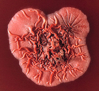
Histoplasmosis is a fungal infection caused by Histoplasma capsulatum. Symptoms of this infection vary greatly, but the disease affects primarily the lungs. Occasionally, other organs are affected; called disseminated histoplasmosis, it can be fatal if left untreated.

<i>Talaromyces marneffei</i> Species of fungus

Talaromyces marneffei, formerly called Penicillium marneffei, was identified in 1956. The organism is endemic to southeast Asia where it is an important cause of opportunistic infections in those with HIV/AIDS-related immunodeficiency. Incidence of T. marneffei infections has increased due to a rise in HIV infection rates in the region.

<span class="mw-page-title-main">Opportunistic infection</span> Infection caused by pathogens that take advantage of an opportunity not normally available

An opportunistic infection is an infection caused by pathogens that take advantage of an opportunity not normally available. These opportunities can stem from a variety of sources, such as a weakened immune system, an altered microbiome, or breached integumentary barriers. Many of these pathogens do not necessarily cause disease in a healthy host that has a non-compromised immune system, and can, in some cases, act as commensals until the balance of the immune system is disrupted. Opportunistic infections can also be attributed to pathogens which cause mild illness in healthy individuals but lead to more serious illness when given the opportunity to take advantage of an immunocompromised host.

<i>Histoplasma</i> Genus of fungi

Histoplasma is a genus of fungi in the order Onygenales. Species are known human pathogens producing yeast-like states under pathogenic conditions. They are the causative agents of histoplasmosis in humans and epizootic lymphangitis in horses.

<i>Sporothrix schenckii</i> Species of fungus

Sporothrix schenckii, a fungus that can be found worldwide in the environment, is named for medical student Benjamin Schenck, who in 1896 was the first to isolate it from a human specimen. The species is present in soil as well as in and on living and decomposing plant material such as peat moss. It can infect humans as well as animals and is the causative agent of sporotrichosis, commonly known as "rose handler's disease." The most common route of infection is the introduction of spores to the body through a cut or puncture wound in the skin. Infection commonly occurs in otherwise healthy individuals but is rarely life-threatening and can be treated with antifungals. In the environment it is found growing as filamentous hyphae. In host tissue it is found as a yeast. The transition between the hyphal and yeast forms is temperature dependent making S. schenckii a thermally dimorphic fungus.

<i>Histoplasma capsulatum</i> Species of fungus

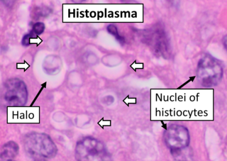
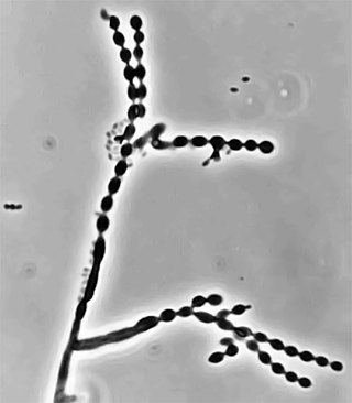
Histoplasma capsulatum is a species of dimorphic fungus. Its sexual form is called Ajellomyces capsulatus. It can cause pulmonary and disseminated histoplasmosis.

Acremonium strictum is an environmentally widespread saprotroph species found in soil, plant debris, and rotting mushrooms. Isolates have been collected in North and Central America, Asia, Europe and Egypt. A. strictum is an agent of hyalohyphomycosis and has been identified as an increasingly frequent human pathogen in immunosuppressed individuals, causing localized, disseminated and invasive infections. Although extremely rare, A. strictum can infect immunocompetent individuals, as well as neonates. Due to the growing number of infections caused by A. strictum in the past few years, the need for new medical techniques in the identification of the fungus as well as for the treatment of human infections has risen considerably.

Exophiala jeanselmei is a saprotrophic fungus in the family Herpotrichiellaceae. Four varieties have been discovered: Exophiala jeanselmei var. heteromorpha, E. jeanselmei var. lecanii-corni, E. jeanselmei var. jeanselmei, and E. jeanselmei var. castellanii. Other species in the genus Exophiala such as E. dermatitidis and E. spinifera have been reported to have similar annellidic conidiogenesis and may therefore be difficult to differentiate.

<span class="mw-page-title-main">Dimorphic fungus</span> Fungi that can exist as mold or yeast

Dimorphic fungi are fungi that can exist in the form of both mold and yeast. This is usually brought about by change in temperature and the fungi are also described as thermally dimorphic fungi. An example is Talaromyces marneffei, a human pathogen that grows as a mold at room temperature, and as a yeast at human body temperature.

Pathogenic fungi are fungi that cause disease in humans or other organisms. Although fungi are eukaryotic, many pathogenic fungi are microorganisms. Approximately 300 fungi are known to be pathogenic to humans; their study is called "medical mycology". Fungal infections are estimated to kill more people than either tuberculosis or malaria—about two million people per year.

Saksenaea vasiformis is an infectious fungus associated with cutaneous or subcutaneous lesions following trauma. It causes opportunistic infections as the entry of the fungus is through open spaces of cutaneous barrier ranging in severity from mild to severe or fatal. It lives in soils worldwide, but is considered as a rare human pathogen since only 38 cases were reported as of 2012. Saksenaea vasiformis usually fails to sporulate on the routine culture media, creating a challenge for early diagnosis, which is essential for a good prognosis. Infections are usually treated using a combination of amphotericin B and surgery. Saksenaea vasiformis is one of the few fungi known to cause necrotizing fasciitis or "flesh-eating disease".

African histoplasmosis is a fungal infection caused by Histoplasma capsulatumvar. duboisii, or Histoplama duboisii (Hcd). Disease has been most often reported in Uganda, Nigeria, Zaire and Senegal, as Hcd is exclusive to Africa. In human disease it manifests differently than histoplasmosis, most often involving the skin and bones and rarely involving the lungs. Also unlike Hcc, Hcd has been reported to rarely present in those with HIV, likely due to underreporting. However, this along with the differences in Hcc and Hcd have been disputed.

<i>Ochroconis gallopava</i> Species of fungus

Ochroconis gallopava, also called Dactylaria gallopava or Dactylaria constricta var. gallopava, is a member of genus Dactylaria. Ochroconis gallopava is a thermotolerant, darkly pigmented fungus that causes various infections in fowls, turkeys, poults, and immunocompromised humans first reported in 1986. Since then, the fungus has been increasingly reported as an agent of human disease especially in recipients of solid organ transplants. Ochroconis gallopava infection has a long onset and can involve a variety of body sites. Treatment of infection often involves a combination of antifungal drug therapy and surgical excision.

Phaeohyphomycosis is a diverse group of fungal infections, caused by dematiaceous fungi whose morphologic characteristics in tissue include hyphae, yeast-like cells, or a combination of these. It can be associated with an array of melanistic filamentous fungi including Alternaria species, Exophiala jeanselmei, and Rhinocladiella mackenziei.

Scedosporiosis is the general name for any mycosis – i.e., fungal infection – caused by a fungus from the genus Scedosporium. Current population-based studies suggest Scedosporium prolificans and Scedosporium apiospermum to be among the most common infecting agents from the genus, although infections caused by other members thereof are not unheard of. The latter is an asexual form (anamorph) of another fungus, Pseudallescheria boydii. The former is a "black yeast", currently not characterized as well, although both of them have been described as saprophytes.

<i>Cladophialophora carrionii</i> Species of fungus

Cladophialophora carrionii is a melanized fungus in the genus Cladophialophora that is associated with decaying plant material like cacti and wood. It is one of the most frequent species of Cladophialophora implicated in human disease. Cladophialophora carrionii is a causative agent of chromoblastomycosis, a subcutaneous infection that occurs in sub-tropical areas such as Madagascar, Australia and northwestern Venezuela. Transmission occurs through traumatic implantation of plant material colonized by C. carrionii, mainly infecting rural workers. When C. carrionii infects its host, it transforms from a mycelial state to a muriform state to better tolerate the extreme conditions in the host's body.

<i>Phialophora verrucosa</i> Species of fungus

Phialophora verrucosa is a pathogenic, dematiaceous fungus that is a common cause of chromoblastomycosis. It has also been reported to cause subcutaneous phaeohyphomycosis and mycetoma in very rare cases. In the natural environment, it can be found in rotting wood, soil, wasp nests, and plant debris. P. verrucosa is sometimes referred to as Phialophora americana, a closely related environmental species which, along with P. verrucosa, is also categorized in the P. carrionii clade.

Arthrographis kalrae is an ascomycetous fungus responsible for human nail infections described in 1938 by Cochet as A. langeronii. A. kalrae is considered a weak pathogen of animals including human restricted to the outermost keratinized layers of tissue. Infections caused by this species are normally responsive to commonly used antifungal drugs with only very rare exceptions.

Emmonsiosis, also known as emergomycosis, is a systemic fungal infection that can affect the lungs, generally always affects the skin and can become widespread. The lesions in the skin look like small red bumps and patches with a dip, ulcer and dead tissue in the centre.

Sporothrix brasiliensis is a fungus that is commonly found in soil. It is an emerging fungal pathogen that is causing disease in humans and cats mainly in Brazil and other countries in South America.

References

  1. 1 2 3 4 5 6 7 8 9 10 11 12 13 14 15 16 17 18 Loulergue, P; Bastides, F; Baudouin, V; Chandenier, J; Mariani-Kurkdjian, P; Dupont, B; Viard, JP; Dromer, F; Lortholary, O (November 2007). "Literature review and case histories of Histoplasma capsulatum var. duboisii infections in HIV-infected patients". Emerging Infectious Diseases. 13 (11): 1647–52. doi:10.3201/eid1311.070665. PMC   3375808 . PMID   18217546.
  2. 1 2 3 4 5 6 7 8 9 10 11 12 13 14 15 Rippon, John Willard (1988). Medical mycology: the pathogenic fungi and the pathogenic actinomycetes (3rd ed.). Philadelphia, PA: Saunders. ISBN   978-0721624440.
  3. 1 2 3 4 5 6 7 8 9 Gugnani, HC (2000). "Histoplasmosis in Africa: a review". The Indian Journal of Chest Diseases & Allied Sciences. 42 (4): 271–7. PMID   15597674.
  4. 1 2 3 Dubois, Albert; Janssens, P.G.; Brutsaert, P.; Vanbreuseghem, R. (1952). "Un cas d'histoplasmose africaine. Avec une note mycologique sur Histoplasma duboisii n. sp". Ann. Soc. Belge Méd. Trop. 32: 569–583.
  5. Ainsworth, G.C. (1959). "J. T. Duncan, F.R.C.S.Ir., D.T.M. & H.". Transactions of the British Mycological Society. 42 (1): 121–122. doi:10.1016/S0007-1536(59)80077-2.
  6. 1 2 Schwartz, Jan (1971). "African Histoplasmosis". In Baker, Roger Denio (ed.). The pathologic anatomy of mycoses: Human infection with fungi, actinomycetes and algae. Berlin: Springer-Verlag. pp. 139–146.
  7. Viviani, Maria Anna (1999). "Obituary: Edouard Drouhet". Mycopathologia. 148 (3): 111–112. doi: 10.1023/A:1007168827983 . S2CID   29419348.
  8. Pine, L; Kaufman, L; Boone, CJ (1964). "Comparative fluorescent antibody staining of Histoplasma capsulatum and Histoplasma duboisii with a specific anti-yeast phase H. capsulatum conjugate". Mycopathologia et Mycologia Applicata. 24 (4): 315–326. doi:10.1007/bf02053642. PMID   14285775. S2CID   869546.
  9. "Histoplasma duboisii". Mycobank.org. Retrieved 6 November 2015.
  10. 1 2 3 4 5 6 7 8 Garcia-Guiñon, A; Torres-Rodríguez, JM; Ndidongarte, DT; Cortadellas, F; Labrín, L (June 2009). "Disseminated histoplasmosis by Histoplasma capsulatum var. duboisii in a paediatric patient from the Chad Republic, Africa". European Journal of Clinical Microbiology & Infectious Diseases. 28 (6): 697–9. doi:10.1007/s10096-008-0668-2. PMID   19005708. S2CID   30017610.
  11. 1 2 3 4 5 6 7 8 9 10 11 12 13 14 15 16 17 18 19 20 21 Ravindran, S; Sobhanakumari, K; Celine, M; Palakkal, S (April 2015). "African histoplasmosis: the first report of an indigenous case in India". International Journal of Dermatology. 54 (4): 451–5. doi:10.1111/ijd.12683. PMID   25514986. S2CID   38826382.
  12. 1 2 3 4 5 Gugnani, HC; Muotoe-Okafor, FA; Kaufman, L; Dupont, B (September 1994). "A natural focus of Histoplasma capsulatum var. duboisii is a bat cave". Mycopathologia. 127 (3): 151–7. doi:10.1007/BF01102915. PMID   7808510. S2CID   2163739.
  13. 1 2 3 Gugnani, HC; Muotoe-Okafor, F (December 1997). "African histoplasmosis: a review". Revista Iberoamericana de Micologia. 14 (4): 155–9. PMID   15538817.
  14. 1 2 3 4 5 6 7 Tsiodras, S.; Drogari-Apiranthitou, M.; Pilichos, K.; Leventakos, K.; Kelesidis, T.; Buitrago, M. J.; Petrikkos, G.; Panayiotides, I. (1 February 2012). "An Unusual Cutaneous Tumor: African Histoplasmosis following Mudbaths: Case Report and Review". American Journal of Tropical Medicine and Hygiene. 86 (2): 261–263. doi:10.4269/ajtmh.2012.11-0557. PMC   3269278 . PMID   22302860.
  15. Klassen-Fischer, M; McEvoy, P; Neafie, RC; Nelson, AM (15 February 2004). "Accurate diagnosis of infection with Histoplasma capsulatum var. duboisii". Clinical Infectious Diseases. 38 (4): 595, author reply 595–6. doi: 10.1086/381455 . PMID   14765358.
  16. 1 2 Kwon-Chung, K.J; Bennett, J.E. (1992). Medical mycology. Philadelphia: Lea & Febiger. ISBN   978-0812114638.
  17. Nethercott, JR; Schachter, RK; Givan, KF; Ryder, DE (April 1978). "Histoplasmosis due to Histoplasma capsulatum var duboisii in a Canadian immigrant". Archives of Dermatology. 114 (4): 595–598. doi:10.1001/archderm.114.4.595. PMID   646378.
  18. Kasuga, T; Taylor, JW; White, TJ (March 1999). "Phylogenetic relationships of varieties and geographical groups of the human pathogenic fungus Histoplasma capsulatum Darling". Journal of Clinical Microbiology. 37 (3): 653–63. doi:10.1128/JCM.37.3.653-663.1999. PMC   84508 . PMID   9986828.
  19. Manfredi, R; Mazzoni, A; Nanetti, A; Chiodo, F (December 1994). "Histoplasmosis capsulati and duboisii in Europe: the impact of the HIV pandemic, travel and immigration". European Journal of Epidemiology. 10 (6): 675–81. doi:10.1007/bf01719280. PMID   7672046. S2CID   22037907.