Intravascular ultrasound

Last updated
Intravascular ultrasound
ICD-9-CM 00.2
OPS-301 code 3-05e
MedlinePlus 007266

Intravascular ultrasound (IVUS) or intravascular echocardiography is a medical imaging methodology using a specially designed catheter with a miniaturized ultrasound probe attached to the distal end of the catheter. The proximal end of the catheter is attached to computerized ultrasound equipment. It allows the application of ultrasound technology, such as piezoelectric transducer or CMUT, to see from inside blood vessels out through the surrounding blood column, visualizing the endothelium (inner wall) of blood vessels. [1]

Contents

The arteries of the heart (the coronary arteries) are the most frequent imaging target for IVUS. IVUS is used in the coronary arteries to determine the amount of atheromatous plaque built up at any particular point in the epicardial coronary artery. Intravascular ultrasound provides a unique method to study the regression or progression of atherosclerotic lesions in vivo. [2] The progressive accumulation of plaque within the artery wall over decades is the setup for vulnerable plaque which, in turn, leads to heart attack and stenosis (narrowing) of the artery (known as coronary artery lesions). IVUS is of use to determine both plaque volume within the wall of the artery and/or the degree of stenosis of the artery lumen. It can be especially useful in situations in which angiographic imaging is considered unreliable; such as for the lumen of ostial lesions or where angiographic images do not visualize lumen segments adequately, such as regions with multiple overlapping arterial segments. It is also used to assess the effects of treatments of stenosis such as with hydraulic angioplasty expansion of the artery, with or without stents, and the results of medical therapy over time.

Advantages over angiography

Arguably the most valuable use of IVUS is to visualize plaque, which cannot be seen by angiography. Over time this technique has evolved into an extremely useful research tool for modern invasive cardiology, [3] and it has been increasingly used in research to better understand the behavior of the atherosclerosis process in living people.

IVUS enables accurately visualizing not only the lumen of the coronary arteries but also the atheroma (membrane/cholesterol loaded white blood cells) "hidden" within the wall. IVUS has thus enabled advances in clinical research providing a more thorough perspective and better understanding.

In the early 1990s, IVUS research on the re-stenosis problem after angioplasty lead to recognition that most of the re-stenosis problem (as visualized by an angiography examination) was not true re-stenosis. Instead it was simply a remodeling of the atheromatous plaque, which was still protruding into the lumen of the artery after completion of angioplasty; the stenosis only appearing to be reduced because blood and contrast could now flow around and through some of the plaque. The angiographic dye column appeared widened adequately; yet considerable plaque was within the newly widened lumen and the lumen remained partially obstructed. This recognition promoted more frequent use of stents to hold the plaque outward against the inner artery walls, out of the lumen.

Additionally, IVUS examinations, as they were done more frequently, served to reveal and confirm the autopsy research findings of the late 1980s, showing that atheromatous plaque tends to cause expansion of the internal elastic lamina, causing the degree of plaque burden to be greatly underestimated by angiography. [4] Angiography only reveals the edge of the atheroma that protrudes into the lumen. [5]

Intravascular ultrasound image of a coronary artery (left), with color-coding on the right, delineating the lumen (yellow), external elastic membrane (blue) and the atherosclerotic plaque burden (green). The percentage stenosis is defined as the area of the lumen (yellow) divided by the area of the external elastic membrane (blue) times 100. As the plaque burden increases, the lumen size will decrease and the degree of stenosis will increase. IVUS of CAD (1).png
Intravascular ultrasound image of a coronary artery (left), with color-coding on the right, delineating the lumen (yellow), external elastic membrane (blue) and the atherosclerotic plaque burden (green). The percentage stenosis is defined as the area of the lumen (yellow) divided by the area of the external elastic membrane (blue) times 100. As the plaque burden increases, the lumen size will decrease and the degree of stenosis will increase.

Perhaps the greatest contribution to understanding, so far, was achieved by clinical research trials completed in the United States in the late 1990s, using combined angiography and IVUS examination, to study which coronary lesions most commonly result in a myocardial infarction. [6] The studies revealed that most myocardial infarctions occur at areas with extensive atheroma within the artery wall, however very little stenosis of the artery opening. [7] The range of lumen stenosis locations at which myocardial infarctions occurred ranged from areas of mild dilation all the way to areas of greater than 95% stenosis. However the average or typical stenosis at which myocardial infarctions occurred were found to be less than 50%, [8] describing plaques long considered insignificant by many. Only 14% of heart attacks occurred at locations with 75% or more stenosis[ citation needed ], the severe stenoses previously thought by many to present the greatest danger to the individual. This research has changed the primary focus for heart attack prevention from severe narrowing to vulnerable plaque.

Current clinical uses of IVUS technology include checking how to treat complex lesions before angioplasty and checking how well an intracoronary stent has been deployed within a coronary artery after angioplasty. If a stent is not expanded flush against the wall of the vessel, turbulent flow may occur between the stent and the wall of the vessel; some fear this might create a nidus for acute thrombosis of the artery.

Disadvantages versus angiography

The primary disadvantages of IVUS being used routinely in a cardiac catheterization laboratory are its expense, the increase in the time of the procedure, and the fact that it is considered an interventional procedure, and should only be performed by angiographers that are trained in interventional cardiology techniques. In addition, there may be additional risk imposed by the use of the IVUS catheter.

The computerized IVUS echocardiographic imaging systems list for $120,000, US, 2007 for a cart based system and ~$70,000 for an installed or integrated solution. The disposable catheters used to do each examination typically cost ~$600, US, 2007. In many hospitals, the IVUS system is placed as part of a bundle deal based on minimum disposable sales volumes. In other words, the cost of the console is paid for by rebates from other purchased products including IVUS catheters. Because no standard exists, IVUS catheters cannot be interchanged between different manufacturers.

Additionally, IVUS adds significant additional examination time and some increased risk to the patient beyond performing a standard diagnostic angiographic examination. This increase is significantly less when IVUS is part of a percutaneous coronary intervention, since much of the setup is the same for the intervention as for the IVUS imaging.

IVUS continues to improve and some manufacturers have proposed building IVUS technology into angioplasty and stent balloon catheters, a potential major advance, but limited by complexity, cost and increased bulk of the catheters.

Comparison versus intravascular optical coherence tomography

Compared to IVUS, intravascular OCT offers an order of magnitude improved resolution for a better visualization of vessel lumen, tissue microstructure and devices (e.g., intracoronary stents). [9] IVUS offers an improved imaging depth for the assessment of lipid or necrotic plaques, while intravascular OCT offers better penetration and enhanced imaging of calcific tissue. Intravascular OCT requires a short injection of contrast (e.g., 2 to 3 seconds) in a similar way to obtain an angiographic image. IVUS does not require a contrast injection as ultrasounds can penetrate through blood.

Method

To visualize an artery or vein, angiographic techniques are used and the physician positions the tip of a guidewire, usually 0.36 mm (0.014") diameter with a very soft and pliable tip and about 200 cm long. The physician steers the guidewire from outside the body, through angiography catheters and into the blood vessel branch to be imaged.

The ultrasound catheter tip is slid in over the guidewire and positioned, using angiography techniques so that the tip is at the farthest away position to be imaged. The sound waves are emitted from the catheter tip, are usually in the 20-40 MHz range, and the catheter also receives and conducts the return echo information out to the external computerized ultrasound equipment which constructs and displays a real time ultrasound image of a thin section of the blood vessel currently surrounding the catheter tip, usually displayed at 30 frames/second image.

The guide wire is kept stationary and the ultrasound catheter tip is slid backwards, usually under motorized control at a pullback speed of 0.5 mm/s. (The motorized pullback tends to be smoother than hand movement by the physician.)

The (a) blood vessel wall inner lining, (b) atheromatous disease within the wall and (c) connective tissues covering the outer surface of the blood vessel are echogenic, i.e. they return echoes making them visible on the ultrasound display.

By contrast, the blood itself and the healthy muscular tissue portion of the blood vessel wall is relatively echolucent, just black circular spaces, in the images.

Heavy calcium deposits in the blood vessel wall both heavily reflect sound, i.e. are very echogenic, but are also distinguishable by shadowing. Heavy calcification blocks sound transmission beyond and so, in the echo images, are seen as both very bright areas but with black shadows behind (from the vantage point of the catheter tip emitting the ultrasound waves).

Uses

IVUS, as outlined above, has been the best technology, so far, to demonstrate the anatomy of the artery wall in living animals and humans. It has led to an explosion of better understanding and research on both (a) the behavior of the atherosclerosis process and (b) the effects of different treatment strategies for changing the evolution of the atherosclerosis disease process. This has been important given that atherosclerosis is the single most frequent disease process for the greatest percentage of individuals living in first world countries.

Intravascular ultrasound in the coronary anatomy

An IVUS image of the ostial left main coronary artery (left). The blue outline delineates the cross-sectional area of the lumen of the artery (A1 in the upper right corner), measuring 6.0 mm . A two-dimensional mapping of the proximal LAD and left main coronary arteries is shown on the right. IVUS-Tracing.png
An IVUS image of the ostial left main coronary artery (left). The blue outline delineates the cross-sectional area of the lumen of the artery (A1 in the upper right corner), measuring 6.0 mm . A two-dimensional mapping of the proximal LAD and left main coronary arteries is shown on the right.

While the routine use of IVUS during percutaneous coronary intervention does not improve short term outcomes, [10] there are a number of situations in which IVUS is of particular use in the treatment of coronary artery disease of the heart. In particular in cases when the degree of stenosis of a coronary artery is unclear, IVUS can directly quantify the percentage of stenosis and give insight into the anatomy of the plaque.

One particular use of IVUS in the coronary anatomy is in the quantification of left main disease in cases where routine coronary angiography gives equivocal results. Many studies in the past have shown that significant left main disease can increase mortality, [11] and that intervention (either coronary artery bypass graft surgery or percutaneous coronary intervention) to reduce mortality is necessary when the left main stenosis is significant.

When using IVUS to determine whether an individual's left main disease is clinically significant, in terms of the desirability of physical intervention, the two most widely used parameters are the degree of stenosis and the minimal lumen area. [12] A cross sectional area of ≤7 mm² in a symptomatic individual[ citation needed ] or ≤6 mm² in an asymptomatic individual [13] is considered to be clinically significant and warrants intervention to improve one-year mortality. However, these exact cutoffs are up for debate and different cutoff cross-sectional areas may be used in practice depending on differing interpretations of the trial data.

Validating the efficacy of new treatments

Because IVUS is widely available in coronary catheterization labs worldwide and can accurately quantify arterial plaque, especially within the coronary arteries, it is increasingly being used to evaluate newer and evolving strategies for the treatment of coronary artery disease, including the statins [14] and other approaches. [15] [16]

See also

Related Research Articles

<span class="mw-page-title-main">Angioplasty</span> Procedure to widen narrow arteries or veins

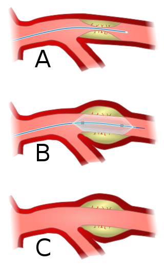
Angioplasty, also known as balloon angioplasty and percutaneous transluminal angioplasty (PTA), is a minimally invasive endovascular procedure used to widen narrowed or obstructed arteries or veins, typically to treat arterial atherosclerosis. A deflated balloon attached to a catheter is passed over a guide-wire into the narrowed vessel and then inflated to a fixed size. The balloon forces expansion of the blood vessel and the surrounding muscular wall, allowing an improved blood flow. A stent may be inserted at the time of ballooning to ensure the vessel remains open, and the balloon is then deflated and withdrawn. Angioplasty has come to include all manner of vascular interventions that are typically performed percutaneously.

<span class="mw-page-title-main">Angiography</span> Medical imaging technique

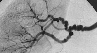
Angiography or arteriography is a medical imaging technique used to visualize the inside, or lumen, of blood vessels and organs of the body, with particular interest in the arteries, veins, and the heart chambers. Modern angiography is performed by injecting a radio-opaque contrast agent into the blood vessel and imaging using X-ray based techniques such as fluoroscopy.

<span class="mw-page-title-main">Coronary catheterization</span> Radiography of heart and blood vessels

A coronary catheterization is a minimally invasive procedure to access the coronary circulation and blood filled chambers of the heart using a catheter. It is performed for both diagnostic and interventional (treatment) purposes.

<span class="mw-page-title-main">Restenosis</span> Recurrence of stenosis, a narrowing of a blood vessel

Restenosis is the recurrence of stenosis, a narrowing of a blood vessel, leading to restricted blood flow. Restenosis usually pertains to an artery or other large blood vessel that has become narrowed, received treatment to clear the blockage and subsequently become renarrowed. This is usually restenosis of an artery, or other blood vessel, or possibly a vessel within an organ.

<span class="mw-page-title-main">Coronary thrombosis</span> Medical condition

Coronary thrombosis is defined as the formation of a blood clot inside a blood vessel of the heart. This blood clot may then restrict blood flow within the heart, leading to heart tissue damage, or a myocardial infarction, also known as a heart attack.

<span class="mw-page-title-main">Atheroma</span> Accumulation of degenerative material in the inner layer of artery walls

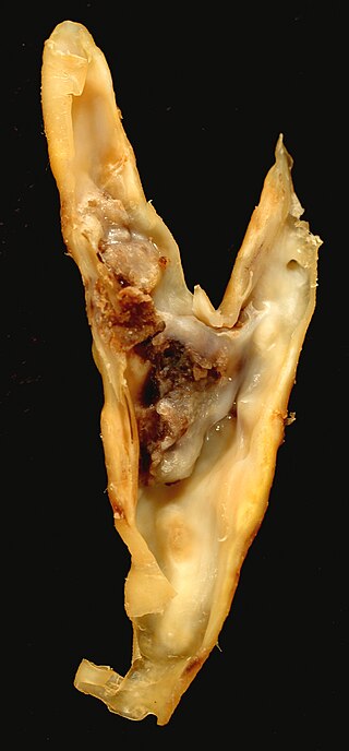
An atheroma, or atheromatous plaque, is an abnormal accumulation of material in the inner layer of an arterial wall.

A vulnerable plaque is a kind of atheromatous plaque – a collection of white blood cells and lipids in the wall of an artery – that is particularly unstable and prone to produce sudden major problems such as a heart attack or stroke.

<span class="mw-page-title-main">Cardiac catheterization</span> Insertion of a catheter into a chamber or vessel of the heart

Cardiac catheterization is the insertion of a catheter into a chamber or vessel of the heart. This is done both for diagnostic and interventional purposes.

<span class="mw-page-title-main">Percutaneous coronary intervention</span> Medical techniques used to manage coronary occlusion

Percutaneous coronary intervention (PCI) is a minimally invasive non-surgical procedure used to treat narrowing of the coronary arteries of the heart found in coronary artery disease. The procedure is used to place and deploy coronary stents, a permanent wire-meshed tube, to open narrowed coronary arteries. PCI is considered 'non-surgical' as it uses a small hole in a peripheral artery (leg/arm) to gain access to the arterial system, an equivalent surgical procedure would involve the opening of the chest wall to gain access to the heart area. The term 'coronary angioplasty with stent' is synonymous with PCI. The procedure visualises the blood vessels via fluoroscopic imaging and contrast dyes. PCI is performed by an interventional cardiologists in a catheterization laboratory setting.

<span class="mw-page-title-main">Fibromuscular dysplasia</span> Human arterial disease

Fibromuscular dysplasia (FMD) is a non-atherosclerotic, non-inflammatory disease of the blood vessels that causes abnormal growth within the wall of an artery. FMD has been found in nearly every arterial bed in the body, although the most commonly affected are the renal and carotid arteries.

Coronary artery anomalies are variations of the coronary circulation, affecting <1% of the general population. Symptoms include chest pain, shortness of breath and syncope, although cardiac arrest may be the first clinical presentation. Several varieties are identified, with a different potential to cause sudden cardiac death.

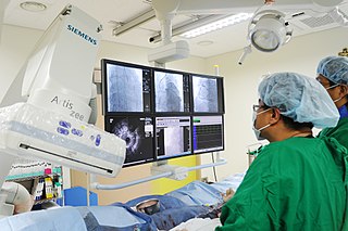
The history of invasive and interventional cardiology is complex, with multiple groups working independently on similar technologies. Invasive and interventional cardiology is currently closely associated with cardiologists, though the development and most of its early research and procedures were performed by diagnostic and interventional radiologists.

Fractional flow reserve (FFR) is a diagnostic technique used in coronary catheterization. FFR measures pressure differences across a coronary artery stenosis to determine the likelihood that the stenosis impedes oxygen delivery to the heart muscle.

<span class="mw-page-title-main">Coronary stent</span> Medical stent implanted into coronary arteries

A coronary stent is a tube-shaped device placed in the coronary arteries that supply blood to the heart, to keep the arteries open in patients suffering from coronary heart disease. The vast majority of stents used in modern interventional cardiology are drug-eluting stents (DES) It is used in a medical procedure called percutaneous coronary intervention (PCI). Coronary stents are divided into two broad types - drug-eluting and bare metal stents, as of 2023 drug-eluting stents were used in more than 90% of all PCI procedures. Stents reduce angina and have been shown to improve survival and decrease adverse events after a patient has suffered a heart attack - medically termed an acute myocardial infarction.

<span class="mw-page-title-main">Spontaneous coronary artery dissection</span> Uncommon cause of heart attacks mostly affecting younger, healthy women

Spontaneous coronary artery dissection (SCAD) is an uncommon but potentially lethal condition in which one of the coronary arteries that supply the heart, spontaneously develops a blood collection, or hematoma, within the artery wall due to a tear in the wall. SCAD is one of the arterial dissections that can occur.

<span class="mw-page-title-main">Hybrid cardiac surgery</span>

A hybrid cardiac surgical procedure in a narrow sense is defined as a procedure that combines a conventional, more invasive surgical part with an interventional part, using some sort of catheter-based procedure guided by fluoroscopy imaging in a hybrid operating room (OR) without interruption. The hybrid technique has a reduced risk of surgical complications and has shown decreased recovery time. It can be used to treat numerous heart diseases and conditions and with the increasing complexity of each case, the hybrid surgical technique is becoming more common.

<span class="mw-page-title-main">Intracoronary optical coherence tomography</span>

Intracoronary optical coherence tomography (OCT) is a catheter-based imaging application of optical coherence tomography. Currently prospective trials demonstrate OCT alters morbidity and/or mortality in coronary stenting and cervical cancer screening as discussed below.

Intravascular imaging is a catheter based system that allows physicians such as interventional cardiologists to acquire images of diseased vessels from inside the artery. Intravascular imaging provides detailed and accurate measurements of vessel lumen morphology, vessel size, extension of diseased artery segments, vessel size and plaque characteristics. Examples of intravascular imaging modalities are intravascular ultrasound (IVUS) and intracoronary optical coherence tomography.

<span class="mw-page-title-main">Intravascular fluorescence</span>

Intravascular fluorescence is a catheter-based molecular imaging technique that uses near-infrared fluorescence to detect artery wall autofluorescence (NIRAF) or fluorescence generated by molecular agents injected intravenously (NIRF). No commercial systems based on intravascular fluorescence are currently on the market, however, significant steps forwards in intravascular fluorescence imaging technology have been made between 2010-2016. It is typically used to detect functional state of artery wall including some known high-risk features of atherosclerosis. It is usually combined with structural imaging modalities such as Intravascular ultrasound and/or Intracoronary optical coherence tomography, to provide functional information in a morphological context.

Blood vessel disorder generally refers to the narrowing, hardening or enlargement of arteries and veins. It is often due to the build-up of fatty deposits in the lumen of blood vessels or infection of the vessel wall. This can occur in various locations such as coronary blood vessels, peripheral arteries and veins. The narrowed arteries would block the blood supply to different organs and tissues. In severe conditions, it may develop into more critical health problems like myocardial infarction, stroke or heart failure, which are some of the major reasons of death.

References

  1. Hector M. Garcia-Garcia, Bill D. Gogas, Patrick W. Serruys & Nico Bruining (2011). "IVUS-based imaging modalities for tissue characterization: similarities and differences". The International Journal of Cardiovascular Imaging. 27 (2): 215–224. doi:10.1007/s10554-010-9789-7. PMC   3078312 . PMID   21327914.{{cite journal}}: CS1 maint: multiple names: authors list (link)
  2. Sgura FA, Di Mario C (June 2001). "[New methods of coronary imaging II. Intracoronary ultrasonography in clinical practice]". Ital Heart J Suppl (in Italian). 2 (6): 579–92. PMID   11460831.
  3. Görge G, Ge J, von Birgelen C, Erbel R (August 1998). "[Intravascular ultrasound--the new gold standard?]". Z Kardiol (in German). 87 (8): 575–85. doi:10.1007/s003920050216. PMID   9782590. S2CID   46488571.
  4. Glagov S, Weisenberg E, Zarins CK, Stankunavicius R, Kolettis GJ (1987). "Compensatory enlargement of human atherosclerotic coronary arteries". N Engl J Med. 316 (22): 1371–5. doi:10.1056/NEJM198705283162204. PMID   3574413.
  5. Zarins CK, Weisenberg E, Kolettis G, Stankunavicius R, Glagov S (1988). "Differential enlargement of artery segments in response to enlarging atherosclerotic plaques". J Vasc Surg. 7 (3): 386–94. doi:10.1016/0741-5214(88)90433-8. PMID   3346952.
  6. Nissen, Steven E.; Yock, Paul (2001-01-30). "Intravascular Ultrasound: Novel Pathophysiological Insights and Current Clinical Applications". Circulation. 103 (4): 604–616. doi: 10.1161/01.CIR.103.4.604 . ISSN   0009-7322. PMID   11157729.
  7. Lusis, Aldons J. (2000-09-14). "Atherosclerosis". Nature. 407 (6801): 233–241. doi:10.1038/35025203. ISSN   0028-0836. PMC   2826222 . PMID   11001066.
  8. Bhardwaj, Rajeev; Kandoria, Arvind; Sharma, Rajesh (2014). "Myocardial infarction in young adults-risk factors and pattern of coronary artery involvement". Nigerian Medical Journal. 55 (1): 44–47. doi: 10.4103/0300-1652.128161 . ISSN   0300-1652. PMC   4071662 . PMID   24970969.
  9. Bezerra, Hiram G.; Costa, Marco A.; Guagliumi, Giulio; Rollins, Andrew M.; Simon, Daniel I. (2009). "Intracoronary Optical Coherence Tomography: A Comprehensive Review". JACC: Cardiovascular Interventions. 2 (11): 1035–1046. doi:10.1016/j.jcin.2009.06.019. ISSN   1936-8798. PMC   4113036 . PMID   19926041.
  10. Schiele F, Meneveau N, Vuillemenot A, Zhang DD, Gupta S, Mercier M, Danchin N, Bertrand B, Bassand JP (1998). "Impact of intravascular ultrasound guidance in stent deployment on 6-month restenosis rate: a multicenter, randomized study comparing two strategies--with and without intravascular ultrasound guidance". J Am Coll Cardiol. 32 (2): 320–8. doi: 10.1016/S0735-1097(98)00249-6 . PMID   9708456.
  11. Abizaid AS, Mintz GS, Abizaid A, Mehran R, Lansky AJ, Pichard AD, Satler LF, Wu H, Kent KM, Leon MB (1999). "One-year follow-up after intravascular ultrasound assessment of moderate left main coronary artery disease in patients with ambiguous angiograms". J Am Coll Cardiol. 34 (3): 707–15. doi: 10.1016/S0735-1097(99)00261-2 . PMID   10483951.
  12. Robert D, Safian MD, Mark S, Freed MD, eds. (2002). "Intravascular Ultrasound". The Manual Of Interventional Cardiology (Third ed.). Royal Oak, Michigan: Physicians' Press. pp.  712. ISBN   978-1-890114-39-8.
  13. Jasti V, Ivan E, Yalamanchili V, Wongpraparut N, Leesar MA (2004). "Correlations between fractional flow reserve and intravascular ultrasound in patients with an ambiguous left main coronary artery stenosis". Circulation. 110 (18): 2831–6. doi: 10.1161/01.CIR.0000146338.62813.E7 . PMID   15492302.
  14. Nissen, SE; Nicholls, SJ; Sipahi, I; Libby, P; Raichlen, JS; Ballantyne, CM; Davignon, J; Erbel, R; et al. (2006). "Effect of very high-intensity statin therapy on regression of coronary atherosclerosis: the ASTEROID trial". JAMA: The Journal of the American Medical Association. 295 (13): 1556–65. doi: 10.1001/jama.295.13.jpc60002 . PMID   16533939.
  15. Nissen SE (2002). "Who is at risk for atherosclerotic disease? Lessons from intravascular ultrasound". Am J Med. 112 (Suppl 8a): 27S–33S. doi:10.1016/S0002-9343(02)01087-2. PMID   12049992.
  16. Nissen SE, Tsunoda T, Tuzcu EM, Schoenhagen P, Cooper CJ, Yasin M, Eaton GM, Lauer MA, Sheldon WS, Grines CL, Halpern S, Crowe T, Blankenship JC, Kerensky R (2003). "Effect of Recombinant ApoA-I Milano on Coronary Atherosclerosis in Patients With Acute Coronary Syndromes". JAMA. 290 (17): 2292–2300. doi: 10.1001/jama.290.17.2292 . PMID   14600188.