Focused assessment with sonography for trauma

Last updated
Focused assessment with sonography in trauma, a.k.a. FAST
Ultrasound image of spleen 110314102702 1031200.jpg
Ultrasound image of a normal spleen that may be seen in part of the eFAST
eMedicine 104363

Focused assessment with sonography in trauma (commonly abbreviated as FAST) is a rapid bedside ultrasound examination performed by surgeons, emergency physicians, and paramedics as a screening test for blood around the heart (pericardial effusion) or abdominal organs (hemoperitoneum) after trauma. [1] There is also the extended FAST (eFAST) which includes some additional ultrasound views to assess for pneumothorax. [2]

Contents

The four classic areas that are examined for free fluid are the perihepatic space (including Morison's pouch or the hepatorenal recess), perisplenic space, pericardium, and the pelvis. With this technique it is possible to identify the presence of intraperitoneal or pericardial free fluid. In the context of traumatic injury, this fluid will usually be due to bleeding.

Indications

Reasons a FAST or eFAST would be performed would be:

  1. Blunt abdominal trauma
  2. Penetrating abdominal trauma
  3. Blunt thoracic trauma
  4. Penetrating thoracic trauma
  5. Undifferentiated shock (low blood pressure) [2]

Contraindications

Since the FAST/eFAST is performed with ultrasound, there is very little risk to the patient as ultrasounds only emit sound waves and record the echo to create a picture. [3] The most common contraindication would be delay of definitive care such as surgical intervention in the setting of obvious trauma or resuscitative efforts in an extreme scenario. [2]

Extended FAST

The eFAST allows for the examination of both lungs by adding bilateral anterior thoracic sonography to the FAST exam. This allows for the detection of a pneumothorax with the absence of normal ‘lung-sliding’ and ‘comet-tail’ artifact (seen on the ultrasound screen). Compared with supine chest radiography, with CT or clinical course as the gold standard, bedside sonography has superior sensitivity (49–99% versus 27–75%), similar specificity (95–100%), and can be performed in under a minute. [4] Several recent prospective studies have validated its use in the setting of trauma resuscitation, and have also shown that ultrasound can provide an accurate estimation of pneumothorax size. [5] [6] Although radiography or CT scanning is generally feasible, immediate bedside detection of a pneumothorax confirms what are often ambiguous physical findings in unstable patients, and guides immediate chest decompression. In addition, in the patient undergoing positive-pressure ventilation, the detection of an otherwise ‘occult’ pneumothorax prior to CT scanning may hasten treatment and subsequently prevent development of a tension pneumothorax, a deadly complication if not treated immediately, and deterioration in the radiology suite (in the CT scanner). [7]

Components of the Examination

There are five components to the eFAST exam:

  1. Right Upper Quadrant of the abdomen (Perihepatic view). Right upper quadrant is examined by working your probe down the midaxillary line starting at the right 8th rib to the 11th rib. This examines for free fluid around the kidney and liver.
  2. Left Upper Quadrant of the abdomen (Perisplenic view). Left upper quadrant is examined by working your probe down the midaxillary line starting at the left 8th rib to the 11th rib. This examines for free fluid around the kidney and spleen.
  3. Pelvic views (Long and transverse axis). The suprapubic view helps assess for free fluid in the pelvic cavity.
  4. Cardiac view. The pericardial component is assessed using the subxiphoid view.
  5. Lung views (Right and Left, Long axis). These final views help determine if a pneumothorax is present. [8]

Findings

eFAST (extended focused assessment with sonography for trauma) allows an emergency physician or a surgeon the ability to determine whether a patient has pneumothorax, hemothorax, pleural effusion, mass/tumor, or a lodged foreign body. The exam allows for visualization of the echogenic tissue, ribs, and lung tissue. Few radiographic signs are important in any trauma and they include the stratosphere sign, the sliding or seashore sign, and the sinusoid sign.

Stratosphere sign is a clinical medical ultrasound finding usually in an eFAST examination that can prove presence of a pneumothorax. The sign is an imaging finding using a 3.5–7.5 MHz ultrasound probe in the fourth and fifth intercostal spaces in the anterior clavicular line using the M-Mode of the machine. This finding is seen in the M-mode tracing as pleura and lung being indistinguishable as linear hyperechogenic lines and is fairly reliable for diagnosis of a pneumothorax. Even though the stratospheric sign can be an indication of pneumothorax its absence is not at all reliable to rule out pneumothorax as definitive diagnosis usually requires X-ray or CT of thorax. [9] [10] [11]

Seashore sign is another eFAST finding usually in the lungs in the M-mode that depicts the glandular echogenicity of the lung abutted by the linear appearance of the visceral pleura. This sign is a normal finding. In absence of a seashore sign or presence of a stratosphere sign, pneumothorax is likely. B-lines or "comet trails" are echogenic bright linear reflections beneath the pleura that are usually lost with any air between the probe and the lung tissue and therefore whose presence with seashore sign indicates absence of a pneumothorax. [9] [11]

Sinusoid sign is another M-mode finding indicating presence of pleural effusion. Due to the cyclical movement of the lung in inspiration and expiration, the motion-time tracing (M-mode) ultrasound shows a sinusoid appearance between the fluid and the line tissue. This finding indicates a possible pleural effusion, empyema, blood in pleural space (hemothorax). [9] [10]

Advantages

FAST is less invasive than diagnostic peritoneal lavage, involves no exposure to radiation and is cheaper compared to computed tomography, but achieves a similar accuracy. [12]

Numerous studies have shown FAST is useful in evaluating trauma patients. [13] [14] [15] [16] It also appears to make emergency department care faster and better. [17] [18]

Interpretation

FAST Algorithm FAST Algorithm.svg
FAST Algorithm

FAST is most useful in trauma patients who are hemodynamically unstable. A positive FAST result is defined as the appearance of a dark ("anechoic") strip in the dependent areas of the peritoneum. In the right upper quadrant this typically appears in Morison's Pouch (between the liver and kidney). This location is most useful as it is the place where fluid will collect with a supine patient. In the left upper quadrant, blood may collect anywhere around the spleen (perisplenic space). In the pelvis, blood generally pools behind the bladder (in the rectovesicular space). A positive result suggests hemoperitoneum; often CT scan will be performed if the patient is stable [20] or a laparotomy if unstable. In those with a negative FAST result, a search for extra-abdominal sources of bleeding may still need to be performed.

See also

Related Research Articles

<span class="mw-page-title-main">Medical ultrasound</span> Diagnostic and therapeutic technique

Medical ultrasound includes diagnostic techniques using ultrasound, as well as therapeutic applications of ultrasound. In diagnosis, it is used to create an image of internal body structures such as tendons, muscles, joints, blood vessels, and internal organs, to measure some characteristics or to generate an informative audible sound. The usage of ultrasound to produce visual images for medicine is called medical ultrasonography or simply sonography, or echography. The practice of examining pregnant women using ultrasound is called obstetric ultrasonography, and was an early development of clinical ultrasonography. The machine used is called an ultrasound machine, a sonograph or an echograph. The visual image formed using this technique is called an ultrasonogram, a sonogram or an echogram.

<span class="mw-page-title-main">Pneumothorax</span> Abnormal collection of air in the pleural space

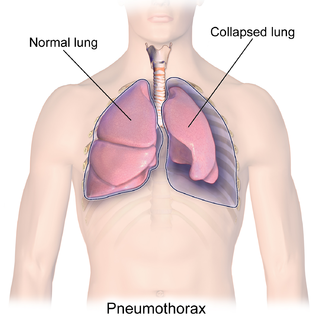
A pneumothorax is an abnormal collection of air in the pleural space between the lung and the chest wall. Symptoms typically include sudden onset of sharp, one-sided chest pain and shortness of breath. In a minority of cases, a one-way valve is formed by an area of damaged tissue, and the amount of air in the space between chest wall and lungs increases; this is called a tension pneumothorax. This can cause a steadily worsening oxygen shortage and low blood pressure. This leads to a type of shock called obstructive shock, which can be fatal unless reversed. Very rarely, both lungs may be affected by a pneumothorax. It is often called a "collapsed lung", although that term may also refer to atelectasis.

<span class="mw-page-title-main">Pleurisy</span> Disease of the lungs

Pleurisy, also known as pleuritis, is inflammation of the membranes that surround the lungs and line the chest cavity (pleurae). This can result in a sharp chest pain while breathing. Occasionally the pain may be a constant dull ache. Other symptoms may include shortness of breath, cough, fever, or weight loss, depending on the underlying cause. Pleurisy can be caused by a variety of conditions, including viral or bacterial infections, autoimmune disorders, and pulmonary embolism.

<span class="mw-page-title-main">Pleural effusion</span> Accumulation of excess fluid in the pleural cavity

A pleural effusion is accumulation of excessive fluid in the pleural space, the potential space that surrounds each lung. Under normal conditions, pleural fluid is secreted by the parietal pleural capillaries at a rate of 0.6 millilitre per kilogram weight per hour, and is cleared by lymphatic absorption leaving behind only 5–15 millilitres of fluid, which helps to maintain a functional vacuum between the parietal and visceral pleurae. Excess fluid within the pleural space can impair inspiration by upsetting the functional vacuum and hydrostatically increasing the resistance against lung expansion, resulting in a fully or partially collapsed lung.

Radiology (X-rays) is used in the diagnosis of tuberculosis. Abnormalities on chest radiographs may be suggestive of, but are never diagnostic of TB, but can be used to rule out pulmonary TB.

<span class="mw-page-title-main">Chest tube</span> Type of surgical drain

A chest tube is a surgical drain that is inserted through the chest wall and into the pleural space or the mediastinum in order to remove clinically undesired substances such as air (pneumothorax), excess fluid, blood (hemothorax), chyle (chylothorax) or pus (empyema) from the intrathoracic space. An intrapleural chest tube is also known as a Bülau drain or an intercostal catheter (ICC), and can either be a thin, flexible silicone tube, or a larger, semi-rigid, fenestrated plastic tube, which often involves a flutter valve or underwater seal.

<span class="mw-page-title-main">Traumatic cardiac arrest</span> Medical condition

Traumatic cardiac arrest (TCA) is a condition in which the heart has ceased to beat due to blunt or penetrating trauma, such as a stab wound to the thoracic area. It is a medical emergency which will always result in death without prompt advanced medical care. Even with prompt medical intervention, survival without neurological complications is rare. In recent years, protocols have been proposed to improve survival rate in patients with traumatic cardiac arrest, though the variable causes of this condition as well as many coexisting injuries can make these protocols difficult to standardize. Traumatic cardiac arrest is a complex form of cardiac arrest often derailing from advanced cardiac life support in the sense that the emergency team must first establish the cause of the traumatic arrest and reverse these effects, for example hypovolemia and haemorrhagic shock due to a penetrating injury.

<span class="mw-page-title-main">Chest radiograph</span> Projection X-ray of the chest

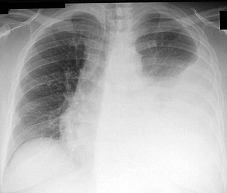
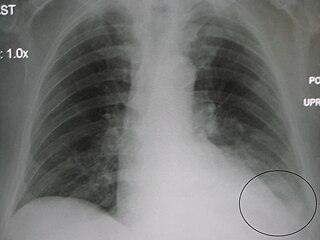
A chest radiograph, called a chest X-ray (CXR), or chest film, is a projection radiograph of the chest used to diagnose conditions affecting the chest, its contents, and nearby structures. Chest radiographs are the most common film taken in medicine.

<span class="mw-page-title-main">Hemothorax</span> Blood accumulation in the pleural cavity

A hemothorax is an accumulation of blood within the pleural cavity. The symptoms of a hemothorax may include chest pain and difficulty breathing, while the clinical signs may include reduced breath sounds on the affected side and a rapid heart rate. Hemothoraces are usually caused by an injury, but they may occur spontaneously due to cancer invading the pleural cavity, as a result of a blood clotting disorder, as an unusual manifestation of endometriosis, in response to Pneumothorax, or rarely in association with other conditions.

<span class="mw-page-title-main">Thoracentesis</span> Removal of fluids/air from the pleural cavity of the lungs

Thoracentesis, also known as thoracocentesis, pleural tap, needle thoracostomy, or needle decompression, is an invasive medical procedure to remove fluid or air from the pleural space for diagnostic or therapeutic purposes. A cannula, or hollow needle, is carefully introduced into the thorax, generally after administration of local anesthesia. The procedure was first performed by Morrill Wyman in 1850 and then described by Henry Ingersoll Bowditch in 1852.

<span class="mw-page-title-main">Pericardial effusion</span> Medical condition

A pericardial effusion is an abnormal accumulation of fluid in the pericardial cavity. The pericardium is a two-part membrane surrounding the heart: the outer fibrous connective membrane and an inner two-layered serous membrane. The two layers of the serous membrane enclose the pericardial cavity between them. This pericardial space contains a small amount of pericardial fluid, normally 15-50 mL in volume. The pericardium, specifically the pericardial fluid provides lubrication, maintains the anatomic position of the heart in the chest, and also serves as a barrier to protect the heart from infection and inflammation in adjacent tissues and organs.

<span class="mw-page-title-main">Blunt trauma</span> Trauma to the body without penetration of the skin

Blunt trauma, also known as blunt force trauma or non-penetrating trauma, describes a physical trauma due to a forceful impact without penetration of the body's surface. Blunt trauma stands in contrast with penetrating trauma, which occurs when an object pierces the skin, enters body tissue, and creates an open wound. Blunt trauma occurs due to direct physical trauma or impactful force to a body part. Such incidents often occur with road traffic collisions, assaults, sports-related injuries, and are notably common among the elderly who experience falls.

Fremitus is a vibration transmitted through the body. In common medical usage, it usually refers to assessment of the lungs by either the vibration intensity felt on the chest wall and/or heard by a stethoscope on the chest wall with certain spoken words, although there are several other types.

<span class="mw-page-title-main">Parapneumonic effusion</span> Medical condition

A parapneumonic effusion is a type of pleural effusion that arises as a result of a pneumonia, lung abscess, or bronchiectasis. There are three types of parapneumonic effusions: uncomplicated effusions, complicated effusions, and empyema. Uncomplicated effusions generally respond well to appropriate antibiotic treatment.

Emergency ultrasound employing point-of-care ultrasound (POCUS) is the application of ultrasound at the point of care to make immediate patient-care decisions. It is performed by the health care professional caring for the injured or ill persons. This point-of-care use of ultrasound is often to evaluate an emergency medical condition, in settings such as an emergency department, critical care unit, ambulance, or combat zone.

Malignant pleural effusion is a condition in which cancer causes an abnormal amount of fluid to collect between the thin layers of tissue (pleura) lining the outside of the lung and the wall of the chest cavity. Lung cancer and breast cancer account for about 50-65% of malignant pleural effusions. Other common causes include pleural mesothelioma and lymphoma.

<span class="mw-page-title-main">Abdominal trauma</span> Medical condition

Abdominal trauma is an injury to the abdomen. Signs and symptoms include abdominal pain, tenderness, rigidity, and bruising of the external abdomen. Complications may include blood loss and infection.

Obstructive shock is one of the four types of shock, caused by a physical obstruction in the flow of blood. Obstruction can occur at the level of the great vessels or the heart itself. Causes include pulmonary embolism, cardiac tamponade, and tension pneumothorax. These are all life-threatening. Symptoms may include shortness of breath, weakness, or altered mental status. Low blood pressure and tachycardia are often seen in shock. Other symptoms depend on the underlying cause.

Prehospital ultrasound is the specialized application of ultrasound by paramedics, to guide immediate care and treatment procedures. Like conventional ultrasound, it is a device that produces cyclic sound pressure to penetrate a medium (flesh) and reveal details about the inner structure of the medium.

A transmediastinal gunshot wound (TMGSW) is a penetrating injury to a person's thorax in which a bullet enters the mediastinum, possibly damaging some of the major structures in this area. Hemodynamic instability has been reported in about 50% of cases with a mortality rate ranging from 20% to 49%. Some studies have shown marked improvement in the mortality rate of patients who survived transfer to the operating room rather than being treated surgically in the ER.

References

  1. "Ultrasound in Trauma - The FAST Exam Focused Assessment with Sonography in Trauma". www.sonoguide.com. Archived from the original on 2008-09-14.
  2. 1 2 3 Bloom, Benjamin A.; Gibbons, Ryan C. (2022). "Focused Assessment with Sonography for Trauma". StatPearls. StatPearls Publishing. PMID   29261902.
  3. "Ultrasound". National Institute of Biomedical Imaging and Bioengineering.
  4. Kirkpatrick AW, Sirois M, Laupland KB, et al., J Trauma, 2004;57(2):288–95.
  5. Zhang M, Liu ZH, Yang JX, et al., Crit Care, 2006;10(4):R112.
  6. Blaivas M, Lyon M, Duggal SA, Acad Emerg Med, 2005;12(9):844–9.
  7. Davis JA, et al. Critical Diagnosis in Bedside Ultrasonography. Diagnostics & Imaging. 2007.
  8. , Patel D, Lewis K, Peterson A, Hafez M. Extended Focused Assessment with Sonography for Trauma (EFAST) Exam. J Med Ins. ;(299.6) doi:https://jomi.com/article/299.6
  9. 1 2 3 Christopher P. Holstege; Alexander B. Baer; Jesse M. Pines; William J. Brady (2011). Visual Diagnosis in Emergency and Critical Care Medicine. Wiley-Blackwell. pp. 95–7. ISBN   9781444397987.
  10. 1 2 Christoph T. Bolliger; F. J. F. Herth; P. Mayo; T. Miyazawa; J. Beamis (2009). Clinical chest ultrasound: from the ICU to the bronchoscopy suite. Karger Publishers. pp. 86–8. ISBN   9783805586429.
  11. 1 2 Steven G. Rothrock (M.D.) (2009). Tarascon Adult Emergency Pocketbook. Tarascon. p. 144.
  12. Rozycki G, Shackford S (1996). "Ultrasound, what every trauma surgeon should know". J Trauma. 40 (1): 1–4. doi:10.1097/00005373-199601000-00001. PMID   8576968.
  13. Dolich MO, McKenney MG, Varela JE, Compton RP, McKenney KL, Cohn SM (Jan 2001). "2,576 ultrasounds for blunt abdominal trauma". Journal of Trauma. 50 (1): 108–12. doi:10.1097/00005373-200101000-00019. PMID   11231679.
  14. Farahmand N, Sirlin CB, Brown MA, Shragg GP, Fortlage D, Hoyt DB, Casola G (May 2005). "Hypotensive patients with blunt abdominal trauma: performance of screening US". Radiology. 235 (2): 436–43. doi:10.1148/radiol.2352040583. PMID   15798158.
  15. Sirlin CB, Brown MA, Andrade-Barreto OA, Deutsch R, Fortlage DA, Hoyt DB, Casola G (Mar 2004). "Blunt abdominal trauma: clinical value of negative screening US scans". Radiology. 230 (3): 661–8. doi:10.1148/radiol.2303021707. PMID   14990832.
  16. Moylan M, Newgard CD, Ma OJ, Sabbaj A, Rogers T, Douglass R (Oct 2007). "Association between a positive ED FAST examination and therapeutic laparotomy in normotensive blunt trauma patients". Journal of Emergency Medicine. 33 (3): 265–71. doi:10.1016/j.jemermed.2007.02.030. PMID   17976554.
  17. Melniker LA, Leibner E, McKenney MG, Lopez P, Briggs WM, Mancuso CA (Sep 2006). "Randomized controlled clinical trial of point-of-care, limited ultrasonography for trauma in the emergency department: the first sonography outcomes assessment program trial". Annals of Emergency Medicine . 48 (3): 227–35. doi:10.1016/j.annemergmed.2006.01.008. PMID   16934640.
  18. Ollerton JE, Sugrue M, Balogh Z, D'Amours SK, Giles A, Wyllie P (Apr 2006). "Prospective study to evaluate the influence of FAST on trauma patient management". Journal of Trauma. 60 (4): 785–91. doi:10.1097/01.ta.0000214583.21492.e8. PMID   16612298.
  19. 1 2 3 "UOTW #18 - Ultrasound of the Week". Ultrasound of the Week. 17 September 2014. Retrieved 27 May 2017.
  20. Scalea T, Rodriguez A, Chiu W, Brenneman F, Fallon W, Kato K, McKenney M, Nerlich M, Ochsner M, Yoshii H (1999). "Focused Assessment with Sonography for Trauma (FAST): results from an international consensus conference". Journal of Trauma. 46 (3): 466–72. doi: 10.1097/00005373-199903000-00022 . PMID   10088853.
Further reading