Flail chest

Last updated
Flail chest
Other namesParadoxical breathing
Flailchest3to9.png
A 3D reconstruction from a CT scan showing a flail chest. Arrows mark the rib fractures.
Specialty Thoracic surgery, Trauma surgery
Complications Pneumothorax

Flail chest is a life-threatening medical condition that occurs when a segment of the rib cage breaks due to trauma and becomes detached from the rest of the chest wall. Two of the symptoms of flail chest are chest pain and shortness of breath. [1]

Contents

It occurs when multiple adjacent ribs are broken in multiple places, separating a segment, so a part of the chest wall moves independently. The number of ribs that must be broken varies by differing definitions: some sources say at least two adjacent ribs are broken in at least two places, [2] some require three or more ribs in two or more places. [3] The flail segment moves in the opposite direction to the rest of the chest wall: because of the ambient pressure in comparison to the pressure inside the lungs, it goes in while the rest of the chest is moving out, and vice versa. This so-called "paradoxical breathing" [4] is painful and increases the work involved in breathing.

Flail chest is usually accompanied by a pulmonary contusion, a bruise of the lung tissue that can interfere with blood oxygenation. [5] Often, it is the contusion, not the flail segment, that is the main cause of respiratory problems in people with both injuries. [6]

Surgery to fix the fractures appears to result in better outcomes. [7]

Signs and symptoms

Two of the symptoms of flail chest are chest pain and shortness of breath. [1]

The characteristic paradoxical motion of the flail segment occurs due to pressure changes associated with respiration that the rib cage normally resists:

Paradoxical motion is a late sign of flail segment; therefore, an absence of paradoxical motion does not mean the patient does not have a flail segment.

The constant motion of the ribs in the flail segment at the site of the fracture is extremely painful, and, untreated, the sharp broken edges of the ribs are likely to eventually puncture the pleural sac and lung, possibly causing a pneumothorax. The concern about "mediastinal flutter" (the shift of the mediastinum with paradoxical diaphragm movement) does not appear to be merited. [8] Pulmonary contusions are commonly associated with flail chest and that can lead to respiratory failure. This is due to the paradoxical motions of the chest wall from the fragments interrupting normal breathing and chest movement. Typical paradoxical motion is associated with stiff lungs, which requires extra work for normal breathing, and increased lung resistance, which makes air flow difficult. [9] The respiratory failure from the flail chest requires mechanical ventilation and a longer stay in an intensive care unit. [10] It is the damage to the lungs from the flail segment that is life-threatening.

Causes

Diagrams depicting the paradoxical motion observed during respiration with a flail segment Flail chest mechaincs.jpg
Diagrams depicting the paradoxical motion observed during respiration with a flail segment

The most common causes of flail chest injuries are vehicle collisions, which account for 76% of flail chest injuries. [11] Another main cause of flail chest injuries is falling. This mainly occurs in the elderly, who are more impacted by the falls as a result of their weak and frail bones, unlike their younger counterparts who can fall without being impacted as severely. Falls account for 14% of flail chest injuries. [1]

Flail chest typically occurs when three or more adjacent ribs are fractured in two or more places, allowing that segment of the thoracic wall to displace and move independently of the rest of the chest wall. Flail chest can also occur when ribs are fractured proximally in conjunction with disarticulation of costal cartilages distally. For the condition to occur, generally there must be a significant force applied over a large surface of the thorax to create the multiple anterior and posterior rib fractures. Rollover and crushing injuries most commonly break ribs at only one point, whereas for flail chest to occur a significant impact is required, breaking the ribs in two or more places. [12] This can be caused by forceful accidents such as the aforementioned vehicle collisions or significant falls. In the elderly, it can be caused by deterioration of bone, although rare. In children, the majority of flail chest injuries result from common blunt force traumas or metabolic bone diseases, including a group of genetic disorders known as osteogenesis imperfecta. [13]

Diagnosis

A chest radiograph of a flail chest associated with right sided pulmonary contusion and subcutaneous emphysema Pulmonary contusion.jpg
A chest radiograph of a flail chest associated with right sided pulmonary contusion and subcutaneous emphysema

Diagnosis is by physical examination performed by a physician. The diagnosis may be assisted or confirmed by use of medical imaging with either plain X ray or CT scan.

Treatment

Treatment of the flail chest initially follows the principles of advanced trauma life support. Further treatment includes:

A person may be intubated with a double lumen tracheal tube. In a double lumen endotracheal tube, each lumen may be connected to a different ventilator. Usually one side of the chest is affected more than the other, so each lung may require drastically different pressures and flows to adequately ventilate.

Surgical fixation can help in significantly reducing the duration of ventilatory support and in conserving the pulmonary function. [15] Surgical intervention has also been shown to reduce the need for tracheostomy, reduces the time spent in the intensive care unit following a traumatic flail chest injury and could reduce the risk of acquiring pneumonia after such an event. [16]

Physiotherapy

In order to begin a rehabilitation program for a flail chest it is important to treat the person's pain so they are able to perform the proper exercises. Due to the underlying conditions that the flail segment has caused onto the respiratory system, chest physiotherapy is important to reduce further complications. Proper positioning of the body is key, including postural alignment for proper drainage of mucous secretions. [17] The therapy will consist of a variety of postural positioning and changes in order to increase normal breathing. Along with postural repositioning, a variety of breathing exercises are also very important in order to allow the chest wall to reposition itself back to normal conditions. Breathing exercises will also include coughing procedures. [17] Furthermore, range of motion exercises are given to reduce the atrophy of the musculature. [18] With progression, resistance exercises are added to the regimen to the shoulder and arm of the side containing the injury. Moreover, trunk exercises will be introduced while sitting and will progress to during standing. [19] Hip flexion exercises can be done to expand the thorax. This is done by lying supine on a flat surface, flexing the knees and hips and bringing them in toward the chest. The knees should come in toward the chest while the person inhales, and exhale when the knees are lowered. [19] This exercise can be done in 3 sets of 6–8 repetitions with a pause in between sets. The person should always make sure to maintain controlled breaths. [19]

Eventually, the person will be progressed to walking and posture correction while walking. [19] Before the person is discharged from the hospital, the person should be able to perform mobility exercises to the core and should have attained good posture.

Prognosis

The death rate of people with flail chest depends on the severity of their condition, ranging from 10 to 25%. [1] A systematic review comparing the safety and effectiveness of surgical fixation versus non-surgical methods for the treatment of flail chest, reported that there was no statistically significant difference in the reported deaths between patients treated surgically and those treated non-surgically i.e. with conservative management methods. The results of the systematic review suggested that surgical intervention reduces the need for tracheostomy, reduces the time spent in the intensive care unit following a traumatic flail chest injury and could reduce the risk of acquiring pneumonia after such an event. [20]

Epidemiology

Approximately 1 out of 13 people admitted to the hospital with fractured ribs are found to have flail chest. [21]

Related Research Articles

<span class="mw-page-title-main">Thorax</span> Frontal part of an animals body, between its head and abdomen

The thorax or chest is a part of the anatomy of humans, mammals, and other tetrapod animals located between the neck and the abdomen. In insects, crustaceans, and the extinct trilobites, the thorax is one of the three main divisions of the creature's body, each of which is in turn composed of multiple segments.

<span class="mw-page-title-main">Pneumothorax</span> Abnormal collection of air in the pleural space

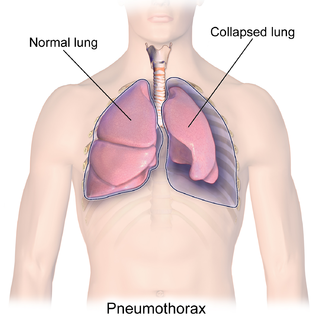
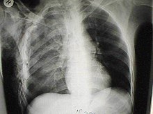
A pneumothorax is an abnormal collection of air in the pleural space between the lung and the chest wall. Symptoms typically include sudden onset of sharp, one-sided chest pain and shortness of breath. In a minority of cases, a one-way valve is formed by an area of damaged tissue, and the amount of air in the space between chest wall and lungs increases; this is called a tension pneumothorax. This can cause a steadily worsening oxygen shortage and low blood pressure. This leads to a type of shock called obstructive shock, which can be fatal unless reversed. Very rarely, both lungs may be affected by a pneumothorax. It is often called a "collapsed lung", although that term may also refer to atelectasis.

<span class="mw-page-title-main">Hemothorax</span> Blood accumulation in the pleural cavity

A hemothorax is an accumulation of blood within the pleural cavity. The symptoms of a hemothorax may include chest pain and difficulty breathing, while the clinical signs may include reduced breath sounds on the affected side and a rapid heart rate. Hemothoraces are usually caused by an injury, but they may occur spontaneously due to cancer invading the pleural cavity, as a result of a blood clotting disorder, as an unusual manifestation of endometriosis, in response to Pneumothorax, or rarely in association with other conditions.

<span class="mw-page-title-main">Chylothorax</span> Medical condition

A chylothorax is an abnormal accumulation of chyle, a type of lipid-rich lymph, in the space surrounding the lung. The lymphatics of the digestive system normally returns lipids absorbed from the small bowel via the thoracic duct, which ascends behind the esophagus to drain into the left brachiocephalic vein. If normal thoracic duct drainage is disrupted, either due to obstruction or rupture, chyle can leak and accumulate within the negative-pressured pleural space. In people on a normal diet, this fluid collection can sometimes be identified by its turbid, milky white appearance, since chyle contains emulsified triglycerides.

<span class="mw-page-title-main">Rib fracture</span> Break in a rib bone

A rib fracture is a break in a rib bone. This typically results in chest pain that is worse with inspiration. Bruising may occur at the site of the break. When several ribs are broken in several places a flail chest results. Potential complications include a pneumothorax, pulmonary contusion, and pneumonia.

<span class="mw-page-title-main">Chest physiotherapy</span> Treatments to improve breathing by indirect removal of mucus from breathing passage

Chest physiotherapy (CPT) are treatments generally performed by physical therapists and respiratory therapists, whereby breathing is improved by the indirect removal of mucus from the breathing passages of a patient. Other terms include respiratory or cardio-thoracic physiotherapy.

<span class="mw-page-title-main">Blunt trauma</span> Physical trauma caused to a body part, either by impact, injury or physical attack

Blunt trauma, also known as blunt force trauma or non-penetrating trauma, describes a physical trauma due to a forceful impact without penetration of the body's surface. Blunt trauma stands in contrast with penetrating trauma, which occurs when an object pierces the skin, enters body tissue, and creates an open wound. Blunt trauma occurs due to direct physical trauma or impactful force to a body part. Such incidents often occur with road traffic collisions, assaults, sports-related injuries, and are notably common among the elderly who experience falls.

<span class="mw-page-title-main">Pneumomediastinum</span> Abnormal presence of gas in the thorax

Pneumomediastinum is pneumatosis in the mediastinum, the central part of the chest cavity. First described in 1819 by René Laennec, the condition can result from physical trauma or other situations that lead to air escaping from the lungs, airways, or bowel into the chest cavity. In underwater divers it is usually the result of pulmonary barotrauma.

<span class="mw-page-title-main">Fat embolism syndrome</span> Medical condition

Fat embolism syndrome occurs when fat enters the blood stream and results in symptoms. Symptoms generally begin within a day. This may include a petechial rash, decreased level of consciousness, and shortness of breath. Other symptoms may include fever and decreased urine output. The risk of death is about 10%.

<span class="mw-page-title-main">Chest injury</span> Medical condition

A chest injury, also known as chest trauma, is any form of physical injury to the chest including the ribs, heart and lungs. Chest injuries account for 25% of all deaths from traumatic injury. Typically chest injuries are caused by blunt mechanisms such as direct, indirect, compression, contusion, deceleration, or blasts caused by motor vehicle collisions or penetrating mechanisms such as stabbings.

Traumatic asphyxia, or Perthes's syndrome, is a medical emergency caused by an intense compression of the thoracic cavity, causing venous back-flow from the right side of the heart into the veins of the neck and the brain.

<span class="mw-page-title-main">Winged scapula</span> Skeletal muscle condition around the shoulder blade

A winged scapula is a skeletal medical condition in which the shoulder blade protrudes from a person's back in an abnormal position.

<span class="mw-page-title-main">Penetrating trauma</span> Type of injury

Penetrating trauma is an open wound injury that occurs when an object pierces the skin and enters a tissue of the body, creating a deep but relatively narrow entry wound. In contrast, a blunt or non-penetrating trauma may have some deep damage, but the overlying skin is not necessarily broken and the wound is still closed to the outside environment. The penetrating object may remain in the tissues, come back out the path it entered, or pass through the full thickness of the tissues and exit from another area.

<span class="mw-page-title-main">Pulmonary contusion</span> Internal bruise of the lungs

A pulmonary contusion, also known as lung contusion, is a bruise of the lung, caused by chest trauma. As a result of damage to capillaries, blood and other fluids accumulate in the lung tissue. The excess fluid interferes with gas exchange, potentially leading to inadequate oxygen levels (hypoxia). Unlike pulmonary laceration, another type of lung injury, pulmonary contusion does not involve a cut or tear of the lung tissue.

<span class="mw-page-title-main">Pulmonary laceration</span> Medical condition

A pulmonary laceration is a chest injury in which lung tissue is torn or cut. An injury that is potentially more serious than pulmonary contusion, pulmonary laceration involves disruption of the architecture of the lung, while pulmonary contusion does not. Pulmonary laceration is commonly caused by penetrating trauma but may also result from forces involved in blunt trauma such as shear stress. A cavity filled with blood, air, or both can form. The injury is diagnosed when collections of air or fluid are found on a CT scan of the chest. Surgery may be required to stitch the laceration, to drain blood, or even to remove injured parts of the lung. The injury commonly heals quickly with few problems if it is given proper treatment; however it may be associated with scarring of the lung or other complications.

Pulmonary hygiene, formerly referred to as pulmonary toilet, is a set of methods used to clear mucus and secretions from the airways. The word pulmonary refers to the lungs. The word toilet, related to the French toilette, refers to body care and hygiene; this root is used in words such as toiletry that also relate to cleansing.

<span class="mw-page-title-main">Sternal fracture</span> Medical condition

A sternal fracture is a fracture of the sternum, located in the center of the chest. The injury, which occurs in 5–8% of people who experience significant blunt chest trauma, may occur in vehicle accidents, when the still-moving chest strikes a steering wheel or dashboard or is injured by a seatbelt. Cardiopulmonary resuscitation (CPR), has also been known to cause thoracic injury, including sternum and rib fractures. Sternal fractures may also occur as a pathological fracture, in people who have weakened bone in their sternum, due to another disease process. Sternal fracture can interfere with breathing by making it more painful; however, its primary significance is that it can indicate the presence of serious associated internal injuries, especially to the heart and lungs.

<span class="mw-page-title-main">Tracheobronchial injury</span> Damage to the tracheobronchial tree

Tracheobronchial injury is damage to the tracheobronchial tree. It can result from blunt or penetrating trauma to the neck or chest, inhalation of harmful fumes or smoke, or aspiration of liquids or objects.

<span class="mw-page-title-main">Diaphragmatic rupture</span> Tear in the thoracic diaphragm, usually caused by physical trauma

Diaphragmatic rupture is a tear of the diaphragm, the muscle across the bottom of the ribcage that plays a crucial role in breathing. Most commonly, acquired diaphragmatic tears result from physical trauma. Diaphragmatic rupture can result from blunt or penetrating trauma and occurs in about 0.5% of all people with trauma.

<span class="mw-page-title-main">Scapular fracture</span> Injury of the shoulder blade

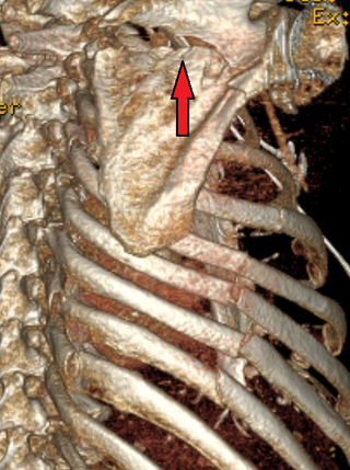
A scapular fracture is a fracture of the scapula, the shoulder blade. The scapula is sturdy and located in a protected place, so it rarely breaks. When it does, it is an indication that the individual was subjected to a considerable amount of force and that severe chest trauma may be present. High-speed vehicle accidents are the most common cause. This could be anywhere from a car accident, motorcycle crash, or high speed bicycle crash but falls and blows to the area can also be responsible for the injury. Signs and symptoms are similar to those of other fractures: they include pain, tenderness, and reduced motion of the affected area although symptoms can take a couple of days to appear. Imaging techniques such as X-ray are used to diagnose scapular fracture, but the injury may not be noticed in part because it is so frequently accompanied by other, severe injuries that demand attention. The injuries that usually accompany scapular fracture generally have the greatest impact on the patient's outcome. However, the injury can also occur by itself; when it does, it does not present a significant threat to life. Treatment involves pain control and immobilizing the affected area, and, later, physical therapy.

References

  1. 1 2 3 4 Athanassiadi, Kalliopi, Michalis Gerzounis, Nikolaos Theakos. Management of 150 flail chest injuries: analysis of risk factors affecting outcome. European Journal of Cardio-thoracic surgery 26. (2004). Pg 373–376.
  2. Emergency Nurses Association (2005). Lorene Newberry; Laura M. Criddle (eds.). Sheehy's Manual of Emergency Care (6th ed.). St Louis, Missouri: Elsevier Mosby. pp. 655–657.
  3. Keel M, Meier C (December 2007). "Chest injuries – what is new?". Current Opinion in Critical Care. 13 (6): 674–9. doi:10.1097/MCC.0b013e3282f1fe71. PMID   17975389. S2CID   19317500.
  4. "OVERVIEW paradoxical breathing". Oxford Reference. Retrieved 7 Sep 2013.
  5. Yamamoto L, Schroeder C, Morley D, Beliveau C (2005). "Thoracic trauma: The deadly dozen". Critical Care Nursing Quarterly . 28 (1): 22–40. doi:10.1097/00002727-200501000-00004. PMID   15732422.
  6. Hemmila MR, Wahl WL (2005). "Management of the injured patient". In Doherty GM (ed.). Current Surgical Diagnosis and Treatment. McGraw-Hill Medical. p. 214. ISBN   0-07-142315-X . Retrieved 2008-07-04.
  7. Coughlin, TA; Ng, JW; Rollins, KE; Forward, DP; Ollivere, BJ (August 2016). "Management of rib fractures in traumatic flail chest: a meta-analysis of randomised controlled trials". The Bone & Joint Journal. 98-B (8): 1119–25. doi:10.1302/0301-620x.98b8.37282. PMID   27482027.
  8. Molnar TF (2007). "(Video Assisted) thoracoscopic surgery: Getting started". J Minim Access Surg. 3 (4): 173–7. doi: 10.4103/0972-9941.38912 . PMC   2749201 . PMID   19789679.
  9. Bemelman M, Poeze M, Blokhuis TJ, Leenen LP (2010). "Historic overview of treatment techniques for rib facturs and flail chest". Eur J Trauma Emerg Surg. 36 (5): 407–415. doi:10.1007/s00068-010-0046-5. PMC   3150827 . PMID   21841952.
  10. Marasco SF, Davies AR, Cooper J, Varma D, Bennett V, Nevill R, Lee G, Bailey M, Fitzgerald M (2013). "Prospective Randomized Controlled Trial of Operative Rib Fixation in Traumatic Flail Chest". Journal of the American College of Surgeons. 216 (5): 924–932. doi:10.1016/j.jamcollsurg.2012.12.024. PMID   23415550.
  11. Borman, JB, L Aharonson-Daniel, B Savitsky, et al. Unilateral flail chest is seldom a lethal injury. Emerg Med J. 23. (2006). P. 903-905.
  12. Bjerke, H Scott (2006-06-16). "Flail Chest". eMedicine. Retrieved 2007-08-01.
  13. Gipson CL, Tobias JD (2006). "Flail Chest in a Neonate Resulting from Nonaccidental Trauma". Southern Medical Journal. 99 (5): 536–538. doi:10.1097/01.smj.0000216471.54786.e5. PMID   16711322.
  14. Mladenovic, Jordanna; Erskine, Ryan N.; Riley, Brooke; Mitchell, Andrew; Abi-fares, Catherine; Basson, Willem; Anstey, Chris; White, Leigh (2022-11-01). "The association between erector spinae plane block timing and reduced rib fracture related respiratory complications: A cohort study". Journal of Clinical Anesthesia. 82: 110940. doi:10.1016/j.jclinane.2022.110940. ISSN   0952-8180. PMID   35917775. S2CID   251206240.
  15. Granetzny A; Abd El-Aal, M; Emam, E; Shalaby, A; Boseila, A (2005). "Surgical versus conservative treatment of flail chest. Evaluation of the pulmonary status". Interact Cardiovasc Thorac Surg. 4 (6): 583–7. doi: 10.1510/icvts.2005.111807 . PMID   17670487.
  16. Apampa, Ayobobola A.; Ali, Ayesha; Kadir, Bryar; Ahmed, Zubair (2021-02-06). "Safety and effectiveness of surgical fixation versus non-surgical methods for the treatment of flail chest in adult populations: a systematic review and meta-analysis". European Journal of Trauma and Emergency Surgery. 48 (2): 1025–1034. doi:10.1007/s00068-021-01606-2. ISSN   1863-9941. PMC   7866961 . PMID   33547910.
  17. 1 2 Ciesla, Nancy D. "Chest Physical Therapy for Patients in the Intensive Care Unit". Journal of the American Physical Therapy Association (1996) 76:609–625.
  18. Kigin, Colleen M. "Chest Physical Therapy for the Postoperative or Traumatic Injury Patient". Journal of the American Physical Therapy Association (1981) 61:1724–1736.
  19. 1 2 3 4 "Post operative physiotherapy management for flail chest or Multiple ribs fracture or Cardio-pulmonary rehabilitation or physiotherapy or physical therapy or flail chest or BPT or MPT or PT or project report or case study or medical field or MGR medical university or Senthil Kumar BPT | PDF | Thorax | Lung". Scribd. Retrieved 2023-10-12.
  20. Apampa, Ayobobola A.; Ali, Ayesha; Kadir, Bryar; Ahmed, Zubair (2022-04-01). "Safety and effectiveness of surgical fixation versus non-surgical methods for the treatment of flail chest in adult populations: a systematic review and meta-analysis". European Journal of Trauma and Emergency Surgery. 48 (2): 1025–1034. doi: 10.1007/s00068-021-01606-2 . ISSN   1863-9941.
  21. Richardson, J. David M.D, Lee Adams M.D, Lewis M. Flint, M.D. Selective Management of Flail Chest and Pulmonary Contusion. Department of Surgery, University of Louisville, School of Medicine, Louisville, Kentucky.