Joint dislocation

Last updated

Joint dislocation
Other names Latin: luxatio
Ankledislocation.JPG
A traumatic dislocation of the tibiotarsal joint of the ankle with distal fibular fracture. Open arrow marks the tibia and the closed arrow marks the talus.
Specialty Orthopedic surgery   OOjs UI icon edit-ltr-progressive.svg

A joint dislocation, also called luxation, occurs when there is an abnormal separation in the joint, where two or more bones meet. [1] A partial dislocation is referred to as a subluxation. Dislocations are often caused by sudden trauma on the joint like an impact or fall. A joint dislocation can cause damage to the surrounding ligaments, tendons, muscles, and nerves. [2] Dislocations can occur in any major joint (shoulder, knees, etc.) or minor joint (toes, fingers, etc.). The most common joint dislocation is a shoulder dislocation. [1]

Contents

Treatment for joint dislocation is usually by closed reduction, that is, skilled manipulation to return the bones to their normal position. Reduction should only be performed by trained medical professionals, because it can cause injury to soft tissue and/or the nerves and vascular structures around the dislocation. [3]

Symptoms and signs

The following symptoms are common with any type of dislocation. [1]

Causes

Joint dislocations are caused by trauma to the joint or when an individual falls on a specific joint. [4] Great and sudden force applied, by either a blow or fall, to the joint can cause the bones in the joint to be displaced or dislocated from normal position. [5] With each dislocation, the ligaments keeping the bones fixed in the correct position can be damaged or loosened, making it easier for the joint to be dislocated in the future. [6]

Some individuals are prone to dislocations due to congenital conditions, such as hypermobility syndrome and Ehlers-Danlos Syndrome. Hypermobility syndrome is genetically inherited disorder that is thought to affect the encoding of the connective tissue protein’s collagen in the ligament of joints. [7] The loosened or stretched ligaments in the joint provide little stability and allow for the joint to be easily dislocated. [1]

Diagnosis

Initial evaluation of a suspected joint dislocation should begin with a thorough patient history, including mechanism of injury, and physical examination. Special attention should be focused on the neurovascular exam both before and after reduction, as injury to these structures may occur during the injury or during the reduction process. [3] Subsequent imaging studies are frequently obtained to assist with diagnosis.

Treatment

A dislocated joint usually can be successfully reduced into its normal position only by a trained medical professional. Trying to reduce a joint without any training could substantially worsen the injury. [15]

X-rays are usually taken to confirm a diagnosis and detect any fractures which may also have occurred at the time of dislocation. A dislocation is easily seen on an X-ray. [16]

Once a diagnosis is confirmed, the joint is usually manipulated back into position. This can be a very painful process, therefore this is typically done either in the emergency department under sedation or in an operating room under a general anaesthetic. [17]

It is important the joint is reduced as soon as possible, as in the state of dislocation, the blood supply to the joint (or distal anatomy) may be compromised. This is especially true in the case of a dislocated ankle, due to the anatomy of the blood supply to the foot. [18]

Shoulder injuries can also be surgically stabilized, depending on the severity, using arthroscopic surgery. [16] The most common treatment method for a dislocation of the Glenohumeral Joint (GH Joint/Shoulder Joint) is exercise based management. [19] Another method of treatment is to place the injured arm in a sling or in another immobilizing device in order to keep the joint stable. [20] A 2012 Cochrane review, found no statistically significant difference in healing or long-term joint mobility between simple shoulder dislocations treated conservatively versus surgically. [21]

Some joints are more at risk of becoming dislocated again after an initial injury. This is due to the weakening of the muscles and ligaments which hold the joint in place. The shoulder is a prime example of this. Any shoulder dislocation should be followed up with thorough physiotherapy. [16]

On field reduction is crucial for joint dislocations. As they are extremely common in sports events, managing them correctly at the game at the time of injury, can reduce long term issues. They require prompt evaluation, diagnosis, reduction, and postreduction management before the person can be evaluated at a medical facility. [20]

After care

After a dislocation, injured joints are usually held in place by a splint (for straight joints like fingers and toes) or a bandage (for complex joints like shoulders). Additionally, the joint muscles, tendons and ligaments must also be strengthened. This is usually done through a course of physiotherapy, which will also help reduce the chances of repeated dislocations of the same joint. [22]

For glenohumeral instability, the therapeutic program depends on specific characteristics of the instability pattern, severity, recurrence and direction with adaptations made based on the needs of the patient. In general, the therapeutic program should focus on restoration of strength, normalization of range of motion and optimization of flexibility and muscular performance. Throughout all stages of the rehabilitation program, it is important to take all related joints and structures into consideration. [23]

Epidemiology

See also

Related Research Articles

<span class="mw-page-title-main">Knee</span> Leg joint in primates

In humans and other primates, the knee joins the thigh with the leg and consists of two joints: one between the femur and tibia, and one between the femur and patella. It is the largest joint in the human body. The knee is a modified hinge joint, which permits flexion and extension as well as slight internal and external rotation. The knee is vulnerable to injury and to the development of osteoarthritis.

<span class="mw-page-title-main">Shoulder problem</span> Medical condition

Shoulder problems including pain, are one of the more common reasons for physician visits for musculoskeletal symptoms. The shoulder is the most movable joint in the body. However, it is an unstable joint because of the range of motion allowed. This instability increases the likelihood of joint injury, often leading to a degenerative process in which tissues break down and no longer function well.

<span class="mw-page-title-main">Ankle</span> Region where the foot and the leg meet

The ankle, the talocrural region or the jumping bone (informal) is the area where the foot and the leg meet. The ankle includes three joints: the ankle joint proper or talocrural joint, the subtalar joint, and the inferior tibiofibular joint. The movements produced at this joint are dorsiflexion and plantarflexion of the foot. In common usage, the term ankle refers exclusively to the ankle region. In medical terminology, "ankle" can refer broadly to the region or specifically to the talocrural joint.

<span class="mw-page-title-main">Bone fracture</span> Physical damage to the continuity of a bone

A bone fracture is a medical condition in which there is a partial or complete break in the continuity of any bone in the body. In more severe cases, the bone may be broken into several fragments, known as a comminuted fracture. A bone fracture may be the result of high force impact or stress, or a minimal trauma injury as a result of certain medical conditions that weaken the bones, such as osteoporosis, osteopenia, bone cancer, or osteogenesis imperfecta, where the fracture is then properly termed a pathologic fracture.

<span class="mw-page-title-main">Lisfranc injury</span> Type of fracture/dislocation injury of the foot

A Lisfranc injury, also known as Lisfranc fracture, is an injury of the foot in which one or more of the metatarsal bones are displaced from the tarsus.

<span class="mw-page-title-main">Shoulder joint</span> Synovial ball and socket joint in the shoulder

The shoulder joint is structurally classified as a synovial ball-and-socket joint and functionally as a diarthrosis and multiaxial joint. It involves an articulation between the glenoid fossa of the scapula and the head of the humerus. Due to the very loose joint capsule that gives a limited interface of the humerus and scapula, it is the most mobile joint of the human body.

<span class="mw-page-title-main">Ankle fracture</span> Medical condition

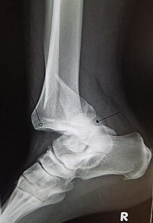
An ankle fracture is a break of one or more of the bones that make up the ankle joint. Symptoms may include pain, swelling, bruising, and an inability to walk on the injured leg. Complications may include an associated high ankle sprain, compartment syndrome, stiffness, malunion, and post-traumatic arthritis.

<span class="mw-page-title-main">Hip dislocation</span> Orthopedic injury

A hip dislocation is when the thighbone (femur) separates from the hip bone (pelvis). Specifically it is when the ball–shaped head of the femur separates from its cup–shaped socket in the hip bone, known as the acetabulum. The joint of the femur and pelvis is very stable, secured by both bony and soft-tissue constraints. With that, dislocation would require significant force which typically results from significant trauma such as from a motor vehicle collision or from a fall from elevation. Hip dislocations can also occur following a hip replacement or from a developmental abnormality known as hip dysplasia.

<span class="mw-page-title-main">Hypermobility (joints)</span> Human condition: joints that stretch further than normal

Hypermobility, also known as double-jointedness, describes joints that stretch farther than normal. For example, some hypermobile people can bend their thumbs backwards to their wrists and bend their knee joints backwards, put their leg behind the head or perform other contortionist "tricks". It can affect one or more joints throughout the body.

<span class="mw-page-title-main">Separated shoulder</span> Medical condition

A separated shoulder, also known as acromioclavicular joint injury, is a common injury to the acromioclavicular joint. The AC joint is located at the outer end of the clavicle where it attaches to the acromion of the scapula. Symptoms include non-radiating pain which may make it difficult to move the shoulder. The presence of swelling or bruising and a deformity in the shoulder is also common depending on how severe the dislocation is.

<span class="mw-page-title-main">Dislocated shoulder</span> Injury

A dislocated shoulder is a condition in which the head of the humerus is detached from the glenoid fossa. Symptoms include shoulder pain and instability. Complications may include a Bankart lesion, Hill-Sachs lesion, rotator cuff tear, or injury to the axillary nerve.

<span class="mw-page-title-main">Calcaneal fracture</span> Medical condition

A calcaneal fracture is a break of the calcaneus. Symptoms may include pain, bruising, trouble walking, and deformity of the heel. It may be associated with breaks of the hip or back.

Shoulder surgery is a means of treating injured shoulders. Many surgeries have been developed to repair the muscles, connective tissue, or damaged joints that can arise from traumatic or overuse injuries to the shoulder.

<span class="mw-page-title-main">Bankart lesion</span> Medical condition

A Bankart lesion is a type of shoulder injury that occurs following a dislocated shoulder. It is an injury of the anterior (inferior) glenoid labrum of the shoulder. When this happens, a pocket at the front of the glenoid forms that allows the humeral head to dislocate into it. It is an indication for surgery and often accompanied by a Hill-Sachs lesion, damage to the posterior humeral head.

<span class="mw-page-title-main">Hill–Sachs lesion</span> Cortical depression in the posterolateral head of the humerus

A Hill–Sachs lesion, or Hill–Sachs fracture, is a cortical depression in the posterolateral head of the humerus. It results from forceful impaction of the humeral head against the anteroinferior glenoid rim when the shoulder is dislocated anteriorly.

<span class="mw-page-title-main">Jammed finger</span> Medical condition

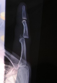
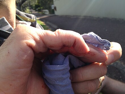
Jammed finger is a colloquialism referring to a variety of injuries to the joints of the fingers, resulting from axial loading beyond that which the ligaments can withstand. Common parts of the finger susceptible to this type of injury are ligaments, joints, and bones. The severity of the damage to the finger increases with the magnitude of the force exerted by the external object on the fingertip. Toes may become jammed as well, with similar results.

<span class="mw-page-title-main">Patellar dislocation</span> Medical condition

A patellar dislocation is a knee injury in which the patella (kneecap) slips out of its normal position. Often the knee is partly bent, painful and swollen. The patella is also often felt and seen out of place. Complications may include a patella fracture or arthritis.

<span class="mw-page-title-main">Index of trauma and orthopaedics articles</span>

Orthopedic surgery is the branch of surgery concerned with conditions involving the musculoskeletal system. Orthopedic surgeons use both surgical and nonsurgical means to treat musculoskeletal injuries, sports injuries, degenerative diseases, infections, bone tumours, and congenital limb deformities. Trauma surgery and traumatology is a sub-specialty dealing with the operative management of fractures, major trauma and the multiply-injured patient.

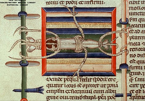
The treatment of broken bones and dislocated joints can be traced as far back as the Ancient Greeks. Hippocrates is credited with a method of reduction of a dislocated shoulder. 16th century Spanish texts talk about the Aztecs use of reduction of fractures using fir branches. The modern discipline of orthopaedics in trauma care developed during the course of World War I, but it was not until after World War II that orthopaedics became the dominant field treating fractures in much of the world. Today, the discipline encompasses conditions such as bone fractures and bone loss, as well as spinal pathology and joint disease.

<span class="mw-page-title-main">Knee dislocation</span> Medical condition

A knee dislocation is an injury in which there is disruption of the knee joint between the tibia and the femur. Symptoms include pain and instability of the knee. Complications may include injury to an artery, most commonly the popliteal artery behind the knee, or compartment syndrome.

References

  1. 1 2 3 4 Dislocations. Lucile Packard Children’s Hospital at Stanford. Retrieved 3 March 2013. Archived 28 May 2013 at the Wayback Machine
  2. Smith, R. L., & Brunolli, J. J. (1990). Shoulder kinesthesia after anterior glenohumeral joint dislocation. Journal of Orthopaedic & Sports Physical Therapy, 11(11), 507–513.
  3. 1 2 Skelley NW, McCormick JJ, Smith MV (May 2014). "In-game Management of Common Joint Dislocations". Sports Health. 6 (3): 246–255. doi:10.1177/1941738113499721. PMC   4000468 . PMID   24790695.
  4. Mayo Clinic: Finger Dislocation Joint Reduction
  5. U.S. National Library of Medicine – Dislocation
  6. Pubmed Health: Dislocation – Joint dislocation
  7. Ruemper, A. & Watkins, K. (2012). Correlations between general joint hypermobility and joint hypermobility syndrome and injury in contemporary dance students. Journal of Dance Medicine & Science, 16(4): 161–166.
  8. Chong M, Karataglis D, Learmonth D (September 2006). "Survey of the Management of Acute Traumatic First-Time Anterior Shoulder Dislocation Among Trauma Clinicians in the UK". Annals of the Royal College of Surgeons of England. 88 (5): 454–458. doi:10.1308/003588406X117115. ISSN   0035-8843. PMC   1964698 . PMID   17002849.
  9. Gaillard F. "Acromioclavicular injury | Radiology Reference Article | Radiopaedia.org". radiopaedia.org. Retrieved 21 February 2018.
  10. "Introduction to Trauma X-ray - Dislocation injury". www.radiologymasterclass.co.uk. Retrieved 15 February 2018.
  11. Abbasi S, Molaie H, Hafezimoghadam P, Zare MA, Abbasi M, Rezai M, Farsi D (August 2013). "Diagnostic accuracy of ultrasonographic examination in the management of shoulder dislocation in the emergency department". Annals of Emergency Medicine. 62 (2): 170–175. doi: 10.1016/j.annemergmed.2013.01.022 . ISSN   1097-6760. PMID   23489654.
  12. Heers G, Hedtmann A (2005). "Correlation of ultrasonographic findings to Tossy's and Rockwood's classification of acromioclavicular joint injuries". Ultrasound in Medicine & Biology. 31 (6): 725–732. doi:10.1016/j.ultrasmedbio.2005.03.002. PMID   15936487.
  13. Gaillard F. "Developmental dysplasia of the hip | Radiology Reference Article | Radiopaedia.org". radiopaedia.org. Retrieved 21 February 2018.
  14. "UpToDate". www.uptodate.com. Retrieved 21 February 2018.
  15. Bankart, A. (2004). The pathology and treatment of recurrent dislocation of the shoulder-joint. Acta Orthop Belg. 70: 515–519
  16. 1 2 3 Dias, J., Steingold, R., Richardson, R., Tesfayohannes, B., Gregg, P. (1987). The conservative treatment of acromioclavicular dislocation. British Editorial Society of Bone and Joint Surgery. 69(5): 719–722.
  17. Holdsworth, F. (1970). Fractures, dislocations, and fracture dislocations of the spine. The Journal of Bone and Joint Surgery. 52 (8): 1534–1551.
  18. Ganz, R., Gill, T., Gautier, E., Ganz, K., Krugel, N., Berlemann, U. (2001). Surgical dislocation of the adult hip. The Journal of Bone and Joint Surgery. 83(8): 1119–1124.
  19. Warby SA, Pizzari T, Ford JJ, Hahne AJ, Watson L (1 January 2014). "The effect of exercise-based management for multidirectional instability of the glenohumeral joint: a systematic review". Journal of Shoulder and Elbow Surgery. 23 (1): 128–142. doi: 10.1016/j.jse.2013.08.006 . PMID   24331125.
  20. 1 2 Skelley NW, McCormick JJ, Smith MV (4 April 2017). "In-game Management of Common Joint Dislocations". Sports Health. 6 (3): 246–255. doi:10.1177/1941738113499721. ISSN   1941-7381. PMC   4000468 . PMID   24790695.
  21. Taylor F, Sims M, Theis J, Herbison GP (18 April 2012). Cochrane Bone, Joint and Muscle Trauma Group (ed.). "Interventions for treating acute elbow dislocations in adults". Cochrane Database of Systematic Reviews. 2012 (4): CD007908. doi:10.1002/14651858.CD007908.pub2. PMC   6465046 . PMID   22513954.
  22. Itoi, E., Hatakeyama, Y., Kido, T., Sato, T., Minagawa, H., Wakabayashi, I., Kobayashi, M. (2003). Journal of Shoulder and Elbow Surgery. 12(5): 413–415.
  23. Cools AM, Borms D, Castelein B, Vanderstukken F, Johansson FR (1 February 2016). "Evidence-based rehabilitation of athletes with glenohumeral instability". Knee Surgery, Sports Traumatology, Arthroscopy. 24 (2): 382–389. doi:10.1007/s00167-015-3940-x. ISSN   0942-2056. PMID   26704789. S2CID   21227767.
  24. 1 2 3 Khiami F, Gérometta A, Loriaut P (2015). "Management of recent first-time anterior shoulder dislocations". Orthopaedics & Traumatology: Surgery & Research. 101 (1): S51–S57. doi: 10.1016/j.otsr.2014.06.027 . PMID   25596982.
  25. 1 2 Olds M, Ellis R, Donaldson K, Parmar P, Kersten P (1 July 2015). "Risk factors which predispose first-time traumatic anterior shoulder dislocations to recurrent instability in adults: a systematic review and meta-analysis". Br J Sports Med. 49 (14): 913–922. doi:10.1136/bjsports-2014-094342. ISSN   0306-3674. PMC   4687692 . PMID   25900943.
  26. Hsiao M, Owens BD, Burks R, Sturdivant RX, Cameron KL (1 October 2010). "Incidence of Acute Traumatic Patellar Dislocation Among Active-Duty United States Military Service Members". The American Journal of Sports Medicine. 38 (10): 1997–2004. doi:10.1177/0363546510371423. ISSN   0363-5465. PMID   20616375. S2CID   19131206.
  27. Fithian DC, Paxton EW, Stone ML, Silva P, Davis DK, Elias DA, White LM (1 July 2004). "Epidemiology and Natural History of Acute Patellar Dislocation". The American Journal of Sports Medicine. 32 (5): 1114–1121. doi:10.1177/0363546503260788. ISSN   0363-5465. PMID   15262631. S2CID   11899852.
  28. Ramponi D (2016). "Patellar Dislocations and Reduction Procedure". Advanced Emergency Nursing Journal . 38 (2): 89–92. doi:10.1097/tme.0000000000000104. PMID   27139130. S2CID   42552493.
  29. Elbow Dislocation
  30. "Carpal dislocations". Archived from the original on 24 December 2014. Retrieved 5 March 2013.
  31. Finger Dislocation Joint Reduction
  32. 1 2 3 Golan E, Kang KK, Culbertson M, Choueka J (2016). "The Epidemiology of Finger Dislocations Presenting for Emergency Care Within the United States". HAND. 11 (2): 192–6. doi:10.1177/1558944715627232. PMC   4920528 . PMID   27390562.
  33. 1 2 3 4 Clegg TE, Roberts CS, Greene JW, Prather BA (2010). "Hip dislocations—Epidemiology, treatment, and outcomes". Injury. 41 (4): 329–334. doi:10.1016/j.injury.2009.08.007. PMID   19796765.
  34. Ruhlmann F, Poujardieu C, Vernois J, Gayet LE (2017). "Isolated Acute Traumatic Subtalar Dislocations: Review of 13 Cases at a Mean Follow-Up of 6 Years and Literature Review". The Journal of Foot and Ankle Surgery (Review). 56 (1): 201–207. doi:10.1053/j.jfas.2016.01.044. PMID   26947001. S2CID   31290747.
  35. García-Regal J, Centeno-Ruano AJ (2013). "[Talocalcaneonavicular dislocation without associated fractures]". Acta Ortopedica Mexicana (Review) (in Spanish). 27 (3): 201–4. PMID   24707608.
  36. Prada-Cañizares A, Auñón-Martín I, Vilá Y Rico J, Pretell-Mazzini J (May 2016). "Subtalar dislocation: management and prognosis for an uncommon orthopaedic condition". International Orthopaedics (Review). 40 (5): 999–1007. doi:10.1007/s00264-015-2910-8. PMID   26208589. S2CID   6090499.
  37. Michael A. Foy, Phillip S. Fagg (5 December 2011). Medicolegal Reporting in Orthopaedic Trauma E-Book. Elsevier Health Sciences. pp. 320–. ISBN   978-0-7020-4886-9.
  38. For a graphic representation of displacements that may lead to a total talar dislocation see: Robert W. Bucholz (29 March 2012). Rockwood and Green's Fractures in Adults: Two Volumes Plus Integrated Content Website (Rockwood, Green, and Wilkins' Fractures). Lippincott Williams & Wilkins. p. 2061. ISBN   978-1-4511-6144-1.
  39. Ringleb SI, Dhakal A, Anderson CD, Bawab S, Paranjape R (1 October 2011). "Effects of lateral ligament sectioning on the stability of the ankle and subtalar joint". Journal of Orthopaedic Research. 29 (10): 1459–1464. doi: 10.1002/jor.21407 . ISSN   1554-527X. PMID   21445995.
  40. Wight L, Owen D, Goldbloom D, Knupp M (October 2017). "Pure Ankle Dislocation: A systematic review of the literature and estimation of incidence". Injury (Review). 48 (10): 2027–2034. doi:10.1016/j.injury.2017.08.011. PMID   28826653.
  41. Earwaker J (1992). "Posttraumatic calcification of the annular ligament of the radius". Skeletal Radiol. 21 (3): 149–54. doi:10.1007/BF00242127. PMID   1604339. S2CID   43615869.