Leadzyme

Last updated
Secondary structure of a leadzyme sequence obtained using mfold. It consists of an asymmetric internal loop made up of six nucleotides.The arrow indicates the cleavage site. Secondary Structure of Leadzyme.png
Secondary structure of a leadzyme sequence obtained using mfold. It consists of an asymmetric internal loop made up of six nucleotides.The arrow indicates the cleavage site.
The figure shows cartoon representations of the ground and pre-catalytic conformations of leadzyme. The green spheres represent Mg ions and the red spheres represent Sr2+ ions.Figure rendered in pymol using coordinates from pdb file 1NUV. The crystal structures of leadzyme.png
The figure shows cartoon representations of the ground and pre-catalytic conformations of leadzyme. The green spheres represent Mg ions and the red spheres represent Sr2+ ions.Figure rendered in pymol using coordinates from pdb file 1NUV.

Leadzyme is a small ribozyme (catalytic RNA), which catalyzes the cleavage of a specific phosphodiester bond. It was discovered using an in-vitro evolution study where the researchers were selecting for RNAs that specifically cleaved themselves in the presence of lead. [1] [2] However, since then, it has been discovered in several natural systems. [3] [4] Leadzyme was found to be efficient and dynamic [5] in the presence of micromolar concentrations of lead ions. [6] Unlike in other small self-cleaving ribozymes, other divalent metal ions cannot replace Pb2+ in the leadzyme. [7] Due to obligatory requirement for a lead, the ribozyme is called a metalloribozyme.

Contents

Leadzyme has been subjected to extensive biochemical and structural characterization. [8] The minimal secondary structure of leadzyme is surprisingly very simple . It comprises an asymmetric internal loop composed of six nucleotides and a helical region on each side of the internal loop. The cleavage site of leadzyme is located within a four-nucleotide long asymmetric internal loop that also consists of RNA helices on its both sides. This is shown in top figure on right, which is the secondary structure of leadzyme generated using mfold. The structures of leadzyme have also been solved using X-ray crystallography and NMR. [9] [10] The crystal structures of the two conformations of leadzyme are shown in the lower figure on right.

Catalytic mechanism of leadzyme

Leadzyme is thought to perform catalysis using a two-step mechanism. [11] In the first step of the reaction, the phosphodiester bond is cleaved into two products: 5’ product terminating in 2’3’ cyclic phosphate and the 3’ product in 5’ hydroxyl. This step is similar to other small self-cleaving ribozymes such as the Hammerhead ribozyme and HDV ribozyme. [12] Both of those ribozymes generate a product, which contain a 2’, 3’ -cyclic phosphate. However, in leadzyme this product is just an intermediate. In the second step of this reaction pathway, the 2’ 3’ -cyclic phosphate undergoes hydrolysis to form 3’ monophosphate. This mode of catalysis is similar to how ribonucleases (proteins) function rather than any known small self-cleaving ribozyme.

The leadzyme is thought to have a highly dynamic structure. [13] [14] Many studies including NMR, X-ray crystallography and molecular modeling have revealed slightly different structures. Recently using time-resolved spectroscopy, it was shown that the active site of leadzyme is very dynamic. [15] It samples a lot of different conformations in solution and that the delta G of the interconversion between different conformations is very low. Consistent with these studies, a high-resolution crystal structure also revealed two distinct conformations of the leadzyme with different binding sites for Mg2+ and Sr2+ (Pb2+ substitutes) in the two conformations. [16] In the ground state, leadzyme binds a single Sr2+ ion at nucleotides G43, G45 and A45. This binding site is away from the scissile bond (cleavage site) and thus does not explain the involvement of the Pb2+ in the catalysis. However, in the second conformation, termed the ‘pre-catalytic’ state, the ribozyme shows two Sr2+ binding sites. G43 and G42 interact with one Sr2+ whereas the second Sr2+ interacts with the A45, C23 and G24. This second Sr2+ binding site also potentially interacts with the 2’-OH of the C23 via a water molecule. This second binding site explains how Pb2+ could facilitate catalysis by abstracting the 2-OH proton and prepare it for an in-line nucleophillic attack on the scissile phosphate. This is also supported by the fact the reaction of the leadzyme is pH dependent. Thus, Pb2+ could be acting as a Lewis acid and activating the 2-OH of C23. The crystal structure is consistent with a two-metal ion mechanism that has been proposed for leadzyme catalysis. [17]

Lead toxicity through leadzyme

Toxic metals like lead are environmental and health hazards and can enter biological systems upon exposure. Lead is a persistent metal and can accumulate in human body over time due to its frequent usage in industries and presence in our environment. Inhalation of lead can have effects that can be range from subtle symptoms to serious illnesses. It is possible that presence of lead in our biological systems can induce catalysis by lead ions. [18] Since leadzyme is a relatively simple motif i.e., it has a simple fold, it appears that there are many sequences in the genomes of many natural systems which can potentially fold into a leadzyme structure. A simple search for this RNA motif in the genomes of humans, Drosophila melanogaster , Caenorhabditis elegans and Arabidopsis thaliana revealed that on average this motif is present with the frequency of 2-9 motifs for 1 Mbp of DNA sequence. [19] They also showed that leadzyme motif is very common in the mRNA sequences of these organisms as well. Thus, these sequences could potentially self-cleave in the presence of lead ions. The targeting of these RNA motifs by lead in mRNAs and other RNAs may explain lead-mediated toxicity resulting in cell death. [20]

Related Research Articles

Metalloprotein Protein that contains a metal ion cofactor

Metalloprotein is a generic term for a protein that contains a metal ion cofactor. A large proportion of all proteins are part of this category. For instance, at least 1000 human proteins contain zinc-binding protein domains although there may be up to 3000 human zinc metalloproteins.

Phenylalanine hydroxylase

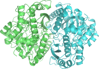
Phenylalanine hydroxylase (PAH) (EC 1.14.16.1) is an enzyme that catalyzes the hydroxylation of the aromatic side-chain of phenylalanine to generate tyrosine. PAH is one of three members of the biopterin-dependent aromatic amino acid hydroxylases, a class of monooxygenase that uses tetrahydrobiopterin (BH4, a pteridine cofactor) and a non-heme iron for catalysis. During the reaction, molecular oxygen is heterolytically cleaved with sequential incorporation of one oxygen atom into BH4 and phenylalanine substrate.

Ribozyme

Ribozymes are RNA molecules that have the ability to catalyze specific biochemical reactions, including RNA splicing in gene expression, similar to the action of protein enzymes. The 1982 discovery of ribozymes demonstrated that RNA can be both genetic material and a biological catalyst, and contributed to the RNA world hypothesis, which suggests that RNA may have been important in the evolution of prebiotic self-replicating systems. The most common activities of natural or in vitro-evolved ribozymes are the cleavage or ligation of RNA and DNA and peptide bond formation. Within the ribosome, ribozymes function as part of the large subunit ribosomal RNA to link amino acids during protein synthesis. They also participate in a variety of RNA processing reactions, including RNA splicing, viral replication, and transfer RNA biosynthesis. Examples of ribozymes include the hammerhead ribozyme, the VS ribozyme, Leadzyme and the hairpin ribozyme.

Matrix metalloproteinases (MMPs), also known as matrix metallopeptidases or matrixins, are metalloproteinases that are calcium-dependent zinc-containing endopeptidases; other family members are adamalysins, serralysins, and astacins. The MMPs belong to a larger family of proteases known as the metzincin superfamily.

Deoxyribozymes, also called DNA enzymes, DNAzymes, or catalytic DNA, are DNA oligonucleotides that are capable of performing a specific chemical reaction, often but not always catalytic. This is similar to the action of other biological enzymes, such as proteins or ribozymes . However, in contrast to the abundance of protein enzymes in biological systems and the discovery of biological ribozymes in the 1980s, there is only little evidence for naturally occurring deoxyribozymes. Deoxyribozymes should not be confused with DNA aptamers which are oligonucleotides that selectively bind a target ligand, but do not catalyze a subsequent chemical reaction.

Enolase

Enolase, also known as phosphopyruvate hydratase, is a metalloenzyme responsible for the catalysis of the conversion of 2-phosphoglycerate (2-PG) to phosphoenolpyruvate (PEP), the ninth and penultimate step of glycolysis. The chemical reaction catalyzed by enolase is:

Enzyme catalysis

Enzyme catalysis is the increase in the rate of a process by a biological molecule, an "enzyme". Most enzymes are proteins, and most such processes are chemical reactions. Within the enzyme, generally catalysis occurs at a localized site, called the active site.

<i>Bam</i>HI

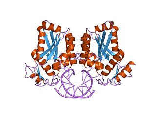
BamHI is a type II restriction endonuclease, having the capacity for recognizing short sequences of DNA and specifically cleaving them at a target site. This exhibit focuses on the structure-function relations of BamHI as described by Newman, et al. (1995). BamHI binds at the recognition sequence 5'-GGATCC-3', and cleaves these sequences just after the 5'-guanine on each strand. This cleavage results in sticky ends which are 4 bp long. In its unbound form, BamHI displays a central b sheet, which resides in between α-helices.

Hammerhead ribozyme

The hammerhead ribozyme is an RNA motif that catalyzes reversible cleavage and ligation reactions at a specific site within an RNA molecule. It is one of several catalytic RNAs (ribozymes) known to occur in nature. It serves as a model system for research on the structure and properties of RNA, and is used for targeted RNA cleavage experiments, some with proposed therapeutic applications. Named for the resemblance of early secondary structure diagrams to a hammerhead shark, hammerhead ribozymes were originally discovered in two classes of plant virus-like RNAs: satellite RNAs and viroids. They have subsequently been found to be widely dispersed within many forms of life.

Hairpin ribozyme

The hairpin ribozyme is a small section of RNA that can act as a ribozyme. Like the hammerhead ribozyme it is found in RNA satellites of plant viruses. It was first identified in the minus strand of the tobacco ringspot virus (TRSV) satellite RNA where it catalyzes self-cleavage and joining (ligation) reactions to process the products of rolling circle virus replication into linear and circular satellite RNA molecules. The hairpin ribozyme is similar to the hammerhead ribozyme in that it does not require a metal ion for the reaction.

VS ribozyme

The Varkud satellite (VS) ribozyme is an RNA enzyme that carries out the cleavage of a phosphodiester bond.

GlmS glucosamine-6-phosphate activated ribozyme

The glucosamine-6-phosphate riboswitch ribozyme is an RNA structure that resides in the 5' untranslated region (UTR) of the mRNA transcript of the glmS gene. This RNA regulates the glmS gene by responding to concentrations of a specific metabolite, glucosamine-6-phosphate (GlcN6P), in addition to catalyzing a self-cleaving chemical reaction upon activation. This cleavage leads to the degradation of the mRNA that contains the ribozyme, and lowers production of GlcN6P. The glmS gene encodes for an enzyme glutamine-fructose-6-phosphate amidotransferase, which catalyzes the formation of GlcN6P, a compound essential for cell wall biosynthesis, from fructose-6-phosphate and glutamine. Thus, when GlcN6P levels are high, the glmS ribozyme is activated and the mRNA transcript is degraded but in the absence of GlcN6P the gene continues to be translated into glutamine-fructose-6-phosphate amidotransferase and GlcN6P is produced. GlcN6P is a cofactor for this cleavage reaction, as it directly participates as an acid-base catalyst. This RNA is the first riboswitch also found to be a self-cleaving ribozyme and, like many others, was discovered using a bioinformatics approach.

Hepatitis delta virus ribozyme

The hepatitis delta virus (HDV) ribozyme is a non-coding RNA found in the hepatitis delta virus that is necessary for viral replication and is the only known human virus that utilizes ribozyme activity to infect its host. The ribozyme acts to process the RNA transcripts to unit lengths in a self-cleavage reaction during replication of the hepatitis delta virus, which is thought to propagate by a double rolling circle mechanism. The ribozyme is active in vivo in the absence of any protein factors and was the fastest known naturally occurring self-cleaving RNA at the time of its discovery.

Nucleic acid tertiary structure

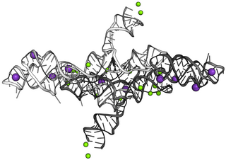
Nucleic acid tertiary structure is the three-dimensional shape of a nucleic acid polymer. RNA and DNA molecules are capable of diverse functions ranging from molecular recognition to catalysis. Such functions require a precise three-dimensional tertiary structure. While such structures are diverse and seemingly complex, they are composed of recurring, easily recognizable tertiary structure motifs that serve as molecular building blocks. Some of the most common motifs for RNA and DNA tertiary structure are described below, but this information is based on a limited number of solved structures. Many more tertiary structural motifs will be revealed as new RNA and DNA molecules are structurally characterized.

Ribonuclease E is a bacterial ribonuclease that participates in the processing of ribosomal RNA and the chemical degradation of bulk cellular RNA.

Twister ribozyme

The twister ribozyme is a catalytic RNA structure capable of self-cleavage. The nucleolytic activity of this ribozyme has been demonstrated both in vivo and in vitro and has one of the fastest catalytic rates of naturally occurring ribozymes with similar function. The twister ribozyme is considered to be a member of the small self-cleaving ribozyme family which includes the hammerhead, hairpin, hepatitis delta virus (HDV), Varkud satellite (VS), and glmS ribozymes.

RNA hydrolysis

RNA hydrolysis is a reaction in which a phosphodiester bond in the sugar-phosphate backbone of RNA is broken, cleaving the RNA molecule. RNA is susceptible to this base-catalyzed hydrolysis because the ribose sugar in RNA has a hydroxyl group at the 2’ position. This feature makes RNA chemically unstable compared to DNA, which does not have this 2’ -OH group and thus is not susceptible to base-catalyzed hydrolysis.

Mercury(II) reductase

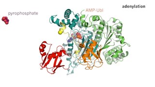
Mercury(II) reductase (EC 1.16.1.1), commonly known as MerA, is an oxidoreductase enzyme and flavoprotein that catalyzes the reduction of Hg2+ to Hg0. Mercury(II) reductase is found in the cytoplasm of many eubacteria in both aerobic and anaerobic environments and serves to convert toxic mercury ions into relatively inert elemental mercury.

Twister sister ribozyme

The twister sister ribozyme (TS) is an RNA structure that catalyzes its own cleavage at a specific site. In other words, it is a self-cleaving ribozyme. The twister sister ribozyme was discovered by a bioinformatics strategy as an RNA Associated with Genes Associated with Twister and Hammerhead ribozymes, or RAGATH.

The pistol ribozyme is an RNA structure that catalyzes its own cleavage at a specific site. In other words, it is a self-cleaving ribozyme. The pistol ribozyme was discovered through comparative genomic analysis. Subsequent biochemical analysis determined further biochemical characteristics of the ribozyme. This understanding was further advanced by an atomic-resolution crystal structure of a pistol ribozyme

References

  1. Pan, T.; Uhlenbeck, O. C. (1992). "A small metalloribozyme with a two-step mechanism". Nature. 358: 560–563. doi:10.1038/358560a0. PMID   1501711.
  2. Pan, T.; Uhlenbeck, O. C. (1992). "In vitro selection of RNAs that undergo autolytic cleavage with lead(2+)". Biochemistry. 31: 3887–3895. doi:10.1021/bi00131a001.
  3. Barciszewska, M. Z.; Wyszko, E.; Bald, R.; Erdmann, V. A.; Barciszewski, J. (2003). "5S rRNA Is a Leadzyme. A Molecular Basis for Lead Toxicity". Journal of Biochemistry. 133: 309–315. doi:10.1093/jb/mvg042.
  4. Kikovska, E.; Mikkelsen, N.-E.; Kirsebom, L. A. (2005). "The naturally trans-acting ribozyme RNase P RNA has leadzyme properties". Nucleic Acids Research. 33: 6920–6930. doi:10.1093/nar/gki993. PMC   1310964 . PMID   16332695.
  5. Kadakkuzha, B. M.; Zhao, L.; Xia, T. (2009). "Conformational Distribution and Ultrafast Base Dynamics of Leadzyme". Biochemistry. 48: 3807–3809. doi:10.1021/bi900256q. PMID   19301929.
  6. Pan, T.; Uhlenbeck, O. C. (1992). "A small metalloribozyme with a two-step mechanism". Nature. 358: 560–563. doi:10.1038/358560a0. PMID   1501711.
  7. Arciszewska, M. Z.; et al. (2005). "Lead toxicity through the leadzyme". Mutation Research/Reviews in Mutation Research. 589: 103–110. doi:10.1016/j.mrrev.2004.11.002.
  8. Sigel, Astrid; Operschall, Bert P.; Sigel, Helmut (2017). "Chapter 11. Complex Formation of Lead(II) with Nucleotides and Their Constituents". In Astrid, S.; Helmut, S.; Sigel, R. K. O. (eds.). Lead: Its Effects on Environment and Health. Metal Ions in Life Sciences. 17. de Gruyter. pp. 319–402. doi:10.1515/9783110434330-011.
  9. Wedekind, J. E.; McKay, D. B. (1999). "Crystal structure of a lead-dependent ribozyme revealing metal binding sites relevant to catalysis". Nature Structural Biology. 6: 261. doi:10.1038/6700.
  10. Wedekind, J. E.; McKay, D. B. (2003). "Crystal Structure of the Leadzyme at 1.8 Å Resolution:  Metal Ion Binding and the Implications for Catalytic Mechanism and Allo Site Ion Regulation†". Biochemistry. 42: 9554–9563. doi:10.1021/bi0300783. PMID   12911297.
  11. Pan, T.; Uhlenbeck, O. C. (1992). "A small metalloribozyme with a two-step mechanism". Nature. 358: 560–563. doi:10.1038/358560a0. PMID   1501711.
  12. Ferré-D'Amaré, A. R.; Scott, W. G. (2010). "Small Self-cleaving Ribozymes". Cold Spring Harbor Perspectives in Biology. 2: a003574. doi:10.1101/cshperspect.a003574. PMC   2944367 . PMID   20843979.
  13. Hoogstraten, C. G.; Legault, P.; Pardi, A. (1998). "NMR solution structure of the lead-dependent ribozyme: evidence for dynamics in RNA catalysis". Journal of Molecular Biology. 284: 337–350. doi:10.1006/jmbi.1998.2182. PMID   9813122.
  14. Kadakkuzha, B. M.; Zhao, L.; Xia, T. (2009). "Conformational Distribution and Ultrafast Base Dynamics of Leadzyme". Biochemistry. 48: 3807–3809. doi:10.1021/bi900256q. PMID   19301929.
  15. Kadakkuzha, B. M.; Zhao, L.; Xia, T. (2009). "Conformational Distribution and Ultrafast Base Dynamics of Leadzyme". Biochemistry. 48: 3807–3809. doi:10.1021/bi900256q. PMID   19301929.
  16. Wedekind, J. E.; McKay, D. B. (2003). "Crystal Structure of the Leadzyme at 1.8 Å Resolution:  Metal Ion Binding and the Implications for Catalytic Mechanism and Allo Site Ion Regulation†". Biochemistry. 42: 9554–9563. doi:10.1021/bi0300783. PMID   12911297.
  17. Ohmichi, T.; Sugimoto, N. (1997). "Role of Nd3+ and Pb2+ on the RNA Cleavage Reaction by a Small Ribozyme†". Biochemistry. 36: 3514–3521. doi:10.1021/bi962030d. PMID   9132001.
  18. Barciszewska, M. Z.; et al. (2005). "Lead toxicity through the leadzyme". Mutation Research/Reviews in Mutation Research. 589: 103–110. doi:10.1016/j.mrrev.2004.11.002.
  19. Barciszewska, M. Z.; et al. (2005). "Lead toxicity through the leadzyme". Mutation Research/Reviews in Mutation Research. 589: 103–110. doi:10.1016/j.mrrev.2004.11.002.
  20. Barciszewska, M. Z.; et al. (2005). "Lead toxicity through the leadzyme". Mutation Research/Reviews in Mutation Research. 589: 103–110. doi:10.1016/j.mrrev.2004.11.002.

Further reading