Hammerhead ribozyme

Last updated
Stylized rendering of the full-length
hammerhead ribozyme RNA molecule Hammerhead ribozyme ribbons.png
Stylized rendering of the full-length
hammerhead ribozyme RNA molecule

The hammerhead ribozyme is an RNA motif that catalyzes reversible cleavage and ligation reactions at a specific site within an RNA molecule. It is one of several catalytic RNAs (ribozymes) known to occur in nature. It serves as a model system for research on the structure and properties of RNA, and is used for targeted RNA cleavage experiments, some with proposed therapeutic applications. Named for the resemblance of early secondary structure diagrams to a hammerhead shark, [1] hammerhead ribozymes were originally discovered in two classes of plant virus-like RNAs: satellite RNAs and viroids. They are also known in some classes of retrotransposons, including the retrozymes. [2] The hammerhead ribozyme motif has been ubiquitously reported in lineages across the tree of life. [3] [4]

Contents

The self-cleavage reactions, first reported in 1986, [5] [6] are part of a rolling circle replication mechanism. The hammerhead sequence is sufficient for self-cleavage [7] and acts by forming a conserved three-dimensional tertiary structure.

Catalysis

In its natural state, a hammerhead RNA motif is a single strand of RNA. Although the cleavage takes place in the absence of protein enzymes, the hammerhead RNA itself is not a catalyst in its natural state, as it is consumed by the reaction (i.e. performs self-cleavage) and therefore cannot catalyze multiple turnovers.

Trans-acting hammerhead constructs can be engineered such that they consist of two interacting RNA strands, with one strand composing a hammerhead ribozyme that cleaves the other strand. [8] [ citation needed ] The strand that gets cleaved can be supplied in excess, and multiple turnover can be demonstrated and shown to obey Michaelis-Menten kinetics, [8] [ citation needed ] typical of protein enzyme kinetics. Such constructs are typically employed for in vitro experiments, and the term "hammerhead RNA" has become in practice synonymous with the more frequently used "hammerhead ribozyme".

The minimal trans-acting hammerhead ribozyme sequence that is catalytically active consists of three base-paired stems flanking a central core of 15 conserved (mostly invariant) nucleotides, as shown. The conserved central bases, with few exceptions, are essential for ribozyme's catalytic activity. Such hammerhead ribozyme constructs exhibit in vitro a turnover rate (kcat) of about 1 molecule/minute and a Km on the order of 10 nanomolar.

The hammerhead ribozyme is arguably the best-characterized ribozyme. Its small size, thoroughly-investigated cleavage chemistry, known crystal structure, and its biological relevance make the hammerhead ribozyme particularly well-suited for biochemical and biophysical investigations into the fundamental nature of RNA catalysis.

Hammerhead ribozymes may play an important role as therapeutic agents; as enzymes which tailor defined RNA sequences, as biosensors, and for applications in functional genomics and gene discovery. [9]

Species distribution

Hammerhead ribozyme HH9
RF02275-rscape.svg
Predicted secondary structure and sequence conservation of the HH9 ribozyme found conserved from lizard to human genomes
Identifiers
SymbolHH9
Rfam RF02275
Other data
RNA type Gene; ribozyme
Domain(s) Eukaryota
SO SO:0000380
PDB structures PDBe

In 1986, the first hammerhead ribozymes were found in RNA plant pathogens like viroids and viral satellites. [5] [6] One year later, a hammerhead ribozyme was also reported in the satellite DNA of newt genomes. [10] New examples of this ribozyme were then found in the genomes of unrelated organisms like schistosomes, [11] cave crickets, [12] Arabidopsis thaliana [13] and a few mammals like rodents and the platypus. [14] In 2010, it was found that the hammerhead ribozyme occurs in a wide variety of bacterial and eukaryal genomes, [15] and even in humans. [16] Similar reports confirmed and extended these observations, [17] [18] [19] unveiling the hammerhead ribozyme as a ubiquitous catalytic RNA in all life kingdoms. [20]

Most eukaryotic hammerhead ribozymes are related to a kind of short interspersed retroelements (SINEs) called retrozymes, which express as small circular RNAs. [21] However, an exceptional group of strikingly conserved hammerheads can be found in the genomes of all amniotes. [16] These hammerhead ribozymes (the so-called HH9 and HH10) occur in the introns of a few specific genes and point to a preserved biological role during pre-mRNA biosynthesis. [22] In 2021, novel Hepatitis D virus genomes of circular RNA from diverse animals were found to encode hammerhead ribozymes similar to those present in plant viroids and viral satellites. [23] A massive bioinformatic search of deltavirus-like agents around the globe has uncovered hundreds of examples of circular RNA genomes carrying hammerhead motifs, indicating that not only this ribozyme but small circular RNAs with ribozymes are ubiquitous molecules in the biosphere. [24]

Chemistry of catalysis

The hammerhead ribozyme carries out a very simple chemical reaction that results in the breakage of the substrate strand of RNA, specifically at C17, the cleavage-site nucleotide. Although RNA cleavage is often referred to as hydrolysis, the mechanism employed does not in fact involve the addition of water. Rather, the cleavage reaction is simply an isomerization that consists of rearrangement of the linking phosphodiester bond. It is the same reaction, chemically, that occurs with random base-mediated RNA degradation, except that it is highly site-specific and the rate is accelerated 10,000-fold or more.

Cleavage by phosphodiester isomerization

In-line transition-state for the hammerhead ribozyme reaction. A general base (B) abstracts a proton from the 2'-O, and a general acid (A), supplies a proton to the 5'-O leaving group as negative charge accumulates. The reaction product is a 2',3'-cyclic phosphate. The bonds breaking (shown in red) and forming (dashed lines) must be in the axial positions and reside approximately 180deg apart, as shown. Electron movements are represented by curly arrows. Hammerhead ribozyme electron transfer mechanism.svg
In-line transition-state for the hammerhead ribozyme reaction. A general base (B) abstracts a proton from the 2'-O, and a general acid (A), supplies a proton to the 5'-O leaving group as negative charge accumulates. The reaction product is a 2',3'-cyclic phosphate. The bonds breaking (shown in red) and forming (dashed lines) must be in the axial positions and reside approximately 180° apart, as shown. Electron movements are represented by curly arrows.

The cleavage reaction is a phosphodiester isomerization reaction that is initiated by abstraction of the cleavage-site ribose 2’-hydroxyl proton from the 2’-oxygen, which then becomes the attacking nucleophile in an “in-line” or SN2(P)-like reaction, although it is not known whether this proton is removed prior to or during the chemical step of the hammerhead cleavage reaction. (The cleavage reaction is technically not bimolecular, but behaves in the same way a genuine SN2(P) reaction does; it undergoes inversion of configuration subsequent to forming an associative transition-state consisting of a pentacoordinated oxyphosphrane.) The attacking and leaving group oxygens will both occupy the two axial positions in the trigonal bipyramidal transition-state structure as is required for an SN2-like reaction mechanism.

The 5’-product, as a result of this cleavage reaction mechanism, possesses a 2’,3’-cyclic phosphate terminus, and the 3’-product possesses a 5’-OH terminus, as with nonenzymatic alkaline cleavage of RNA. The reaction is therefore reversible, as the scissile phosphate remains a phosphodiester, and may thus act as a substrate for hammerhead RNA-mediated ligation without a requirement for ATP or a similar exogenous energy source. [25] The hammerhead ribozyme-catalyzed reaction, unlike the formally identical non-enzymatic alkaline cleavage of RNA, is a highly sequence-specific cleavage reaction with a typical turnover rate of approximately 1 molecule of substrate per molecule of enzyme per minute at pH 7.5 in 10 mM Mg2+ (so-called “standard reaction conditions” for the minimal hammerhead RNA sequence), depending upon the sequence of the particular hammerhead ribozyme construct measured. This represents an approximately 10,000-fold rate enhancement over the nonenzymatic cleavage of RNA.

Requirement for divalent metal ions

All ribozymes were originally thought to be metallo-enzymes. It was assumed that divalent metal ions like Mg2+ were thought to have two roles: Promote proper folding of RNA and to form the catalytic core. [26] Since RNA itself did not contain enough variation in the functional groups, metal ions were thought to play a role at the active site, as was known about proteins. The proposed mechanism for the Mg2+ ion was: the deprotonation of the 2'-OH group by a Magnesium.aqua.hydroxy complex bound by the pro-R oxygen at the phosphate-cleavage site, followed by nucleophilic attack of the resultant 2'-alkaoxide on the scissile phosphate forming a pentacoordinate phosphate intermediate. The last step is the departing of the 5' leaving group, yielding a 2',3'-cyclic phosphate with an inverted configuration. [27]

It was presumed that hexahydrated magnesium ions, which exist in equilibrium with magnesium hydroxide, could play the roles of general acid and general base, in a way analogous to those played by two histidines in RNase A. An additional role for divalent metal ions has also been proposed in the form of electrostatic stabilization of the transition-state.

Not a metallo-enzyme

In 1998 it was discovered [28] that the hammerhead ribozyme, as well as the VS ribozyme and hairpin ribozyme, do not require the presence of metal ions for catalysis, provided a sufficiently high concentration of monovalent cation is present to permit the RNA to fold. This discovery suggested that the RNA itself, rather than serving as an inert, passive scaffold for the binding of chemically active divalent metal ions, is instead itself intimately involved in the chemistry of catalysis. The latest structural results, described below, indeed confirm that two invariant nucleotides, G12 and G8, are positioned consistent with roles as the general base and general acid in the hammerhead cleavage reaction.

Strictly speaking, therefore, the hammerhead ribozyme cannot be a metallo-enzyme.

Primary and secondary structure

Secondary structures and sequences of the minimal (A) and full-length (B) hammerhead ribozymes. Conserved and invariant nucleotides are shown explicitly. Watson-Crick base-paired helical stems are represented as ladder-like drawings. The red arrow depicts the cleavage site, 3' to C17, on each construct. Minimal and full length hammerhead sequences.png
Secondary structures and sequences of the minimal (A) and full-length (B) hammerhead ribozymes. Conserved and invariant nucleotides are shown explicitly. Watson-Crick base-paired helical stems are represented as ladder-like drawings. The red arrow depicts the cleavage site, 3' to C17, on each construct.

Minimal ribozyme

The minimal hammerhead sequence that is required for the self-cleavage reaction includes approximately 13 conserved or invariant "core" nucleotides, most of which are not involved in forming canonical Watson-Crick base-pairs. The core region is flanked by Stems I, II and III, which are in general made of canonical Watson-Crick base-pairs but are otherwise not constrained with respect to sequence. The catalytic turnover rate of minimal hammerhead ribozymes is ~ 1/min (a range of 0.1/min to 10/min is commonly observed, depending upon the nonconserved sequences and the lengths of the three helical stems) under standard reaction conditions of high Mg2+ (~10 mM), pH 7.5 and 25 °C. Much of the experimental work carried out on hammerhead ribozymes has used a minimal construct.

Type I, type II and type III hammerhead RNA

Hammerhead ribozyme (type I)
RF00163-rscape.svg
Predicted secondary structure and sequence conservation of Hammerhead_1
Identifiers
SymbolHammerhead_1
Rfam RF00163
Other data
RNA type Gene; ribozyme
Domain(s) Viroids; Eukaryota
SO SO:0000380
PDB structures PDBe
Hammerhead ribozyme (type III)
RF00008-rscape.svg
Predicted secondary structure and sequence conservation of Hammerhead ribozyme (type III)
Identifiers
SymbolHammerhead_3
Rfam RF00008
Other data
RNA type Gene; ribozyme
Domain(s) Viroids; Eukaryota
SO SO:0000380
PDB structures PDBe

Structurally the hammerhead ribozyme is composed of three base paired helices, separated by short linkers of conserved sequences. These helices are called I, II and III. Hammerhead ribozymes can be classified into three types based on which helix the 5' and 3' ends are found in. If the 5' and 3' ends of the sequence contribute to stem I then it is a type I hammerhead ribozyme, to stem II is a type II and to stem III then it is a type III hammerhead ribozyme. Of the three possible topological types, type I can be found in the genomes of prokaryotes, eukaryotes and RNA plant pathogens, whereas type II have been only described in prokaryotes [18] [19] and type III are mostly found in plants, plant pathogens and prokaryotes. [20] [22]

Full-length ribozyme

The full-length hammerhead ribozyme consists of additional sequence elements in stems I and II that permit additional tertiary contacts to form. The tertiary interactions stabilize the active conformation of the ribozyme, resulting in cleavage rates up to 1000-fold greater than those for corresponding minimal hammerhead sequences. [29] [30]

Tertiary structure

Minimal

The crystal structure of a minimal hammerhead ribozyme Minimal hammerhead ribozyme structure.png
The crystal structure of a minimal hammerhead ribozyme

The minimal hammerhead ribozyme has been exhaustively studied by biochemists and enzymologists as well as by X-ray crystallographers, NMR spectroscopists, and other practitioners of biophysical techniques. The first detailed three-dimensional structural information for a hammerhead ribozyme appeared in 1994 in the form of an X-ray crystal structure of a hammerhead ribozyme bound to a DNA substrate analogue, published in Nature by Pley, Flaherty and McKay. [31] Subsequently, an all-RNA minimal hammerhead ribozyme structure was published by Scott, Finch and Klug in Cell in early 1995. [32]

The minimal hammerhead ribozyme is composed of three base paired helices, separated by short linkers of conserved sequence as shown in the crystal structure. [32] These helices are called I, II and III. The conserved uridine-turn links helix I to helix II and usually contains the sequence CUGA. Helix II and III are linked by a sequence GAAA. The cleavage reaction occurs between helix III and I, and is usually a C.

The structure of a full-length ribozyme shows that there are extensive interactions between the loop of stem II and stem I. [33]

Similar structures are observed in other ribozymes such as hatchet ribozymes.

Structure-function

Despite the observations of hammerhead ribozyme catalysis in a crystal of the minimal hammerhead sequence in which the crystal lattice packing contacts by necessity confined the global positions of the distal termini of all three flanking helical stems, many biochemical experiments designed to probe transition-state interactions and the chemistry of catalysis appeared to be irreconcilable with the crystal structures.

For example, the invariant core residues G5, G8, G12 and C3 in the minimal hammerhead ribozyme were each observed to be so fragile that changing even a single exocyclic functional group on any one of these nucleotides results in a dramatic reduction or abolition of catalytic activity, yet few of these appeared to form hydrogen bonds involving the Watson-Crick faces of these nucleotide bases in any of the minimal hammerhead structures, apart from a G-5 interaction in the product structure.

A particularly striking and only recently observed example consisted of G8 and G12, which were identified as possible participants in acid/base catalysis. Once it was demonstrated that the hammerhead RNA does not require divalent metal ions for catalysis, it gradually became apparent that the RNA itself, rather than passively bound divalent metal ions, must play a direct chemical role in any acid-base chemistry in the hammerhead ribozyme active site. It was however completely unclear how G12 and G8 could accomplish this, given the original structures of the minimal hammerhead ribozyme.

Other concerns included an NOE between U4 and U7 of the cleaved hammerhead ribozyme that had also been observed during NMR characterization, which suggested that these nucleotide bases must approach one another closer than about 6 Å, although close approach of U7 to U4 did not appear to be possible from the crystal structure. Finally, as previously discussed, the attacking nucleophile in the original structures, the 2’-OH of C17, was not in a position amenable to in-line attack upon the adjacent scissile phosphate.

Perhaps most worrisome were experiments that suggested the A-9 and scissile phosphates must come within about 4 Å of one another in the transition-state, based upon double phosphorothioate substitution and soft metal ion rescue experiments; the distance between these phosphates in the minimal hammerhead crystal structure was about 18 Å, with no clear mechanism for close approach if the Stem II and Stem I A-form helices were treated as rigid bodies. Taken together, these results appeared to suggest that a fairly large-scale conformational change must have taken place in order to reach the transition-state within the minimal hammerhead ribozyme structure.

For these reasons, the two sets of experiments (biochemical vs. crystallographic) appeared not only to be at odds, but to be completely and hopelessly irreconcilable, generating a substantial amount of discord in the field. No compelling evidence for dismissing either set of experimental results was ever made successfully, although many claims to the contrary were made in favor of each.

Full-length

Three-dimensional structure of the full-length hammerhead ribozyme Full length hammerhead ribozyme.png
Three-dimensional structure of the full-length hammerhead ribozyme

In 2006 a 2.2 Å resolution crystal structure of the full-length hammerhead ribozyme was obtained. This new structure (shown on the right) appears to resolve the most worrisome of the previous discrepancies. In particular, C17 is now positioned for in-line attack, and the invariant residues C3, G5, G8 and G12 all appear involved in vital interactions relevant to catalysis. Moreover, the A9 and scissile phosphates are observed to be 4.3 Å apart, consistent with the idea that, when modified, these phosphates could bind a single thiophilic metal ion. The structure also reveals how two invariant residues, G-12 and G-8, are positioned within the active site consistent with their previously proposed role in acid/base catalysis. G12 is within hydrogen bonding distance to the 2’–O of C17, the nucleophile in the cleavage reaction, and the ribose of G8 hydrogen bonds to the leaving group 5’-O. (see below), while the nucleotide base of G8 forms a Watson-Crick pair with the invariant C3. This arrangement permits one to suggest that G12 is the general base in the cleavage reaction, and that G8 may function as the general acid, consistent with previous biochemical observations. G5 hydrogen bonds to the furanose oxygen of C17, helping to position it for in-line attack. U4 and U7, as a consequence of the base-pair formation between G8 and C3, are now positioned such that an NOE between their bases is easily explained.

The crystal structure of the full-length hammerhead ribozyme thus clearly addresses all of the major concerns that appeared irreconcilable with the previous crystal structures of the minimal hammerhead ribozyme.

Structure and catalysis

The active site of the full-length hammerhead ribozyme. G12 is positioned consistent with a role as a general base in the cleavage reaction, and the 2'-OH of G8 is positioned for acid catalysis. Potentially "active" hydrogen bonds are shown as orange dotted lines. The 2'-O of C17 is shown to be aligned for nucleophilic attack along the blue dotted line trajectory. Acid base hammerhead ribozyme.png
The active site of the full-length hammerhead ribozyme. G12 is positioned consistent with a role as a general base in the cleavage reaction, and the 2'-OH of G8 is positioned for acid catalysis. Potentially "active" hydrogen bonds are shown as orange dotted lines. The 2'-O of C17 is shown to be aligned for nucleophilic attack along the blue dotted line trajectory.

The tertiary interactions in the full-length hammerhead ribozyme stabilize what strongly appears to be the active conformation. The nucleophile, the 2'-oxygen of the cleavage-site nucleotide, C17, is aligned almost perfectly for an in-line attack (the SN2(P) reaction). G12 is positioned within hydrogen bonding distance of this nucleophile, and therefore would be able to abstract a proton from the 2'-oxygen if G12 itself becomes deprotonated. The 2'-OH of G8 forms a hydrogen bond to the 5'-leaving group oxygen, and therefore potentially may supply a proton as negative charge accumulates on the 5'-oxygen of the ribose of A1.1.

The most likely explanation is then that G12, in the deprotonated form, is the general base, and the ribose of G8 is the general acid. The apparent kinetic pKa [ clarification needed ] of the hammerhead ribozyme is 8.5, whereas the pKa of guanosine is about 9.5. It is possible that the pKa of G12 is perturbed from 9.5 to 8.5 in the hammerhead catalytic core; this hypothesis is currently the subject of intense investigation.

Possible transition-state interactions as extrapolated from the crystal structure. Hammerhead ribozyme mechanism.png
Possible transition-state interactions as extrapolated from the crystal structure.

If the invariant G8 is changed to C8, hammerhead catalysis is abolished. However, a G8C + C3G double-mutant that maintains the G8-C3 base pair found in the full-length hammerhead restores most of the catalytic activity. The 2'-OH of G8 has also been observed to be essential for catalysis; replacement of G8 with deoxyG8 greatly reduces the rate of catalysis, suggesting the 2'-OH is indeed crucial to the catalytic mechanism.

The close approach of the A9 and scissile phosphates requires the presence of a high concentration of positive charge. This is probably the source of the observation that divalent metal ions are required at low ionic strength, but can be dispensed with at higher concentrations of monovalent cations.

The reaction thus likely involves abstraction of the 2'-proton from C17, followed by nucleophilic attack upon the adjacent phosphate. As the bond between the scissile phosphorus and the 5'-O leaving group begins to break, a proton is supplied from the ribose of G8, which then likely reprotonates at the expense of a water molecule observed to hydrogen bond to it in the crystal structure.

Therapeutic applications

Modified hammerhead ribozymes are being tested as therapeutic agents. [34] Synthetic RNAs containing sequences complementary to the mutant SOD1 mRNA and sequences necessary to form the hammerhead catalytic structure are being studied as a possible therapy for amyotrophic lateral sclerosis. Work is also underway to find out whether they could be used to engineer HIV-resistant lines of T-cells. Modified hammerhead ribozyme adenoviruses have been shown to be potent in treating cancer both in vitro and in vivo. [35]

The therapeutic use of trans-cleaving hammerhead ribozymes has been severely hampered by its low-level activity in vivo. The true catalytic potential of trans-cleaving hammerhead ribozymes may be recouped in vivo and therapeutic derivatives are likely to complement other nucleic acid hybridizing therapeutic strategies. Already there are hammerhead ribozymes which are close to clinical application. [9]

Related Research Articles

<span class="mw-page-title-main">Ribozyme</span> Type of RNA molecules

Ribozymes are RNA molecules that have the ability to catalyze specific biochemical reactions, including RNA splicing in gene expression, similar to the action of protein enzymes. The 1982 discovery of ribozymes demonstrated that RNA can be both genetic material and a biological catalyst, and contributed to the RNA world hypothesis, which suggests that RNA may have been important in the evolution of prebiotic self-replicating systems.

Deoxyribozymes, also called DNA enzymes, DNAzymes, or catalytic DNA, are DNA oligonucleotides that are capable of performing a specific chemical reaction, often but not always catalytic. This is similar to the action of other biological enzymes, such as proteins or ribozymes . However, in contrast to the abundance of protein enzymes in biological systems and the discovery of biological ribozymes in the 1980s, there is only little evidence for naturally occurring deoxyribozymes. Deoxyribozymes should not be confused with DNA aptamers which are oligonucleotides that selectively bind a target ligand, but do not catalyze a subsequent chemical reaction.

<span class="mw-page-title-main">Stem-loop</span> Intramolecular base-pairing pattern in RNA and DNA

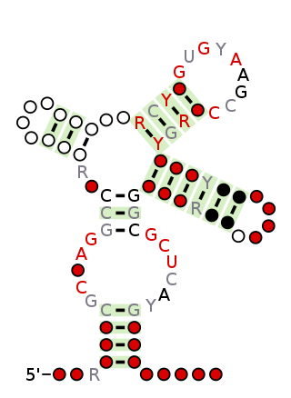
Stem-loop intramolecular base pairing is a pattern that can occur in single-stranded RNA. The structure is also known as a hairpin or hairpin loop. It occurs when two regions of the same strand, usually complementary in nucleotide sequence when read in opposite directions, base-pair to form a double helix that ends in an unpaired loop. The resulting structure is a key building block of many RNA secondary structures. As an important secondary structure of RNA, it can direct RNA folding, protect structural stability for messenger RNA (mRNA), provide recognition sites for RNA binding proteins, and serve as a substrate for enzymatic reactions.

<span class="mw-page-title-main">Group II intron</span> Class of self-catalyzing ribozymes

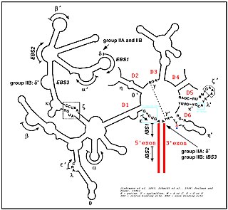
Group II introns are a large class of self-catalytic ribozymes and mobile genetic elements found within the genes of all three domains of life. Ribozyme activity can occur under high-salt conditions in vitro. However, assistance from proteins is required for in vivo splicing. In contrast to group I introns, intron excision occurs in the absence of GTP and involves the formation of a lariat, with an A-residue branchpoint strongly resembling that found in lariats formed during splicing of nuclear pre-mRNA. It is hypothesized that pre-mRNA splicing may have evolved from group II introns, due to the similar catalytic mechanism as well as the structural similarity of the Group II Domain V substructure to the U6/U2 extended snRNA. Finally, their ability to site-specifically insert into DNA sites has been exploited as a tool for biotechnology. For example, group II introns can be modified to make site-specific genome insertions and deliver cargo DNA such as reporter genes or lox sites

<i>Avsunviroidae</i> Family of viruses

The Avsunviroidae are a family of viroids. There are four species in three genera. They consist of RNA genomes between 246 and 375 nucleotides in length. They are single-stranded covalent circles and have intramolecular base pairing. All members lack a central conserved region.

<span class="mw-page-title-main">Hairpin ribozyme</span> Enzymatic section of RNA

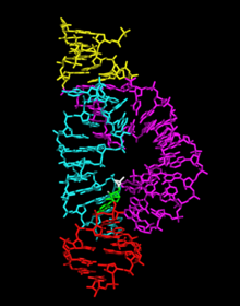
The hairpin ribozyme is a small section of RNA that can act as a ribozyme. Like the hammerhead ribozyme it is found in RNA satellites of plant viruses. It was first identified in the minus strand of the tobacco ringspot virus (TRSV) satellite RNA where it catalyzes self-cleavage and joining (ligation) reactions to process the products of rolling circle virus replication into linear and circular satellite RNA molecules. The hairpin ribozyme is similar to the hammerhead ribozyme in that it does not require a metal ion for the reaction.

<span class="mw-page-title-main">Leadzyme</span>

Leadzyme is a small ribozyme (catalytic RNA), which catalyzes the cleavage of a specific phosphodiester bond. It was discovered using an in-vitro evolution study where the researchers were selecting for RNAs that specifically cleaved themselves in the presence of lead. However, since then, it has been discovered in several natural systems. Leadzyme was found to be efficient and dynamic in the presence of micromolar concentrations of lead ions. Unlike in other small self-cleaving ribozymes, other divalent metal ions cannot replace Pb2+ in the leadzyme. Due to obligatory requirement for a lead, the ribozyme is called a metalloribozyme.

<span class="mw-page-title-main">VS ribozyme</span>

The Varkud satellite (VS) ribozyme is an RNA enzyme that carries out the cleavage of a phosphodiester bond.

<span class="mw-page-title-main">GlmS glucosamine-6-phosphate activated ribozyme</span>

The glucosamine-6-phosphate riboswitch ribozyme is an RNA structure that resides in the 5' untranslated region (UTR) of the mRNA transcript of the glmS gene. This RNA regulates the glmS gene by responding to concentrations of a specific metabolite, glucosamine-6-phosphate (GlcN6P), in addition to catalyzing a self-cleaving chemical reaction upon activation. This cleavage leads to the degradation of the mRNA that contains the ribozyme, and lowers production of GlcN6P. The glmS gene encodes for an enzyme glutamine-fructose-6-phosphate amidotransferase, which catalyzes the formation of GlcN6P, a compound essential for cell wall biosynthesis, from fructose-6-phosphate and glutamine. Thus, when GlcN6P levels are high, the glmS ribozyme is activated and the mRNA transcript is degraded but in the absence of GlcN6P the gene continues to be translated into glutamine-fructose-6-phosphate amidotransferase and GlcN6P is produced. GlcN6P is a cofactor for this cleavage reaction, as it directly participates as an acid-base catalyst. This RNA is the first riboswitch also found to be a self-cleaving ribozyme and, like many others, was discovered using a bioinformatics approach.

<span class="mw-page-title-main">Hepatitis delta virus ribozyme</span>

The hepatitis delta virus (HDV) ribozyme is a non-coding RNA found in the hepatitis delta virus that is necessary for viral replication and is the only known human virus that utilizes ribozyme activity to infect its host. The ribozyme acts to process the RNA transcripts to unit lengths in a self-cleavage reaction during replication of the hepatitis delta virus, which is thought to propagate by a double rolling circle mechanism. The ribozyme is active in vivo in the absence of any protein factors and was the fastest known naturally occurring self-cleaving RNA at the time of its discovery.

The Lariat capping ribozyme is a ~180 nt ribozyme with an apparent resemblance to a group I ribozyme. It is found within a complex type of group I introns also termed twin-ribozyme introns. Rather than splicing, it catalyses a branching reaction in which the 2'OH of an internal residue is involved in a nucleophilic attack at a nearby phosphodiester bond. As a result, the RNA is cleaved at an internal processing site (IPS), leaving a 3'OH and a downstream product with a 3 nt lariat at its 5' end. The lariat has the first and the third nucleotide joined by a 2',5' phosphodiester bond and is referred to as 'the lariat cap' because it caps an intron-encoded mRNA. The resulting lariat cap seems to contribute by increasing the half-life of the HE mRNA, thus conferring an evolutionary advantage to the HE.

<span class="mw-page-title-main">Nucleic acid tertiary structure</span> Three-dimensional shape of a nucleic acid polymer

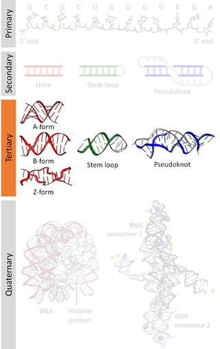
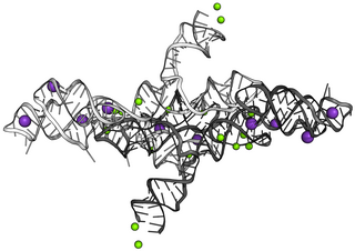
Nucleic acid tertiary structure is the three-dimensional shape of a nucleic acid polymer. RNA and DNA molecules are capable of diverse functions ranging from molecular recognition to catalysis. Such functions require a precise three-dimensional structure. While such structures are diverse and seemingly complex, they are composed of recurring, easily recognizable tertiary structural motifs that serve as molecular building blocks. Some of the most common motifs for RNA and DNA tertiary structure are described below, but this information is based on a limited number of solved structures. Many more tertiary structural motifs will be revealed as new RNA and DNA molecules are structurally characterized.

In molecular biology, a scissile bond is a covalent chemical bond that can be broken by an enzyme. Examples would be the cleaved bond in the self-cleaving hammerhead ribozyme or the peptide bond of a substrate cleaved by a peptidase.

<span class="mw-page-title-main">Twister ribozyme</span> Ribozyme capable of self-cleavage

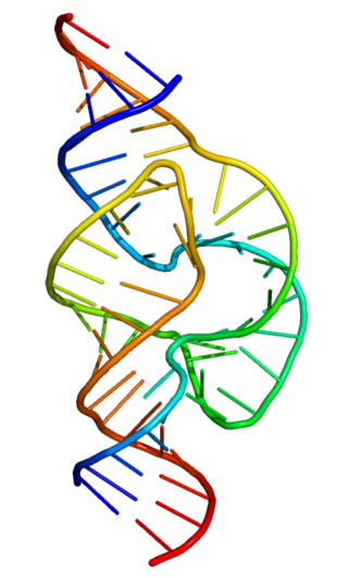
The twister ribozyme is a catalytic RNA structure capable of self-cleavage. The nucleolytic activity of this ribozyme has been demonstrated both in vivo and in vitro and has one of the fastest catalytic rates of naturally occurring ribozymes with similar function. The twister ribozyme is considered to be a member of the small self-cleaving ribozyme family which includes the hammerhead, hairpin, hepatitis delta virus (HDV), Varkud satellite (VS), and glmS ribozymes.

<span class="mw-page-title-main">RNA hydrolysis</span>

RNA hydrolysis is a reaction in which a phosphodiester bond in the sugar-phosphate backbone of RNA is broken, cleaving the RNA molecule. RNA is susceptible to this base-catalyzed hydrolysis because the ribose sugar in RNA has a hydroxyl group at the 2’ position. This feature makes RNA chemically unstable compared to DNA, which does not have this 2’ -OH group and thus is not susceptible to base-catalyzed hydrolysis.

<span class="mw-page-title-main">Twister sister ribozyme</span> RNA structure

The twister sister ribozyme (TS) is an RNA structure that catalyzes its own cleavage at a specific site. In other words, it is a self-cleaving ribozyme. The twister sister ribozyme was discovered by a bioinformatics strategy as an RNA Associated with Genes Associated with Twister and Hammerhead ribozymes, or RAGATH.

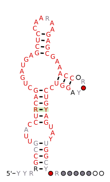
The pistol ribozyme is an RNA structure that catalyzes its own cleavage at a specific site. In other words, it is a self-cleaving ribozyme. The pistol ribozyme was discovered through comparative genomic analysis. Subsequent biochemical analysis determined further biochemical characteristics of the ribozyme. This understanding was further advanced by an atomic-resolution crystal structure of a pistol ribozyme

<span class="mw-page-title-main">Hatchet ribozyme</span> Self-cleaving ribozyme

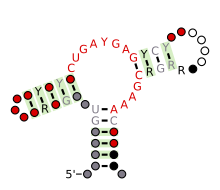
Background: The hatchet ribozyme is an RNA structure that catalyzes its own cleavage at a specific site. In other words, it is a self-cleaving ribozyme. Hatchet ribozymes were discovered by a bioinformatics strategy as RNAs Associated with Genes Associated with Twister and Hammerhead ribozymes, or RAGATH.

<span class="mw-page-title-main">Hovlinc</span>

Hovlinc RNA is a self-cleaving ribozyme of about 168 nucleotides found in a very long noncoding RNA (vlincRNA) in humans, chimpanzees, and gorillas. The word "hovlinc" comes from "hominin vlincRNA-located" RNA. Hovlinc is only a fourth known case of a ribozyme in human. Self-cleavage activity of Hovlinc has been shown in human, chimpanzees and bonobos, but is absent in gorillas, raising questions about Hovlinc's biological function and evolution.

Retrozymes are a family of retrotransposons first discovered in the genomes of plants but now also known in genomes of animals. Retrozymes contain a hammerhead ribozyme (HHR) in their sequences, although they do not possess any coding regions. Retrozymes are nonautonomous retroelements, and so borrow proteins from other elements to move into new regions of a genome. Retrozymes are actively transcribed into covalently closed circular RNAs and are detected in both polarities, which may indicate the use of rolling circle replication in their lifecycle.

References

  1. Forster AC, Symons RH (1987). "Self-cleavage of plus and minus RNAs of a virusoid and a structural model for the active sites". Cell. 49 (2): 211–220. doi:10.1016/0092-8674(87)90562-9. PMID   2436805. S2CID   33415709.
  2. Cervera, Amelia; Urbina, Denisse; de la Peña, Marcos (2016). "Retrozymes are a unique family of non-autonomous retrotransposons with hammerhead ribozymes that propagate in plants through circular RNAs". Genome Biology. 17 (1): 135. doi: 10.1186/s13059-016-1002-4 . ISSN   1474-760X. PMC   4918200 . PMID   27339130.
  3. de la Peña, Marcos; García-Robles, Inmaculada (2010). "Ubiquitous presence of the hammerhead ribozyme motif along the tree of life". RNA. 16 (10): 1943–1950. doi:10.1261/rna.2130310. ISSN   1355-8382. PMC   2941103 . PMID   20705646.
  4. Hammann, Christian; Luptak, Andrej; Perreault, Jonathan; de la Peña, Marcos (2012). "The ubiquitous hammerhead ribozyme". RNA. 18 (5): 871–885. doi:10.1261/rna.031401.111. ISSN   1355-8382. PMC   3334697 . PMID   22454536.
  5. 1 2 Prody GA, Bakos JT, Buzayan JM, Schneider IR, Bruening G (1986). "Autolytic Processing of Dimeric Plant Virus Satellite RNA". Science. 231 (4745): 1577–1580. Bibcode:1986Sci...231.1577P. doi:10.1126/science.231.4745.1577. PMID   17833317. S2CID   21563490.
  6. 1 2 Hutchins CJ, Rathjen PD, Forster AC, Symons RH (1986). "Self-cleavage of plus and minus RNA transcripts of avocado sunblotch viroid". Nucleic Acids Res. 14 (9): 3627–3640. doi:10.1093/nar/14.9.3627. PMC   339804 . PMID   3714492.
  7. Forster AC, Symons RH (1987). "Self-cleavage of virusoid RNA is performed by the proposed 55-nucleotide active site". Cell. 50 (1): 9–16. doi:10.1016/0092-8674(87)90657-X. PMID   3594567. S2CID   7231363.
  8. 1 2 Usman, Nassim; Beigelman, Leonid; McSwiggen, James A (1996-08-01). "Hammerhead ribozyme engineering". Current Opinion in Structural Biology. 6 (4): 527–533. doi:10.1016/S0959-440X(96)80119-9. PMID   8794164.
  9. 1 2 Hean J, Weinberg MS (2008). "The Hammerhead Ribozyme Revisited: New Biological Insights for the Development of Therapeutic Agents and for Reverse Genomics Applications". RNA and the Regulation of Gene Expression: A Hidden Layer of Complexity. Caister Academic Press. ISBN   978-1-904455-25-7.
  10. Epstein LM, Gall JG (1987). "Self-cleaving transcripts of satellite DNA from the newt". Cell. 48 (3): 535–543. doi:10.1016/0092-8674(87)90204-2. PMID   2433049. S2CID   24110291.
  11. Ferbeyre G, Smith JM, Cedergren R (1998). "Schistosome satellite DNA encodes active hammerhead ribozymes". Mol. Cell. Biol. 18 (7): 3880–3888. doi:10.1128/MCB.18.7.3880. PMC   108972 . PMID   9632772.
  12. Rojas AA, Vazquez-Tello A, Ferbeyre G, Venanzetti F, Bachmann L, Paquin B, Sbordoni V, Cedergren R (2000). "Hammerhead-mediated processing of satellite pDo500 family transcripts from Dolichopoda cave crickets". Nucleic Acids Res. 28 (20): 4037–4043. doi:10.1093/nar/28.20.4037. PMC   110794 . PMID   11024185.
  13. Przybilski R, Gräf S, Lescoute A, Nellen W, Westhof E, Steger G, Hammann C (2005). "Functional hammerhead ribozymes naturally encoded in the genome of Arabidopsis thaliana". Plant Cell. 17 (7): 1877–1885. doi:10.1105/tpc.105.032730. PMC   1167538 . PMID   15937227.
  14. Martick M, Horan LH, Noller HF, Scott WG (2008). "A discontinuous hammerhead ribozyme embedded in a mammalian messenger RNA". Nature. 454 (7206): 899–902. Bibcode:2008Natur.454..899M. doi:10.1038/nature07117. PMC   2612532 . PMID   18615019.
  15. De la Peña M, García-Robles I (2010). "Ubiquitous presence of the hammerhead ribozyme motif along the tree of life". RNA. 16 (10): 1943–1950. doi:10.1261/rna.2130310. PMC   2941103 . PMID   20705646.
  16. 1 2 De la Peña M, García-Robles I (2010). "Intronic hammerhead ribozymes are ultraconserved in the human genome". EMBO Rep. 11 (9): 711–716. doi:10.1038/embor.2010.100. PMC   2933863 . PMID   20651741.
  17. Seehafer C, Kalweit A, Steger G, Gräf S, Hammann C (2011). "From alpaca to zebrafish: hammerhead ribozymes wherever you look". RNA. 17 (1): 21–26. doi:10.1261/rna.2429911. PMC   3004062 . PMID   21081661.
  18. 1 2 Jimenez RM, Delwart E, Lupták A (2011). "Structure-based search reveals hammerhead ribozymes in the human microbiome". J Biol Chem. 286 (10): 7737–7743. doi: 10.1074/jbc.C110.209288 . PMC   3048661 . PMID   21257745.
  19. 1 2 Perreault J, Weinberg Z, Roth A, Popescu O, Chartrand P, Ferbeyre G, Breaker RR (May 2011). "Identification of hammerhead ribozymes in all domains of life reveals novel structural variations". PLOS Computational Biology. 7 (5): e1002031. Bibcode:2011PLSCB...7E2031P. doi: 10.1371/journal.pcbi.1002031 . PMC   3088659 . PMID   21573207.
  20. 1 2 Hammann C, Luptak A, Perreault J, de la Peña M (2012). "The ubiquitous hammerhead ribozyme". RNA. 18 (5): 871–885. doi:10.1261/rna.031401.111. PMC   3334697 . PMID   22454536.
  21. Cervera A, de la Peña M (2020). "Small circRNAs with self-cleaving ribozymes are highly expressed in diverse metazoan transcriptomes". Nucleic Acids Research. 48 (9): (9):5054–5064. doi:10.1093/nar/gkaa187. PMC   7229834 . PMID   32198887.
  22. 1 2 García-Robles I, Sánchez-Navarro J, de la Peña M (2012). "Intronic hammerhead ribozymes in mRNA biogenesis". Biol Chem. 393 (11): 1317–1326. doi:10.1515/hsz-2012-0223. hdl: 10251/34564 . PMID   23109545. S2CID   16402212.
  23. de la Peña M, Ceprian R, Casey J, Cervera A (2021). "Hepatitis delta virus-like circular RNAs from diverse metazoans encode conserved hammerhead ribozymes". Virus Evolution. 7 (1): veab016. doi:10.1093/ve/veab016. PMC   7936874 . PMID   33708415.
  24. Edgar RC, Taylor J, Lin V, Altman T, Barbera P, Meleshko D, Lohr D, Novakovsky G, Buchfink B, Al-Shayeb B, Banfield JF, de la Peña M, Korobeynikov A, Chikhi R, Babaian A (2022). "Petabase-scale sequence alignment catalyses viral discovery". Nature. 602 (7895): 142–147. Bibcode:2022Natur.602..142E. doi: 10.1038/s41586-021-04332-2 . PMID   35082445. S2CID   246297430.
  25. Canny MD, Jucker FM, Pardi A (2007). "Efficient ligation of the Schistosoma hammerhead ribozyme". Biochemistry. 46 (12): 3826–3834. doi:10.1021/bi062077r. PMC   3203546 . PMID   17319693.
  26. Lott, William B.; Pontius, Brian W.; von Hippel, Peter H. (1998-01-20). "A two-metal ion mechanism operates in the hammerhead ribozyme-mediated cleavage of an RNA substrate". Proceedings of the National Academy of Sciences of the United States of America. 95 (2): 542–547. Bibcode:1998PNAS...95..542L. doi: 10.1073/pnas.95.2.542 . ISSN   0027-8424. PMC   18456 . PMID   9435228.
  27. Birikh, K. R.; Heaton, P. A.; Eckstein, F. (1997-04-01). "The structure, function and application of the hammerhead ribozyme". European Journal of Biochemistry. 245 (1): 1–16. doi: 10.1111/j.1432-1033.1997.t01-3-00001.x . ISSN   0014-2956. PMID   9128718.
  28. J.B. Murray; A.A. Seyhan; N.G. Walter; J.M. Burke; W.G. Scott (1998). "The hammerhead, hairpin and VS ribozymes are catalytically proficient in monovalent cations alone" (PDF). Chem Biol. 5 (10): 587–595. doi:10.1016/S1074-5521(98)90116-8. PMID   9818150. S2CID   17025877.
  29. Khvorova A, Lescoute A, Westhof E, Jayasena SD (2003). "Sequence elements outside the hammerhead ribozyme catalytic core enable intracellular activity". Nature Structural & Molecular Biology. 10 (9): 708–712. doi:10.1038/nsb959. PMID   12881719. S2CID   52859654.
  30. De la Peña M; Gago S.; Flores R. (2003). "Peripheral regions of natural hammerhead ribozymes greatly increase their self-cleavage activity". EMBO J. 22 (20): 5561–5570. doi:10.1093/emboj/cdg530. PMC   213784 . PMID   14532128.
  31. Pley HW, Flaherty KM, McKay DB (1994). "Three-dimensional structure of a hammerhead ribozyme". Nature. 372 (6501): 68–74. Bibcode:1994Natur.372...68P. doi:10.1038/372068a0. PMID   7969422. S2CID   4333072.
  32. 1 2 Scott WG, Finch JT, Klug A (1995). "The crystal structure of an all-RNA hammerhead ribozyme: a proposed mechanism for RNA catalytic cleavage" (PDF). Cell. 81 (7): 991–1002. doi:10.1016/S0092-8674(05)80004-2. PMID   7541315. S2CID   6965437.
  33. Martick M, Scott WG (2006). "Tertiary contacts distant from the active site prime a ribozyme for catalysis". Cell. 126 (2): 309–320. doi:10.1016/j.cell.2006.06.036. PMC   4447102 . PMID   16859740.
  34. Citti L, Rainaldi G (2005). "Synthetic hammerhead ribozymes as therapeutic tools to control disease genes". Current Gene Therapy. 5 (1): 11–24. CiteSeerX   10.1.1.333.3872 . doi:10.2174/1566523052997541. PMID   15638708.
  35. Fei Q, Zhang H, Fu L, Dai X, Gao B, Ni M, Ge C, Li J, Ding X, Ke Y, Yao X, Zhu J (June 2008). "Experimental cancer gene therapy by multiple anti-survivin hammerhead ribozymes". Acta Biochimica et Biophysica Sinica. 40 (6): 466–77. doi:10.1111/j.1745-7270.2008.00430.x. PMID   18535745. S2CID   31532433.