Hatchet ribozyme

Last updated
Hatchet
RF02678.svg
Consensus secondary structure and sequence conservation of Hatchet ribozyme
Identifiers
SymbolHatchet
Rfam RF02678
Other data
RNA type Gene; Ribozyme
GO GO:0003824
SO SO:0000374
PDB structures PDBe

Background: The hatchet ribozyme is an RNA structure that catalyzes its own cleavage at a specific site. In other words, it is a self-cleaving ribozyme. Hatchet ribozymes were discovered by a bioinformatics strategy [1] as RNAs Associated with Genes Associated with Twister and Hammerhead ribozymes, or RAGATH.

Contents

Subsequent biochemical analysis supports the conclusion of a ribozyme function, and determined further characteristics of the chemical reaction catalyzed by the ribozyme. [2]

Nucleolytic ribozymes are small RNAs that adopt compact folds capable of site-specific cleavage/ligation reactions. 14 unique nucleolytic ribozymes have been identified to date, including recently discovered twister, pistol, twister-sister, and hatchet ribozymes that were identified based on application of comparative sequence and structural algorithms.

The consensus sequence and secondary structure of this class includes 13 highly conserved and numerous other modestly conserved nucleotides inter-dispersed among bulges linking four base-paired substructures. A representative hatchet ribozyme requires divalent cations such as Mg2+ to promote RNA strand scission with a maximum rate constant of ~4/min. As with all other small self-cleaving ribozymes discovered to date, hatchet ribozymes employ a general mechanism for catalysis consisting of a nucleophilic attack of a ribose 2-oxygen atom on the adjacent phosphorus center. Kinetic characteristics of the reaction demonstrate that members of this ribozyme class have an essential requirement for divalent metal cations and that they have a complex active site which employs multiple catalytic strategies to accelerate RNA cleavage by internal phosphoester transfer. [3]

Mechanism

Nucleolytic ribozymes like the Hatchet Ribozyme adopt an SN2-like mechanism that results in site-specific phosphodiester bond cleavage. An activated 2-OH of the ribose 5 to the scissile phosphate adopts an in-line alignment to target the adjacent to-be-cleaved P-O5 phosphodiester bond, resulting in formation of 2,3-cyclic phosphate and 5-OH groups. X-ray crystallographic structural studies on the hammerhead, hairpin, GlmS, hepatitis delta virus (HDV), Varkud satellite, and pistol ribozymes have defined the overall RNA fold, the catalytic pocket arrangement, the in-line alignment, and the key residues that contribute to the cleavage reaction. The cleavage site is located at the 5' end of its consensus secondary motif. [4]

In addition, the removal of the nucleophilic hydroxyl renders the ribozyme inactive as it is not able to create the cleavage site. More specifically, if the 2'-ribose or 2'-OH is replaced with a 2'-deoxyribose or 2'-H, there are no electrons available to perform the nucleophilic attack on the adjacent phosphate group. This results in no phosphoester bond being formed, which again inactivates the ribozyme's enzymatic cleavage ability.

Secondary Structure

In 2019, researchers crystallized a 2.1 Å product of the Hatchet Ribozyme. The consensus sequence is depicted in the image to the right. Most hatchet ribozymes and ribozymes in general adopt a P0 configuration. P0 is an additional hairpin loop located at the 5' end of the cleavage site, though it does not contribute to catalytic activity or functionality unlike Hammerhead ribozymes which have a short consensus sequence near P1, or the 5' end, that promotes high speed catalytic activity. About 90% of the sequence is conserved and similar to other ribozymes in this class. [1]

Based on the RNA sequence, the resulting DNA sequence which ends up coding for the Hatchet Ribozyme is as follows from 5'-3' because in DNA uracil is replaced by thymine.

TTAGCAAGAATGACTATAGTCACTG TTTGTACACCCCGAATAGATTAGAA GCCTAATCATAATCACGTCTGCAAT TTTGGTACA

Due to this sequence construct, after self catalyzed cleavage, it leaves an 8 nucleotide residue upstream on the 3'-end of the RNA. [5]

Tertiary Structure

Each ribozyme may have different motifs and thus different tertiary structures:

The Tertiary structure of the Hatchet Ribozyme with the motif of HT-UUCG is through dimerization. The dimer is formed through the swapping of the 3' ends of the pairing strands which is also in equilibrium with the dimer formed product of HT-GAAA. Therefore, the RNA sequence shifts between monomer and dimer configurations. To view the 3-D shape of the ribozyme see Figure S1A and B. [4] Two molecules of the HT-GAAA ribozyme can actually form a pseudosymmetric dimer with both monomers of the ribozyme exhibiting relatively well-defined electron density. The tertiary fold consists of four stem substructures which covalently stack upon each other forming the helical and loop structures, called P1, P2, P3, and P4, L1, L2 and L3 respectively (though not shown in the figure above). The actual cleavage site is positioned between the junction of P1 and P2 adjacent to P3 and L2. P1 is composed of three or six base pairs roughly 40% and 60% of the time respectively in its natural state, suggesting that length corresponds to catalytic function. [3]

There is also a conserved palindromic sequencing between base U70' and A67', which likely triggers the formation of the dimer due to Watson-Crick base pair interactions.

The tertiary structure also has long range implications within itself based on interactions between its loops. [4]

Effect of pH and Mg2+

Ribozyme catalysis experiments were done by the addition of MgCl2 and stopped for measurement at each time point by the addition of a stop solution containing urea and EDTA.

A plot of the kobs values measured at pH 7.5 with increasing concentrations of Mg2+. There is a sharp increase in ribozyme function that plateaus as the concentration approaches 10 mM. The steep slope observed at lower Mg2+ concentrations suggests that more than one metal ion is necessary for each RNA to achieve maximal ribozyme activity. Moreover, this suggests that the construct requires higher than normal physiological concentrations of Mg2+ to become completely saturated with Mg2+ as the cofactor. It is possible that native unimolecular constructs, also carrying P0, might achieve saturation at concentrations of Mg2+ that are closer to normal physiological levels.

The effect of pH on ribozyme rate constant in reactions containing 10 mM Mg2+ was also experimentally measured. pH-dependent ribozyme activity increases linearly with a slope of 1 until reaching a kobs, of a Michaelis-Menten plot, plateau of ~4/min near a pH value of 7.5. Any higher pH has the same catalytic effect and more acidic pH's begin denaturing the ribozyme and thus reducing catalytic function. Both the pH dependency and the maximum rate constant have interesting implications for the possible catalytic strategies used by this ribozyme class. [3]

The effects of various mono- and divalent metal ions on hatchet ribozyme activity

The Hatchet ribozyme construct remains completely inactive when incubated in the absence of Mg2+ in reactions containing only other monovalent cations at 1 M (Na+, K+, Rb+, Li+, Cs+), 2.5 M (Na+, K+), or 3 M (Li+). In contrast, other divalent metal ions such as Mn2+, Co2+, Zn2+, and Cd2+ support ribozyme function with varying levels of efficiency. Furthermore, two metal ions (Zn2+, Cd2+) function only at low concentrations, and three metal ions (Ba2+, Ni2+, and Cu2+) inhibit activity at 0.5 mM, even when Mg2+ is present. These results indicate that hatchet ribozymes are relatively restrictive in their use of cations to promote catalysis, perhaps indicating that one or more specialized binding sites that accommodate a limited number of divalent cations are present in the RNA structure or perhaps even at the active site. Inhibition by certain divalent metal ions could be due to the displacement of critical Mg2+ ions or by general disruption of RNA folding. [3]

Significance/Applications

One standard application is to use flanking self-cleaving ribozymes to generate precisely cut out sequences of functional RNA molecules (i.e. shRNA, saiRNA, sgRNA). This is especially useful for in vivo expression of gene editing systems (i.e. CRISPR/Cas sgRNA) and inhibitory systems. [6]

Another method is for in vivo transcription of siRNA. This design uses multiple self-cleaving ribozymes, which are all transcribed from the same gene. After cleavage, both parts of the precursor siRNA (siRNA 1 and 2) can form a double strand and act as intended. To see the setup, see saiRNA graphic [7]

Lastly, if you want to combine self-cleaving ribozymes with protein sequences, it is important to know that the self-cleaving mechanism of the ribozymes will modify the mRNA. A 5' ribozyme will modify the downstream 5' end of the pre-mRNA, disabling the cell from creating a 5' cap. This decreases the stability of the pre-mRNA and prevents it from being fully functional mature mRNA. On the other side, a 3' ribozyme would prevent polyadenylation of the upstream pre-mRNA, again decreasing stability and preventing maturation. Both interfere with translation as well. [5]

Related Research Articles

<span class="mw-page-title-main">Metalloprotein</span> Protein that contains a metal ion cofactor

Metalloprotein is a generic term for a protein that contains a metal ion cofactor. A large proportion of all proteins are part of this category. For instance, at least 1000 human proteins contain zinc-binding protein domains although there may be up to 3000 human zinc metalloproteins.

<span class="mw-page-title-main">Ribozyme</span> Type of RNA molecules

Ribozymes are RNA molecules that have the ability to catalyze specific biochemical reactions, including RNA splicing in gene expression, similar to the action of protein enzymes. The 1982 discovery of ribozymes demonstrated that RNA can be both genetic material and a biological catalyst, and contributed to the RNA world hypothesis, which suggests that RNA may have been important in the evolution of prebiotic self-replicating systems.

Deoxyribozymes, also called DNA enzymes, DNAzymes, or catalytic DNA, are DNA oligonucleotides that are capable of performing a specific chemical reaction, often but not always catalytic. This is similar to the action of other biological enzymes, such as proteins or ribozymes . However, in contrast to the abundance of protein enzymes in biological systems and the discovery of biological ribozymes in the 1980s, there is only little evidence for naturally occurring deoxyribozymes. Deoxyribozymes should not be confused with DNA aptamers which are oligonucleotides that selectively bind a target ligand, but do not catalyze a subsequent chemical reaction.

<i>Hin</i>dIII Enzyme

HindIII (pronounced "Hin D Three") is a type II site-specific deoxyribonuclease restriction enzyme isolated from Haemophilus influenzae that cleaves the DNA palindromic sequence AAGCTT in the presence of the cofactor Mg2+ via hydrolysis.

<i>Bam</i>HI Restriction enzyme

BamHI is a type II restriction endonuclease, having the capacity for recognizing short sequences of DNA and specifically cleaving them at a target site. This exhibit focuses on the structure-function relations of BamHI as described by Newman, et al. (1995). BamHI binds at the recognition sequence 5'-GGATCC-3', and cleaves these sequences just after the 5'-guanine on each strand. This cleavage results in sticky ends which are 4 bp long. In its unbound form, BamHI displays a central b sheet, which resides in between α-helices.

<span class="mw-page-title-main">Hammerhead ribozyme</span>

The hammerhead ribozyme is an RNA motif that catalyzes reversible cleavage and ligation reactions at a specific site within an RNA molecule. It is one of several catalytic RNAs (ribozymes) known to occur in nature. It serves as a model system for research on the structure and properties of RNA, and is used for targeted RNA cleavage experiments, some with proposed therapeutic applications. Named for the resemblance of early secondary structure diagrams to a hammerhead shark, hammerhead ribozymes were originally discovered in two classes of plant virus-like RNAs: satellite RNAs and viroids. They are also known in some classes of retrotransposons, including the retrozymes. The hammerhead ribozyme motif has been ubiquitously reported in lineages across the tree of life.

<span class="mw-page-title-main">Hairpin ribozyme</span> Enzymatic section of RNA

The hairpin ribozyme is a small section of RNA that can act as a ribozyme. Like the hammerhead ribozyme it is found in RNA satellites of plant viruses. It was first identified in the minus strand of the tobacco ringspot virus (TRSV) satellite RNA where it catalyzes self-cleavage and joining (ligation) reactions to process the products of rolling circle virus replication into linear and circular satellite RNA molecules. The hairpin ribozyme is similar to the hammerhead ribozyme in that it does not require a metal ion for the reaction.

<span class="mw-page-title-main">Leadzyme</span>

Leadzyme is a small ribozyme (catalytic RNA), which catalyzes the cleavage of a specific phosphodiester bond. It was discovered using an in-vitro evolution study where the researchers were selecting for RNAs that specifically cleaved themselves in the presence of lead. However, since then, it has been discovered in several natural systems. Leadzyme was found to be efficient and dynamic in the presence of micromolar concentrations of lead ions. Unlike in other small self-cleaving ribozymes, other divalent metal ions cannot replace Pb2+ in the leadzyme. Due to obligatory requirement for a lead, the ribozyme is called a metalloribozyme.

<span class="mw-page-title-main">VS ribozyme</span>

The Varkud satellite (VS) ribozyme is an RNA enzyme that carries out the cleavage of a phosphodiester bond.

<span class="mw-page-title-main">GlmS glucosamine-6-phosphate activated ribozyme</span>

The glucosamine-6-phosphate riboswitch ribozyme is an RNA structure that resides in the 5' untranslated region (UTR) of the mRNA transcript of the glmS gene. This RNA regulates the glmS gene by responding to concentrations of a specific metabolite, glucosamine-6-phosphate (GlcN6P), in addition to catalyzing a self-cleaving chemical reaction upon activation. This cleavage leads to the degradation of the mRNA that contains the ribozyme, and lowers production of GlcN6P. The glmS gene encodes for an enzyme glutamine-fructose-6-phosphate amidotransferase, which catalyzes the formation of GlcN6P, a compound essential for cell wall biosynthesis, from fructose-6-phosphate and glutamine. Thus, when GlcN6P levels are high, the glmS ribozyme is activated and the mRNA transcript is degraded but in the absence of GlcN6P the gene continues to be translated into glutamine-fructose-6-phosphate amidotransferase and GlcN6P is produced. GlcN6P is a cofactor for this cleavage reaction, as it directly participates as an acid-base catalyst. This RNA is the first riboswitch also found to be a self-cleaving ribozyme and, like many others, was discovered using a bioinformatics approach.

<span class="mw-page-title-main">Group I catalytic intron</span> Large self-splicing ribozymes

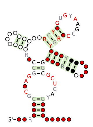
Group I introns are large self-splicing ribozymes. They catalyze their own excision from mRNA, tRNA and rRNA precursors in a wide range of organisms. The core secondary structure consists of nine paired regions (P1-P9). These fold to essentially two domains – the P4-P6 domain and the P3-P9 domain. The secondary structure mark-up for this family represents only this conserved core. Group I introns often have long open reading frames inserted in loop regions.

<span class="mw-page-title-main">Hepatitis delta virus ribozyme</span>

The hepatitis delta virus (HDV) ribozyme is a non-coding RNA found in the hepatitis delta virus that is necessary for viral replication and is the only known human virus that utilizes ribozyme activity to infect its host. The ribozyme acts to process the RNA transcripts to unit lengths in a self-cleavage reaction during replication of the hepatitis delta virus, which is thought to propagate by a double rolling circle mechanism. The ribozyme is active in vivo in the absence of any protein factors and was the fastest known naturally occurring self-cleaving RNA at the time of its discovery.

<span class="mw-page-title-main">ADP-ribose diphosphatase</span>

ADP-ribose diphosphatase (EC 3.6.1.13) is an enzyme that catalyzes a hydrolysis reaction in which water nucleophilically attacks ADP-ribose to produce AMP and D-ribose 5-phosphate. Enzyme hydrolysis occurs by the breakage of a phosphoanhydride bond and is dependent on Mg2+ ions that are held in complex by the enzyme.

The Lariat capping ribozyme is a ~180 nt ribozyme with an apparent resemblance to a group I ribozyme. It is found within a complex type of group I introns also termed twin-ribozyme introns. Rather than splicing, it catalyses a branching reaction in which the 2'OH of an internal residue is involved in a nucleophilic attack at a nearby phosphodiester bond. As a result, the RNA is cleaved at an internal processing site (IPS), leaving a 3'OH and a downstream product with a 3 nt lariat at its 5' end. The lariat has the first and the third nucleotide joined by a 2',5' phosphodiester bond and is referred to as 'the lariat cap' because it caps an intron-encoded mRNA. The resulting lariat cap seems to contribute by increasing the half-life of the HE mRNA, thus conferring an evolutionary advantage to the HE.

<span class="mw-page-title-main">Nucleic acid tertiary structure</span> Three-dimensional shape of a nucleic acid polymer

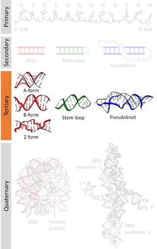
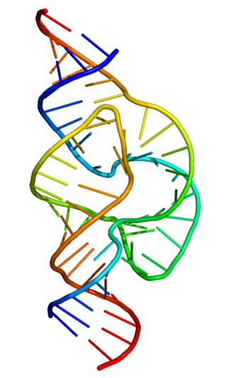
Nucleic acid tertiary structure is the three-dimensional shape of a nucleic acid polymer. RNA and DNA molecules are capable of diverse functions ranging from molecular recognition to catalysis. Such functions require a precise three-dimensional structure. While such structures are diverse and seemingly complex, they are composed of recurring, easily recognizable tertiary structural motifs that serve as molecular building blocks. Some of the most common motifs for RNA and DNA tertiary structure are described below, but this information is based on a limited number of solved structures. Many more tertiary structural motifs will be revealed as new RNA and DNA molecules are structurally characterized.

Ribonuclease E is a bacterial ribonuclease that participates in the processing of ribosomal RNA and the chemical degradation of bulk cellular RNA.

<span class="mw-page-title-main">Twister ribozyme</span> Ribozyme capable of self-cleavage

The twister ribozyme is a catalytic RNA structure capable of self-cleavage. The nucleolytic activity of this ribozyme has been demonstrated both in vivo and in vitro and has one of the fastest catalytic rates of naturally occurring ribozymes with similar function. The twister ribozyme is considered to be a member of the small self-cleaving ribozyme family which includes the hammerhead, hairpin, hepatitis delta virus (HDV), Varkud satellite (VS), and glmS ribozymes.

<span class="mw-page-title-main">RNA hydrolysis</span>

RNA hydrolysis is a reaction in which a phosphodiester bond in the sugar-phosphate backbone of RNA is broken, cleaving the RNA molecule. RNA is susceptible to this base-catalyzed hydrolysis because the ribose sugar in RNA has a hydroxyl group at the 2’ position. This feature makes RNA chemically unstable compared to DNA, which does not have this 2’ -OH group and thus is not susceptible to base-catalyzed hydrolysis.

<span class="mw-page-title-main">Twister sister ribozyme</span> RNA structure

The twister sister ribozyme (TS) is an RNA structure that catalyzes its own cleavage at a specific site. In other words, it is a self-cleaving ribozyme. The twister sister ribozyme was discovered by a bioinformatics strategy as an RNA Associated with Genes Associated with Twister and Hammerhead ribozymes, or RAGATH.

The pistol ribozyme is an RNA structure that catalyzes its own cleavage at a specific site. In other words, it is a self-cleaving ribozyme. The pistol ribozyme was discovered through comparative genomic analysis. Subsequent biochemical analysis determined further biochemical characteristics of the ribozyme. This understanding was further advanced by an atomic-resolution crystal structure of a pistol ribozyme

References

  1. 1 2 Weinberg Z, Kim PB, Chen TH, Li S, Harris KA, Lünse CE, Breaker RR (2015). "New classes of self-cleaving ribozymes revealed by comparative genomics analysis". Nat. Chem. Biol. 11 (8): 606–10. doi:10.1038/nchembio.1846. PMC   4509812 . PMID   26167874.
  2. Li S, Lünse CE, Harris KA, Breaker RR (2015). "Biochemical analysis of hatchet self-cleaving ribozymes". RNA. 21 (11): 1845–51. doi:10.1261/rna.052522.115. PMC   4604424 . PMID   26385510.
  3. 1 2 3 4 Li, Sanshu; Lünse, Christina E.; Harris, Kimberly A.; Breaker, Ronald R. (November 2015). "Biochemical analysis of hatchet self-cleaving ribozymes". RNA. 21 (11): 1845–1851. doi:10.1261/rna.052522.115. ISSN   1355-8382. PMC   4604424 . PMID   26385510.
  4. 1 2 3 Zheng, Luqian; Falschlunger, Christoph; Huang, Kaiyi; Mairhofer, Elisabeth; Yuan, Shuguang; Wang, Juncheng; Patel, Dinshaw J.; Micura, Ronald; Ren, Aiming (2019-05-14). "Hatchet ribozyme structure and implications for cleavage mechanism". Proceedings of the National Academy of Sciences. 116 (22): 10783–10791. Bibcode:2019PNAS..11610783Z. doi: 10.1073/pnas.1902413116 . ISSN   0027-8424. PMC   6561176 . PMID   31088965.
  5. 1 2 "Team:Hamburg/Contribution - 2020.igem.org". 2020.igem.org. Retrieved 2021-11-24.
  6. Gao, Yangbin; Zhao, Yunde (April 2014). "Self-processing of ribozyme-flanked RNAs into guide RNAs in vitro and in vivo for CRISPR-mediated genome editing". Journal of Integrative Plant Biology. 56 (4): 343–349. doi: 10.1111/jipb.12152 . ISSN   1672-9072. PMID   24373158.
  7. "Content". labs.biology.ucsd.edu. Retrieved 2021-11-24.