Lichtheimia corymbifera

Last updated

Lichtheimia corymbifera
Absidia corymbifera.jpg
Scientific classification Red Pencil Icon.png
Kingdom: Fungi
Division: Mucoromycota
Order: Mucorales
Family: Lichtheimiaceae
Genus: Lichtheimia
Species:
L. corymbifera
Binomial name
Lichtheimia corymbifera
(Cohn) Vuill. (1903)
Synonyms

Mucor corymbifer Cohn ex. Lichtheim (1884)
Absidia corymbifera (Cohn) Sacc. & Trotter
Mycocladus corymbifer (Cohn) Vánová (1991)
=L. hyalospora K. Hoffmann, G.Walther & K.Voigt (2009)
=L. sphaerocystis A. Alastruey-Izquierdo & G.Walther(2010)
=L. hongkongensis K.Y. Yuen (2010)

Contents

Lichtheimia corymbifera is a thermophilic fungus in the phylum Zygomycota. It normally lives as a saprotrophic mold, but can also be an opportunistic pathogen known to cause pulmonary, CNS, rhinocerebral, or cutaneous infections in animals and humans with impaired immunity. [1]

Appearance

Absidia corymbifera apophysis.jpeg
Absidia corymbifera sporangium.jpg
Lichtheimia corymbifera apophysis (left) and sporangium (right).

Lichtheimia corymbifera was originally described as Mucor corymbifer in 1884 by Lichtheim from clinical isolations in Wrocław, Poland. [2] :149 At the time of the description, the species epithet, "corymbifer" was attributed to Cohn. In 1903, the fungus was transferred to the mucoralean genus Lichtheimia (honoring Lichtheim) by Jules Vuillemin as L. corymbifera. In 1912 the species was again transferred by Saccardo and Trotter to the genus Absidia as A. corymbifera where it remained for most of the 20th century. Alastruey-Izquierdo and colleagues in 1991 transferred the species to the genus Mycocladus, described originally by Beauverie in 1900. The type of Mycocladus has since been shown to be a co-culture with elements that appear to be conspecific with Absidia van Tieghem (1876). [3] Thus the oldest available name for the fungus is Lichthemia corymbifera. [3] :441 [4] Although conventionally treated in the family Mucoraceae, the erection of a new family to accommodate the genus Lichtheimia, the "Lichtheimiaceae", has been proposed. [3] :445

Lichtheimia corymbifera produces small, dark spores inside pear-shaped (pyriform) sporangia. [5] The species is characterized by a conically shaped columella and a short, pronounced projection, a funnel-shaped apophysis, on the top. The sporangiophores (sporangia-bearing stalks) are hyaline to slightly pigmented, sometimes branched, and arising from stolons in groups of three to seven. The zygospores are naked with equatorial rings, have opposed suspensors, and lack appendages. There is limited production of rhizoids, thus, it is often difficult to identify them without the assistance of a dissecting microscope. [4]

Lichtheimia corymbifera produces more compact, less effusive growth than L. ramosa. As well, the sporangiospores of L. corymbifera are ellipsodial and hyaline when mature, versus those of L. ramosa which are lightly colored and ellipsodial. [6]

Ecology

Originally described in Central Europe, the fungus has been found in the Middle East, Great Britain, North and South America, India, and Africa. [5] It is usually found inhabiting soil or dead plant material, [4] [5] and in association with farm animals, such as cattle and horses, probably because it decomposes hay and grass. [5] Infections in farm animals can cause death relatively quickly if not treated immediately. [5] The fungus was described as a cause of mycotic abortion in cattle in Po Valley, Italy. [7]

The natural environment for L. corymbifera appears to be soil and decaying grasses, with an optimal pH between 3.0-8.0. [5] In addition, it has an optimal soil depth of 30–40 cm, according to a study in India. [5] However, they can also be found in the air or underground and can survive on humans and animals, giving it its infectious quality. [5] Furthermore, L. corymbifera can survive well in humid environments, for example, in swamps, dung, grasslands or sewage, in cotton, grains and straw as well. [5] Lichtheimia corymbifera has optimal growth at human body temperature, 35-37 °C; however, due to its thermophilic nature, it can tolerate temperatures up to 50 °C. [8] Lichthemia corymbifera is a fast-growing species in both culture and in vivo. In culture, the fungus is pale white at first and turns grey with age; the colonies are observed to reach up to 15 mm in thickness. [9] The fungus can also be cultured on a range of other materials including bread. [5]

Epidemiology

Lichtheimia corymbifera accounts for approximately 5% of mucormycoses today, [4] but true prevalence is unclear because the disease is not generally reportable and the diagnosis of mucormycosis is often empirical or based on the recovery of any zygomycetous fungus. [6] Infection can occur in people with normal immune systems but contraction is rare if the person is merely in contact with infected soil. [5] On the other hand, infection is common in individuals with weakened immune systems due to diseases like HIV/AIDS. [5] A 100% mortality rate has been reported for individuals with HIV/AIDS who become infected with L. corymbifera. [5] Other immunodeficiency diseases that predispose to infection are diabetes, blood cancers like leukemia or large skin wounds such as those secondary to burn injury. [5] In patients with leukemia, the mortality rate is 1%. [5] In humans, mucormycosis caused by L. corymbifera typically involves deep infection of the rhinocerebral and bronchorespiratory tract. [10] The most common presentation in farm animals is mycotic abortion. [11]

Infection is promoted in immunosuppressed organisms through spore inhalation or direct contact of L. corymbifera spores with tissues. [12] Upon infection, the fungus invades the blood vessels and there is nearby tissue destruction or blood clot formation. [5] Symptoms can be seen in various organ systems. For example, if the abdomen is infected, diagnosis is difficult since it is similar to an abscess presentation. Hence, diagnosis is often only made at death. [5] Diagnosis is also difficult if the lungs are infected. X-rays are often inconclusive in differentiating between other infections. If the sinus/craniofacial area is infected, symptoms begin with facial pain, progressing to muscle or nerve impairment in the eye area, and ultimately leading to the formation of blood clots in arteries serving the brain. [5]

Treatment

Treatment involves a combination of surgical debridement and antifungal therapy. Lichtheimia corymbifera is most susceptible to the antifungal drug Amphotericin B and Posaconazole, [10] however, negative side effects are possible. These side effects can be relatively mild, resulting in chills and muscle pain but can also include severe forms of nephrotoxicity. [5] Amphotericin B binds to ergosterol, found in fungal cell membranes. This causes ion and sugar leakage progressing to cell death. This species is variably susceptible to Itraconazole and highly resistant to Fluconazole, Ketoconazole, Voriconazole and Echinocandins. [13] Higher doses must be administered for these antifungal drugs to be effective. Other species of Lichtheimia are morphologically and genetically distinct but share highly similar antifungal drug susceptibilities.

Related Research Articles

<span class="mw-page-title-main">Tinea capitis</span> Cutaneous fungal infection of the scalp

Tinea capitis is a cutaneous fungal infection (dermatophytosis) of the scalp. The disease is primarily caused by dermatophytes in the genera Trichophyton and Microsporum that invade the hair shaft. The clinical presentation is typically single or multiple patches of hair loss, sometimes with a 'black dot' pattern, that may be accompanied by inflammation, scaling, pustules, and itching. Uncommon in adults, tinea capitis is predominantly seen in pre-pubertal children, more often boys than girls.
<span class="mw-page-title-main">Zygomycosis</span> Medical condition

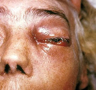
Zygomycosis is the broadest term to refer to infections caused by bread mold fungi of the zygomycota phylum. However, because zygomycota has been identified as polyphyletic, and is not included in modern fungal classification systems, the diseases that zygomycosis can refer to are better called by their specific names: mucormycosis, phycomycosis and basidiobolomycosis. These rare yet serious and potentially life-threatening fungal infections usually affect the face or oropharyngeal cavity. Zygomycosis type infections are most often caused by common fungi found in soil and decaying vegetation. While most individuals are exposed to the fungi on a regular basis, those with immune disorders (immunocompromised) are more prone to fungal infection. These types of infections are also common after natural disasters, such as tornadoes or earthquakes, where people have open wounds that have become filled with soil or vegetative matter.

<span class="mw-page-title-main">Fungal infection</span> Disease caused by fungi to animals or humans

Fungal infection, also known as mycosis, is a disease caused by fungi. Different types are traditionally divided according to the part of the body affected; superficial, subcutaneous, and systemic. Superficial fungal infections include common tinea of the skin, such as tinea of the body, groin, hands, feet and beard, and yeast infections such as pityriasis versicolor. Subcutaneous types include eumycetoma and chromoblastomycosis, which generally affect tissues in and beneath the skin. Systemic fungal infections are more serious and include cryptococcosis, histoplasmosis, pneumocystis pneumonia, aspergillosis and mucormycosis. Signs and symptoms range widely. There is usually a rash with superficial infection. Fungal infection within the skin or under the skin may present with a lump and skin changes. Pneumonia-like symptoms or meningitis may occur with a deeper or systemic infection.

<span class="mw-page-title-main">Dermatophytosis</span> Fungal infection of the skin

Dermatophytosis, also known as ringworm, is a fungal infection of the skin. Typically it results in a red, itchy, scaly, circular rash. Hair loss may occur in the area affected. Symptoms begin four to fourteen days after exposure. Multiple areas can be affected at a given time.
<span class="mw-page-title-main">Sporotrichosis</span> Medical condition

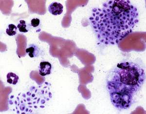
Sporotrichosis, also known as rose handler's disease, is a fungal infection that may be localised to skin, lungs, bone and joint, or become systemic. It presents with firm painless nodules that later ulcerate. Following initial exposure to Sporothrix schenckii, the disease typically progresses over a period of a week to several months. Serious complications may develop in people who have a weakened immune system.

<i>Sporothrix schenckii</i> Species of fungus

Sporothrix schenckii, a fungus that can be found worldwide in the environment, is named for medical student Benjamin Schenck, who in 1896 was the first to isolate it from a human specimen. The species is present in soil as well as in and on living and decomposing plant material such as peat moss. It can infect humans as well as animals and is the causative agent of sporotrichosis, commonly known as "rose handler's disease." The most common route of infection is the introduction of spores to the body through a cut or puncture wound in the skin. Infection commonly occurs in otherwise healthy individuals but is rarely life-threatening and can be treated with antifungals. In the environment it is found growing as filamentous hyphae. In host tissue it is found as a yeast. The transition between the hyphal and yeast forms is temperature dependent making S. schenckii a thermally dimorphic fungus.
<span class="mw-page-title-main">Conidiobolomycosis</span> Fungal infection

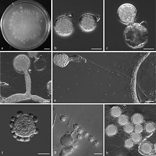
Conidiobolomycosis is a rare long-term fungal infection that is typically found just under the skin of the nose, sinuses, cheeks and upper lips. It may present with a nose bleed or a blocked or runny nose. Typically there is a firm painless swelling which can slowly extend to the nasal bridge and eyes, sometimes causing facial disfigurement.

<i>Setosphaeria rostrata</i> Species of fungus

Setosphaeria rostrata is a heat tolerant fungus with an asexual reproductive form (anamorph) known as Exserohilum rostratum. This fungus is a common plant pathogen, causing leaf spots as well as crown rot and root rot in grasses. It is also found in soils and on textiles in subtropical and tropical regions. Exserohilum rostratum is one of the 35 Exserohilum species implicated uncommonly as opportunistic pathogens of humans where it is an etiologic agent of sinusitis, keratitis, skin lesions and an often fatal meningoencephalitis. Infections caused by this species are most often seen in regions with hot climates like Israel, India and the southern USA.

<i>Acrophialophora fusispora</i> Species of ascomycete fungus found in soil, air and various plants

Acrophialophora fusispora is a poorly studied ascomycete fungus found in soil, air and various plants. A. fusispora is morphologically similar to the genera Paecilomyces and Masonia, but differ in the presence of pigmented conidiophores, verticillate phialides, and frequent sympodial proliferation. Moreover, A. fusispora is distinguished by its pigmented spindle-shaped conidia, covered with spiral bands. The fungus is naturally found in soils of tropical to temperate regions. The fungus has been identified as a plant and animal pathogen, and has recently been recognized as an emerging opportunistic human pathogen. A. fusispora infection in human is rare and has few documented clinical cases, but due to the rarity of the fungus and potential misidentification, the infections may be underdiagnosed. Clinical cases of A. fusispora include cases of keratitis, pulmonary colonization and infection, and cerebral infections. The fungus also has two documented cases of infection in dogs.

<span class="mw-page-title-main">Mucormycosis</span> Fungal infection

Mucormycosis, also known as black fungus, is a serious fungal infection that comes under fulminant fungal sinusitis, usually in people who are immunocompromised. It is curable only when diagnosed early. Symptoms depend on where in the body the infection occurs. It most commonly infects the nose, sinuses, eyes and brain resulting in a runny nose, one-sided facial swelling and pain, headache, fever, blurred vision, bulging or displacement of the eye (proptosis), and tissue death. Other forms of disease may infect the lungs, stomach and intestines, and skin.

Saksenaea vasiformis is an infectious fungus associated with cutaneous or subcutaneous lesions following trauma. It causes opportunistic infections as the entry of the fungus is through open spaces of cutaneous barrier ranging in severity from mild to severe or fatal. It lives in soils worldwide, but is considered as a rare human pathogen since only 38 cases were reported as of 2012. Saksenaea vasiformis usually fails to sporulate on the routine culture media, creating a challenge for early diagnosis, which is essential for a good prognosis. Infections are usually treated using a combination of amphotericin B and surgery. Saksenaea vasiformis is one of the few fungi known to cause necrotizing fasciitis or "flesh-eating disease".

<i>Apophysomyces variabilis</i> Species of fungus

Apophysomyces variabilis is an emerging fungal pathogen that can cause serious and sometimes fatal infection in humans. This fungus is a soil-dwelling saprobe with tropical to subtropical distribution. It is a zygomycete that causes mucormycosis, an infection in humans brought about by fungi in the order Mucorales. Infectious cases have been reported globally in locations including the Americas, Southeast Asia, India, and Australia. Apophysomyces variabilis infections are not transmissible from person to person.

<span class="mw-page-title-main">Jena Microbial Resource Collection</span>

The Jena Microbial Resource Collection (JMRC) is a joint collection of the Leibniz Institute for Natural Product Research and Infection Biology – Hans-Knöll-Institute and the University of Jena. It was founded in October 2010 by merging two earlier collections from the two above-mentioned institutes. It is a research institution, which is not normally open to the public. With about 15,000 fungal and about 35,000 bacterial living isolates, it is the largest collection of this kind in Germany. The microorganisms are maintained as active cultures, with some of them in cryopreservation.

Cunninghamella bertholletiae is a species of zygomycetous fungi in the order Mucorales. It is found globally, with increased prevalence in Mediterranean and subtropical climates. It typically grows as a saprotroph and is found in a wide variety of substrates, including soil, fruits, vegetables, nuts, crops, and human and animal waste. Although infections are still rare, C. betholletiae is emerging as an opportunistic human pathogen, predominantly in immunocompromised people, leukemia patients, and people with uncontrolled diabetes. Cunninghamella bertholletiae infections are often highly invasive, and can be more difficult to treat with antifungal drugs than infections with other species of the Mucorales, making prompt and accurate recognition and diagnosis of mycoses caused by this fungus an important medical concern.

Chaetomium atrobrunneum is a darkly pigmented mould affiliated with the fungal division, Ascomycota. This species is predominantly saprotrophic, although it has been known to infect animals including humans, showing a proclivity for the tissues of the central nervous system. Chaetomium atrobrunneum was described in 1949 from a mouldy military mattress cover obtained from the island of Guadalcanal.

<i>Rhinocladiella mackenziei</i> Species of fungus

Rhinocladiella mackenziei is a deeply pigmented fungus that is a common cause of human cerebral phaeohyphomycosis. Rhinocladiella mackenziei was believed to be endemic solely to the Middle East, due to the first cases of infection being limited to the region. However, cases of R. mackenziei infection are increasingly reported from regions outside the Middle East. This pathogen is unique in that the majority of cases have been reported from immunologically normal people.

Sarocladium kiliense is a saprobic fungus that is occasionally encountered as a opportunistic pathogen of humans, particularly immunocompromised and individuals. The fungus is frequently found in soil and has been linked with skin and systemic infections. This species is also known to cause disease in the green alga, Cladophora glomerata as well as various fruit and vegetable crops grown in warmer climates.

Mycotypha microspora, also known as Microtypha microspora, is a filamentous fungus in the division Zygomycota. It was discovered in a Citrus aurantium peel in 1932 by E. Aline Fenner, who proposed a new genus Mycotypha to accommodate it. Mycotypha africana, which is another species in the genus Mycotypha, is closely related to M. microspora. The fungus has subsequently been isolated from both outdoor and indoor settings around the world, and is typically found in soil and dung. The species rarely causes infections in humans, but has recently been involved in the clinical manifestation of the life-threatening disease mucormycosis.

Chaetomium perlucidum is a neurotropic dematiaceous fungus that is naturally found in the soil, including in agricultural soil, and in the stems of dead plants. The fungus can also be found on the feathers of birds, manure, seeds, and even paper. It is able to thrive at temperatures of 35 and 42 °C.

Lichtheimia ramosa is a saprotrophic zygomycete, typically found in soil or dead plant material. It is a thermotolerant fungus that has also been known to act as an opportunistic pathogen–infecting both humans and animals.

References

  1. Fungal Infections, Lichtheimia corymbifera. (2013). Retrieved from October 10, 2013 from the Leading International Fungal Education (LIFE) Web Site: http://www.life-worldwide.org/fungal-diseases/lichtheimia-corymbifera/.
  2. Lichtheim, L. (1884). "Ueber pathogene Mucorineen und die durch sie erzeugten Mykosen des Kaninchens". Zeitschrift für klinische Medizin. 7: 140–177. Retrieved 7 December 2013.
  3. 1 2 3 Gherbawy, Youssuf; Voigt, Kerstin (2010). Molecular identification of fungi. Berlin: Springer. ISBN   978-3-642-05042-8.
  4. 1 2 3 4 Alastruey-Izquierdo, Ana; Hoffmann, Kerstin; de Hoog, G. Sybren; Rodriguez-Tudela, Juan Luis; Voigt, Kerstin; Bibashi, Evangelia; Walther, Grit (2010). "Species Recognition and Clinical Relevance of the Zygomycetous Genus Lichtheimia (syn. Absidia Pro Parte, Mycocladus)". Journal of Clinical Microbiology. 48 (6): 2154–2170. doi:10.1128/JCM.01744-09. ISSN   0095-1137. PMC   2884488 . PMID   20357218.
  5. 1 2 3 4 5 6 7 8 9 10 11 12 13 14 15 16 17 18 19 Ransohoff. (November 26, 2010). Draft for 2010/2011 EOL University Species Pages Initiative by Katie Ransohoff. In https://mushroomobserver.org/name/show_name_description/1875?_js=on&_new=true. Retrieved November 9, 2013, from The Mushroom Observer.
  6. 1 2 Garcia-Hermoso, D.; Hoinard, D.; Gantier, J.-C.; Grenouillet, F.; Dromer, F.; Dannaoui, E. (2009). "Molecular and Phenotypic Evaluation of Lichtheimia corymbifera (Formerly Absidia corymbifera) Complex Isolates Associated with Human Mucormycosis: Rehabilitation of L. ramosa". Journal of Clinical Microbiology. 47 (12): 3862–3870. doi:10.1128/JCM.02094-08. ISSN   0095-1137. PMC   2786664 . PMID   19759217.
  7. Piancastelli, Chiara; Ghidini, Francesca; Donofrio, Gaetano; Jottini, Stefano; Taddei, Simone; Cavirani, Sandro; Cabassi, Clotilde S (2009). "Isolation and characterization of a strain of Lichtheimia corymbifera (ex Absidia corymbifera) from a case of bovine abortion". Reproductive Biology and Endocrinology. 7 (1): 138. doi:10.1186/1477-7827-7-138. ISSN   1477-7827. PMC   2789724 . PMID   19948023.
  8. Kirk, P.M.; et al. (2001). Ainsworth & Bisby's dictionary of the fungi (9th ed.). Wallingford: CABI. ISBN   0-85199-377-X.
  9. Fungal Infections, Lichtheimia corymbifera. (2013). Retrieved from October 10, 2013 from the Leading International Fungal Education (LIFE) Web Site: http://www.life-worldwide.org/fungal-diseases/lichtheimia-corymbifera/
  10. 1 2 Schrödl, W.; Heydel, T.; Schwartze, V. U.; Hoffmann, K.; Grosse-Herrenthey, A.; Walther, G.; Alastruey-Izquierdo, A.; Rodriguez-Tudela, J. L.; Olias, P.; Jacobsen, I. D.; de Hoog, G. S.; Voigt, K. (2011). "Direct Analysis and Identification of Pathogenic Lichtheimia Species by Matrix-Assisted Laser Desorption Ionization-Time of Flight Analyzer-Mediated Mass Spectrometry". Journal of Clinical Microbiology. 50 (2): 419–427. doi:10.1128/JCM.01070-11. ISSN   0095-1137. PMC   3264171 . PMID   22135259.
  11. Lichtheimia corymbifera. (2013). Retrieved October 15, 2013, from The University of Adelaide, Mycology Online Web Site: "Mycology Online -- Absidia". Archived from the original on 2007-09-20. Retrieved 2007-11-26.
  12. Almaslamani, Muna; Taj-Aldeen, Saad J.; Garcia-Hermoso, Dea; Dannaoui, Eric; Alsoub, Hussam; Alkhal, Abdullatif (2009). "An increasing trend of cutaneous zygomycosis caused by Mycocladus corymbifer(formerly Absidia corymbifera): report of two cases and review of primary cutaneous Mycocladus infections". Medical Mycology. 47 (5): 532–538. doi: 10.1080/13693780802595746 . ISSN   1369-3786. PMID   19184768.
  13. Fungal Infections, Lichtheimia corymbifera. (2013). Retrieved from October 10, 2013 from the Leading International Fungal Education (LIFE) Web Site: http://www.life-worldwide.org/fungal-diseases/lichtheimia-corymbifera/