Lung cancer screening

Last updated

Lung cancer screening
CT scan.jpg
Computed tomography (CT) scanner, the usually recommended screening technique
Purposeidentify early lung cancers before they cause symptoms

Lung cancer screening refers to cancer screening strategies used to identify early lung cancers before they cause symptoms, at a point where they are more likely to be curable. Lung cancer screening is critically important because of the incidence and prevalence of lung cancer. More than 235,000 new cases of lung cancer are expected in the United States in 2021 with approximately 130,000 deaths expected in 2021. [1] In addition, at the time of diagnosis, 57% of lung cancers are discovered in advanced stages (III and IV), meaning they are more widespread or aggressive cancers. [2] Because there is a substantially higher probability of long-term survival following treatment of localized (60%) versus advanced stage (6%) lung cancer, lung cancer screening aims to diagnose the disease in the localized (stage I) stage. [3]

Contents

Results from large randomized studies have recently prompted a large number of professional organizations and governmental agencies in the U.S. to now recommend lung cancer screening in select populations. The 3 main types of lung cancer screening are low-dose, computerized tomographic (LDCT) screening, chest x-rays, and sputum cytology tests. [4] Currently multiple professional organizations, as well as the United States Preventive Services Task Force (USPSTF), the Centers for Medicare and Medicaid Services (CMS) and the European Commission's science advisors [5] concur and endorse low-dose, computerized tomographic screening for individuals at high-risk of lung cancer.

Current guidelines

Low Dose Chest CT (LDCT) Scan

The 2021 U.S. Preventive Services Task Force guidelines recommends annual screening for lung cancer with low-dose computed tomography in adults aged 50 to 80 years who have a 20 pack-year smoking history and currently smoke or have quit within the past 15 years. [6] Screening should be discontinued once a person has not smoked for 15 years or develops a health problem that substantially limits life expectancy or the ability or willingness to have curative lung surgery. [6]

The definition of those considered to be at sufficiently high risk to benefit from screening varies according to different guidelines. The National Comprehensive Cancer Network suggests screening for two high risk groups. [7] Group 1 guidelines include 55–77 years of age, 30 or more pack years of smoking and has quit within the past 14 years, and are a current smoker. Group 2 includes those 50 years of age or older, 20 or more pack years of smoking, and other risk factors excluding second-hand smoke. [7] Other risk factors include contact with cancer-causing agents (radon, arsenic, beryllium, cadmium, chromium, nickel, asbestos, coal smoke, soot, silica, diesel fumes), history of cancer, family history of lung cancer, or a history of COPD or pulmonary fibrosis. [7]

In 2022, the European Commission's Scientific Advice Mechanism concluded that "there is a strong scientific basis for introducing lung screening for current and ex-smokers using the latest technologies, such as low-dose CT scanning". [5]

Risks of screening

Low-dose CT screening has been associated with falsely positive test results which may result in unneeded treatment. [8] In a series of studies assessing the frequence of false positive rates, results reported that rates ranged from 8-49%. [9] The false-positive rate declined when more screening rounds were performed. [9] Other concerns include radiation exposure, the cost of testing alone, and the cost if follow up of tests and screenings. [8] False reassurance from false negative findings, over-diagnosis, short term anxiety/distress, and increased rate of incidental findings are other risks. [10] The currently used low dose CT scan results in a radiation exposure of about 2 millisieverts (equal to roughly 20 two view chest x-rays). [11] It has been estimated that radiation exposure from repeated screening studies could induce cancer formation in a small percentage of screened subjects, so this risk should be mitigated by a (relatively) high prevalence of lung cancer in the population being screened. [8]

Attendance

Deprivation can reduce the numbers of people attending lung cancer screening. A UK study showed that making the screening easily accessible increased take-up. Providing mobile screening units parked in supermarket car parks, for example, in the poorer areas of Manchester was an acceptable way of offering lung checks to high-risk groups such as smokers. A simple test measured obstruction to the flow of air in and out of the lungs. A third of the tests showed airflow obstruction, a sign of chronic obstructive pulmonary disease which is a risk factor for lung cancer and other health conditions. [12] [13]

History

Calcified lung nodule seen on Low Dose Chest CT (circled) CT-Low-Dose-1.25-Lung-Calcified-Nodule.jpg
Calcified lung nodule seen on Low Dose Chest CT (circled)
Calcified lung nodule seen on Standard Dose Chest CT (circled) CT-Standard-Dose-2.50-Lung-Calcified-Nodule.jpg
Calcified lung nodule seen on Standard Dose Chest CT (circled)

Systematic examination of lung cancer screening began in the 1970s when the National Cancer Institute sponsored clinical trials to examine chest x-rays and sputum cytology at Johns Hopkins, Memorial Sloan-Kettering Cancer Center, and Mayo Clinic. [14] [15] [16] In the Mayo Clinic study, termed "The Mayo Lung Project," researchers randomized over 9000 male smokers age 45 and older to receive either chest x-ray and sputum screening three times a year or annual chest x-ray screening. The results showed that more frequent screening resulted in higher resectability rate (more early-stage detection) but made no difference in mortality from lung cancer. Chest x-ray screenings were found to detect 6 times as many new cancers as sputum tests, proving the disutility of sputum tests in lung cancer screening. [17] Unfortunately, results from the Mayo Lung Project and the Hopkins and Memorial Sloan-Kettering studies were eventually discredited due to failure to account for lead time and length time bias. Since none demonstrated reduced lung cancer incidence or mortality between randomized groups, chest x-ray was determined to be an ineffective screening tool. [18]

In the following years, the scientific community shifted its attention to computer tomography (CT). In 1996, results were published of a study of 1369 subjects screened in Japan that revealed that 73% of lung cancers that were missed by chest x-ray were able to be detected by CT scan. [19] Among the earliest United States-based clinical trials was the Early Lung Cancer Action Project (ELCAP), which published its results in 1999. [20] ELCAP screened 1000 volunteers with low-dose CT and chest x-ray. They were able to detect non-calcified nodules in 23% of patients by CT compared with 7% by chest x-ray. While this trial and a similar trial conducted by Mayo Clinic in 2005 demonstrated that CT was able to detect lung cancer at a higher rate than chest x-ray, both these trials used survival improvement, rather than mortality reduction, as an outcome, and thus were unable to prove that the use of CTs in lung cancer screening was actually impacting the number of people dying from lung cancer. [21] [18]

In 2006, results of CT screening on over 31,000 high-risk patients- an expansion study of the Early Lung Cancer Action Project - was published in the New England Journal of Medicine . [22] In this study, 85% of the 484 detected lung cancers were stage I and thus highly treatable. Historically, such stage I patients would have an expected 10-year survival of 88%. Critics of the I-ELCAP study point out that there was no randomization of patients (all received CT scans and there was no comparison group receiving only chest x-rays) and the patients were not actually followed out to 10 years post detection (the median followup was 40 months).

In contrast, a March 2007 study in the Journal of the American Medical Association (JAMA) found no mortality benefit from CT-based lung cancer screening. [23] 3,200 current or former smokers were screened for 4 years and offered 3 or 4 CT scans. Lung cancer diagnoses were 3 times as high, and surgeries were 10 times as high, as predicted by a model, but there were no significant differences between observed and expected numbers of advanced cancers or deaths. [24] Additional controversy arose after a 2008 New York Times reported that the 2006, pro-CT scan study in the New England Journal of Medicine had been funded indirectly by the parent company of the Liggett Group, a tobacco company. [25]

In 2011, the National Lung Screening Trial found that CT screening offers benefits over other screenings. [26] This study was recognized for providing supporting evidence for using CT to screen for lung cancer and for encouraging others to reflect on the merits and drawbacks of other types of screening. [27] This trial led to a recommendation in the United States that CT screening be used on people at high risk for developing lung cancer in an effort to detect the cancer earlier and reduce mortality. [27]

Development of guidelines

After the National Cancer Institute's National Lung Screening Trial publication in 2011, many national organizations revised their guidelines.

In December 2013, the U.S. Preventive Services Task Force (USPSTF) changed its long-standing recommendation that there is insufficient evidence to recommend for or against screening for lung cancer to the following: "The USPSTF recommends annual screening for lung cancer with low-dose computed tomography in adults ages 55 to 80 years who have a 30 pack-year smoking history and currently smoke or have quit within the past 15 years. Screening should be discontinued once a person has not smoked for 15 years or develops a health problem that substantially limits life expectancy or the ability or willingness to have curative lung surgery". [28]

Similarly, clinical practice guidelines previously issued by the American College of Chest Physicians (ACCP) in 2007 recommended against routine screening for lung cancer because of a lack of evidence that such screening was effective. [29] The 2013 ACCP guidelines take into account findings from the National Lung Screening Trial and state: "For smokers and former smokers who are age 55 to 74 and who have smoked for 30 pack-years or more and either continue to smoke or have quit within the past 15 years, we suggest that annual screening with low-dose CT (LDCT) should be offered over both annual screening with CXR or no screening, but only in settings that can deliver the comprehensive care provided to National Lung Screening Trial participants (Grade 2B)". [30] The most recent 2021 guidelines divide their seven recommendations into "strong" and "weak" and the evidence behind it as "moderate-quality" and "low-quality". [31] Their one strong recommendation with moderate-quality evidence is: "For asymptomatic individuals age 55 to 77 who have smoked 30 pack years or more and either continue to smoke or have quit within the past 15 years, we recommend that annual screening with low-dose CT should be offered." [31]

Guidelines were released initially in 2012 by the National Comprehensive Cancer Network, an alliance of now 31 cancer centers in the United States. Their consensus guidelines, which are updated annually, support screening as a process, not a single test, and discuss risks and benefits of screening in high risk individuals within a comprehensive multidisciplinary program. Screening is only recommended for individuals defined as high risk meeting specific criteria. More details can be found in their patient guidelines. [32] While lung cancer screening programs have been supported by the NCCN, [33] International Association for the Study of Lung Cancer (IASLC), [34] American Cancer Society, [35] The American Society of Clinical Oncology (ASCO), [36] and other organizations, the costs of screening may not be covered by medical insurance policies, unless the eligibility criteria specified by the Centers for Medicare and Medicaid Services (CMS) are met. [36] [37] As of 2017 usage of lung cancer screening in the U.S. after Medicare agreed to pay for screening and after guidelines were published was low, with the most uptake in the Midwest. [38] In 2017 a task force published a review of evidence and recommendations for advancing implementation. [38]

The English National Health Service was in 2014 re-examining the evidence for screening. [39] In 2019, the NHS implemented the Targeted Lung Health Checks (TLHC) program in order to target those most at risk of lung cancer. [40]

In 2022, the European Union proposed to update its guidelines on cancer screening to take into account new evidence that had emerged since 2016. A comprehensive evidence review by the European Commission's Scientific Advice Mechanism recommended lung cancer screening for current and ex-smokers, combined with smoking cessation programmes. [5]

Related Research Articles

<span class="mw-page-title-main">Lung cancer</span> Malignant tumor characterized by uncontrolled cell growth in lung tissue

Lung cancer, also known as lung carcinoma, is a malignant tumor that begins in the lung. Lung cancer is caused by genetic damage to the DNA of cells in the airways, often caused by cigarette smoking or inhaling damaging chemicals. Damaged airway cells gain the ability to multiply unchecked, causing the growth of a tumor. Without treatment, tumors spread throughout the lung, damaging lung function. Eventually lung tumors metastasize, spreading to other parts of the body.

<span class="mw-page-title-main">CT scan</span> Medical imaging procedure using X-rays to produce cross-sectional images

A computed tomography scan is a medical imaging technique used to obtain detailed internal images of the body. The personnel that perform CT scans are called radiographers or radiology technologists.

<span class="mw-page-title-main">Pulmonary embolism</span> Blockage of an artery in the lungs

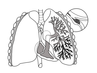
Pulmonary embolism (PE) is a blockage of an artery in the lungs by a substance that has moved from elsewhere in the body through the bloodstream (embolism). Symptoms of a PE may include shortness of breath, chest pain particularly upon breathing in, and coughing up blood. Symptoms of a blood clot in the leg may also be present, such as a red, warm, swollen, and painful leg. Signs of a PE include low blood oxygen levels, rapid breathing, rapid heart rate, and sometimes a mild fever. Severe cases can lead to passing out, abnormally low blood pressure, obstructive shock, and sudden death.

<span class="mw-page-title-main">Mammography</span> Process of using low-energy X-rays to examine the human breast for diagnosis and screening

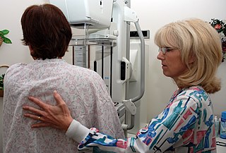
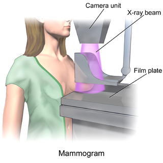
Mammography is the process of using low-energy X-rays to examine the human breast for diagnosis and screening. The goal of mammography is the early detection of breast cancer, typically through detection of characteristic masses or microcalcifications.

In medical or research imaging, an incidental imaging finding is an unanticipated finding which is not related to the original diagnostic inquiry. As with other types of incidental medical findings, they may represent a diagnostic, ethical, and philosophical dilemma because their significance is unclear. While some coincidental findings may lead to beneficial diagnoses, others may lead to overdiagnosis that results in unnecessary testing and treatment, sometimes called the "cascade effect".

<span class="mw-page-title-main">Tuberculosis diagnosis</span> Methods for diagnosing tuberculosis

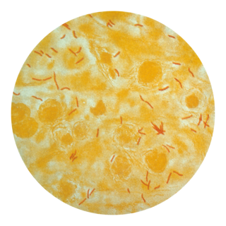
Tuberculosis is diagnosed by finding Mycobacterium tuberculosis bacteria in a clinical specimen taken from the patient. While other investigations may strongly suggest tuberculosis as the diagnosis, they cannot confirm it.

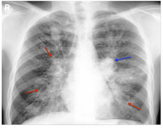
Community-acquired pneumonia (CAP) refers to pneumonia contracted by a person outside of the healthcare system. In contrast, hospital-acquired pneumonia (HAP) is seen in patients who have recently visited a hospital or who live in long-term care facilities. CAP is common, affecting people of all ages, and its symptoms occur as a result of oxygen-absorbing areas of the lung (alveoli) filling with fluid. This inhibits lung function, causing dyspnea, fever, chest pains and cough.

<span class="mw-page-title-main">Allergic bronchopulmonary aspergillosis</span> Medical condition

Allergic bronchopulmonary aspergillosis (ABPA) is a condition characterised by an exaggerated response of the immune system to the fungus Aspergillus. It occurs most often in people with asthma or cystic fibrosis. Aspergillus spores are ubiquitous in soil and are commonly found in the sputum of healthy individuals. A. fumigatus is responsible for a spectrum of lung diseases known as aspergilloses.

<span class="mw-page-title-main">Computed tomography angiography</span> Medical investigation technique

Computed tomography angiography is a computed tomography technique used for angiography—the visualization of arteries and veins—throughout the human body. Using contrast injected into the blood vessels, images are created to look for blockages, aneurysms, dissections, and stenosis. CTA can be used to visualize the vessels of the heart, the aorta and other large blood vessels, the lungs, the kidneys, the head and neck, and the arms and legs. CTA can also be used to localise arterial or venous bleed of the gastrointestinal system.

<span class="mw-page-title-main">CT pulmonary angiogram</span> Medical imaging of blood flow between heart and lungs

A CT pulmonary angiogram (CTPA) is a medical diagnostic test that employs computed tomography (CT) angiography to obtain an image of the pulmonary arteries. Its main use is to diagnose pulmonary embolism (PE). It is a preferred choice of imaging in the diagnosis of PE due to its minimally invasive nature for the patient, whose only requirement for the scan is an intravenous line.

A coronary CT calcium scan is a computed tomography (CT) scan of the heart for the assessment of severity of coronary artery disease. Specifically, it looks for calcium deposits in atherosclerotic plaques in the coronary arteries that can narrow arteries and increase the risk of heart attack. These plaques are the cause of most heart attacks, and become calcified as they develop.

<span class="mw-page-title-main">Tomosynthesis</span>

Tomosynthesis, also digital tomosynthesis (DTS), is a method for performing high-resolution limited-angle tomography at radiation dose levels comparable with projectional radiography. It has been studied for a variety of clinical applications, including vascular imaging, dental imaging, orthopedic imaging, mammographic imaging, musculoskeletal imaging, and chest imaging.

<span class="mw-page-title-main">Lung nodule</span> Medical condition

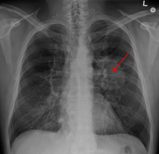
A lung nodule or pulmonary nodule is a relatively small focal density in the lung. A solitary pulmonary nodule (SPN) or coin lesion, is a mass in the lung smaller than three centimeters in diameter. A pulmonary micronodule has a diameter of less than three millimetres. There may also be multiple nodules.

<span class="mw-page-title-main">Cancer screening</span> Method to detect cancer

The objective of cancer screening is to detect cancer before symptoms appear, involving various methods such as blood tests, urine tests, DNA tests, and medical imaging. The purpose of screening is early cancer detection, to make the cancer easier to treat and extending life expectancy. In 2019, cancer was the second leading cause of death globally; more recent data is pending due to the COVID-19 pandemic.

<span class="mw-page-title-main">Adenocarcinoma of the lung</span> Medical condition

Adenocarcinoma of the lung is the most common type of lung cancer, and like other forms of lung cancer, it is characterized by distinct cellular and molecular features. It is classified as one of several non-small cell lung cancers (NSCLC), to distinguish it from small cell lung cancer which has a different behavior and prognosis. Lung adenocarcinoma is further classified into several subtypes and variants. The signs and symptoms of this specific type of lung cancer are similar to other forms of lung cancer, and patients most commonly complain of persistent cough and shortness of breath.

The National Lung Screening Trial was a United States-based clinical trial which recruited research participants between 2002 and 2004. It was sponsored by the National Cancer Institute and conducted by the American College of Radiology Imaging Network and the Lung Screening Study Group. The major objective of the trial was to compare the efficacy of low-dose helical computed tomography and standard chest X-ray as methods of lung cancer screening. The primary study ended in 2010, and the initial findings were published in November 2010, with the main results published in 2011 in the New England Journal of Medicine.

<span class="mw-page-title-main">Breast imaging</span>

In medicine, breast imaging is a sub-speciality of diagnostic radiology that involves imaging of the breasts for screening or diagnostic purposes. There are various methods of breast imaging using a variety of technologies as described in detail below. Traditional screening and diagnostic mammography uses x-ray technology and has been the mainstay of breast imaging for many decades. Breast tomosynthesis is a relatively new digital x-ray mammography technique that produces multiple image slices of the breast similar to, but distinct from, computed tomography (CT). Xeromammography and galactography are somewhat outdated technologies that also use x-ray technology and are now used infrequently in the detection of breast cancer. Breast ultrasound is another technology employed in diagnosis and screening that can help differentiate between fluid filled and solid lesions, an important factor to determine if a lesion may be cancerous. Breast MRI is a technology typically reserved for high-risk patients and patients recently diagnosed with breast cancer. Lastly, scintimammography is used in a subgroup of patients who have abnormal mammograms or whose screening is not reliable on the basis of using traditional mammography or ultrasound.

<span class="mw-page-title-main">Limited-stage small cell lung carcinoma</span> Medical condition

Limited-stage small cell lung carcinoma (LS-SCLC) is a type of small cell lung cancer (SCLC) that is confined to an area which is small enough to be encompassed within a radiation portal. This generally includes cancer to one side of the lung and those might have reached the lymph nodes on the same side of the lung. 33% patients with small cell lung cancer are diagnosed with limited-stage small cell lung carcinoma when it is first found. Common symptoms include but are not limited to persistent cough, chest pain, rust-coloured sputum, shortness of breath, fatigue, weight loss, wheezing, hoarseness and recurrent respiratory tract infections such as pneumonia and bronchitis. Nervous system problems, Cushing syndrome and SIADH can also be associated with small cell lung cancer. Unlike extensive-stage small cell lung cancer, limited-stage small cell lung carcinoma is potentially curable. Standard treatments consist of surgery, platinum-based combination chemotherapy, thoracic irradiation, and prophylactic cranial irradiation. Patient five-year survival rate has significantly increased from 1% with surgery to 26% after the application of combination chemotherapy.

Denise R. Aberle is an American radiologist and oncologist. As a professor of radiology in the David Geffen School of Medicine at UCLA and a professor of bioengineering in the UCLA Henry Samueli School of Engineering and Applied Science, Aberle was elected a member of the National Academy of Medicine and Fellow of the American Institute for Medical and Biological Engineering.

<span class="mw-page-title-main">Smoking-related interstitial fibrosis (SRIF)</span> Abnormal amount of collagen in the lung (fibrosis) caused by cigarette smoking

Smoking-related interstitial fibrosis (SRIF) is an abnormality in the lungs characterized by excessive collagen deposition within the walls of the air sacs. This abnormality can be seen with a microscope and diagnosed by pathologists. It is caused by cigarette smoking.

References

PD-icon.svg This article incorporates public domain material from Agency for Healthcare Research and Quality, U.S. Preventive Services Task Force. United States Department of Health and Human Services . Retrieved 19 June 2017.

  1. American Cancer Society. Cancer Prevention & Early Detection Facts & Figures 2019-2020. Atlanta: American Cancer Society; 2017.
  2. Miller KD, Siegel RL, Lin CC, Mariotto AB, Kramer JL, Rowland JH, Stein KD, Alteri R, Jemal A (2016). "Cancer treatment and survivorship statistics, 2016". CA: A Cancer Journal for Clinicians. 66 (4): 271–289. doi: 10.3322/caac.21349 . PMID   27253694.
  3. "Lung and Bronchus Cancer - Cancer Stat Facts".
  4. "Lung Cancer Screening (PDQ®)–Patient Version - National Cancer Institute". www.cancer.gov. 30 July 2021. Retrieved 20 September 2021.
  5. 1 2 3 SAPEA, Scientific Advice for Policy by European Academies. (2 March 2022). "Improving cancer screening in the European Union" . Retrieved 10 March 2022.{{cite web}}: |last= has generic name (help)
  6. 1 2 "Recommendation: Lung Cancer: Screening | United States Preventive Services Taskforce". www.uspreventiveservicestaskforce.org. Retrieved 10 September 2021.
  7. 1 2 3 "Lung Cancer Screening" (PDF). 2020. Retrieved 10 September 2021.
  8. 1 2 3 American College of Chest Physicians, American Thoracic Society (September 2013), "Five Things Physicians and Patients Should Question", Choosing Wisely , American College of Chest Physicians and American Thoracic Society, retrieved 6 January 2013
  9. 1 2 Jonas D (15 September 2021). "Screening for Lung Cancer With Low-Dose Computed Tomography Updated Evidence Report and Systematic Review for the US Preventive Services Task Force". JAMA Network. 325 (10): 971–987. doi: 10.1001/jama.2021.0377 . PMID   33687468. S2CID   232159404.
  10. Humphrey LL, Deffebach M, Pappas M, Baumann C, Artis K, Mitchell JP, Zakher B, Fu R, Slatore CG (2013). "Screening for Lung Cancer With Low-Dose Computed Tomography: A Systematic Review to Update the U.S. Preventive Services Task Force Recommendation". Annals of Internal Medicine. 159 (6): 411–20. doi: 10.7326/0003-4819-159-6-201309170-00690 . PMID   23897166.
  11. Mascalchi, Mario, and Lapo Sali. “Lung cancer screening with low dose CT and radiation harm-from prediction models to cancer incidence data.” Annals of translational medicine vol. 5,17 (2017): 360. doi:10.21037/atm.2017.06.41
  12. "Lung health checks in supermarket car parks reach older smokers in deprived communities". NIHR Evidence (Plain English summary). 5 August 2020. doi:10.3310/alert_40661. S2CID   243394660.
  13. Balata H, Harvey J, Barber PV, Colligan D, Duerden R, Elton P, Evison M, Greaves M, Howells J, Irion K, Karunaratne D (15 July 2020). "Spirometry performed as part of the Manchester community-based lung cancer screening programme detects a high prevalence of airflow obstruction in individuals without a prior diagnosis of COPD". Thorax. 75 (8): 655–660. doi: 10.1136/thoraxjnl-2019-213584 . ISSN   0040-6376. PMID   32444437. S2CID   218855570.
  14. Stitik FP, Tockman MS (December 1978). "Radiographic screening in the early detection of lung cancer". Radiologic Clinics of North America. 16 (3): 347–366. ISSN   0033-8389. PMID   746141.
  15. Melamed MR, Flehinger BJ, Zaman MB, Heelan RT, Perchick WA, Martini N (July 1984). "Screening for Early Lung Cancer". Chest. 86 (1): 44–53. doi:10.1378/chest.86.1.44. ISSN   0012-3692. PMID   6734291.
  16. Muhm JR, Miller WE, Fontana RS, Sanderson DR, Uhlenhopp MA (1 September 1983). "Lung cancer detected during a screening program using four-month chest radiographs". Radiology. 148 (3): 609–615. doi:10.1148/radiology.148.3.6308709. ISSN   0033-8419. PMID   6308709.
  17. Sanderson DR. "Lung Cancer Screening: The Mayo Study". Chest. 1986;89(4_Supplement)
  18. 1 2 Sharma D, Newman TG, Aronow WS (12 October 2015). "Lung cancer screening: history, current perspectives, and future directions". Archives of Medical Science. 11 (5): 1033–1043. doi:10.5114/aoms.2015.54859 (inactive 31 January 2024). ISSN   1734-1922. PMC   4624749 . PMID   26528348.{{cite journal}}: CS1 maint: DOI inactive as of January 2024 (link)
  19. Kaneko M, Eguchi K, Ohmatsu H, Kakinuma R, Naruke T, Suemasu K, Moriyama N (December 1996). "Peripheral lung cancer: screening and detection with low-dose spiral CT versus radiography". Radiology. 201 (3): 798–802. doi:10.1148/radiology.201.3.8939234. PMID   8939234.[ permanent dead link ]
  20. Henschke CI, McCauley DI, Yankelevitz DF, Naidich DP, McGuinness G, Miettinen OS, Libby DM, Pasmantier MW, Koizumi J, Altorki NK, Smith JP (July 1999). "Early Lung Cancer Action Project: overall design and findings from baseline screening". The Lancet. 354 (9173): 99–105. doi: 10.1016/s0140-6736(99)06093-6 . ISSN   0140-6736. PMID   10408484. S2CID   32737974.
  21. Swensen SJ, Jett JR, Hartman TE, Midthun DE, Mandrekar SJ, Hillman SL, Sykes AM, Aughenbaugh GL, Bungum AO, Allen KL (April 2005). "CT Screening for Lung Cancer: Five-year Prospective Experience". Radiology. 235 (1): 259–265. doi:10.1148/radiol.2351041662. ISSN   0033-8419. PMID   15695622.
  22. Henschke CI, Yankelevitz DF, Libby DM, Pasmantier MW, Smith JP, Miettinen OS (2006). "Survival of patients with stage I lung cancer detected on CT screening". N. Engl. J. Med. 355 (17): 1763–71. doi: 10.1056/NEJMoa060476 . PMID   17065637.
  23. Bach PB, Jett JR, Pastorino U, Tockman MS, Swensen SJ, Begg CB (2007). "Computed tomography screening and lung cancer outcomes". JAMA. 297 (9): 953–61. doi: 10.1001/jama.297.9.953 . PMID   17341709.
  24. Crestanello JA, Allen MS, Jett J, Cassivi SD, Nichols Iii FC, Swensen SJ, Deschamps C, Pairolero PC (2004). "Thoracic surgical operations in patients enrolled in a computed tomographic screening trial". Journal of Thoracic and Cardiovascular Surgery. 128 (2): 254–259. doi: 10.1016/j.jtcvs.2004.02.017 . PMID   15282462.
  25. Cigarette Company Paid for Lung Cancer Study, by Gardiner Harris. Published in the New York Times on March 26, 2008.
  26. Aberle DR, Adams AM, Berg CD, Black WC, Clapp JD, Fagerstrom RM, Gareen IF, Gatsonis C, Marcus PM, Sicks JD (2011). "Reduced Lung-Cancer Mortality with Low-Dose Computed Tomographic Screening". New England Journal of Medicine. 365 (5): 395–409. doi:10.1056/NEJMoa1102873. ISSN   0028-4793. PMC   4356534 . PMID   21714641.
  27. 1 2 Aberle DR, Abtin F, Brown K (2013). "Computed Tomography Screening for Lung Cancer: Has It Finally Arrived? Implications of the National Lung Screening Trial". Journal of Clinical Oncology. 31 (8): 1002–1008. doi:10.1200/JCO.2012.43.3110. ISSN   0732-183X. PMC   3589698 . PMID   23401434.
  28. "Lung Cancer Screening". U.S. Preventative Services Task Force. 2013. Archived from the original on 4 November 2010. Retrieved 21 July 2014.
  29. Alberts WM, American College of Chest Physicians (2007). "Diagnosis and Management of Lung Cancer Executive Summary: ACCP Evidence-Based Clinical Practice Guidelines (2nd Edition)". Chest. 132 (3_suppl): 1S–19S. doi:10.1378/chest.07-1860. PMID   17873156.
  30. Detterbeck FC, Lewis SZ, Diekemper R (1 May 2013). "Executive summary: Diagnosis and management of lung cancer, 3rd ed: American college of chest physicians evidence-based clinical practice guidelines". Chest. 143 (5S): 7S–37S. doi: 10.1378/chest.12-2377 . PMID   23649434.
  31. 1 2 Mazzone PJ, Silvestri GA, Souter LH, Caverly TJ, Kanne JP, Katki HA, Wiener RS, Detterbeck FC (13 July 2021). "Screening for Lung Cancer". Chest. 160 (5): e427–e494. doi:10.1016/j.chest.2021.06.063. ISSN   0012-3692. PMC   8727886 . PMID   34270968. S2CID   236000983.
  32. "NCCN Guidelines for Patients™ | Lung Cancer Screening". Archived from the original on 14 August 2012. Retrieved 2013-04-09.
  33. "NCCN Guidelines". National Comprehensive Cancer Network. Retrieved 7 September 2016.
  34. "Lung Cancer Association | Research | IASLC". Archived from the original on 4 July 2011. Retrieved 2011-06-24.
  35. Simon S (11 January 2013). "New Lung Cancer Screening Guidelines for Heavy Smokers". American Cancer Society. American Cancer Society, Inc. Retrieved 7 September 2016.
  36. 1 2 Wood DE, Kazerooni EA, Baum SL, Eapen GA, Ettinger DS, Hou L, Jackman DM, Klippenstein D, Kumar R, Lackner RP, Leard LE (1 April 2018). "Lung Cancer Screening, Version 3.2018, NCCN Clinical Practice Guidelines in Oncology". Journal of the National Comprehensive Cancer Network. 16 (4): 412–441. doi:10.6004/jnccn.2018.0020. ISSN   1540-1405. PMC   6476336 . PMID   29632061.
  37. "Lung Cancer Screening Coverage".
  38. 1 2 National Academies of Sciences, Engineering, and Medicine (2016). Implementation of Lung Cancer Screening: Proceedings of a Workshop. The National Academies Press. ISBN   9780309451321.{{cite book}}: CS1 maint: multiple names: authors list (link)
  39. Baldwin DR, Hansell DM, Duffy SW, Field JK (7 March 2014). "Lung cancer screening with low dose computed tomography". BMJ (Clinical Research Ed.). 348: g1970. doi:10.1136/bmj.g1970. PMID   24609921. S2CID   39926785.
  40. "Targeted Screening for Lung Cancer with Low Radiation Dose Computed Tomography" (PDF). NHS. Archived from the original (PDF) on 11 May 2021. Retrieved 13 September 2021.