Related Research Articles

Cell adhesion is the process by which cells interact and attach to neighbouring cells through specialised molecules of the cell surface. This process can occur either through direct contact between cell surfaces such as cell junctions or indirect interaction, where cells attach to surrounding extracellular matrix, a gel-like structure containing molecules released by cells into spaces between them. Cells adhesion occurs from the interactions between cell-adhesion molecules (CAMs), transmembrane proteins located on the cell surface. Cell adhesion links cells in different ways and can be involved in signal transduction for cells to detect and respond to changes in the surroundings. Other cellular processes regulated by cell adhesion include cell migration and tissue development in multicellular organisms. Alterations in cell adhesion can disrupt important cellular processes and lead to a variety of diseases, including cancer and arthritis. Cell adhesion is also essential for infectious organisms, such as bacteria or viruses, to cause diseases.

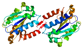
In molecular biology, intercellular adhesion molecules (ICAMs) and vascular cell adhesion molecule-1 (VCAM-1) are part of the immunoglobulin superfamily. They are important in inflammation, immune responses and in intracellular signalling events. The ICAM family consists of five members, designated ICAM-1 to ICAM-5. They are known to bind to leucocyte integrins CD11/CD18 such as LFA-1 and Macrophage-1 antigen, during inflammation and in immune responses. In addition, ICAMs may exist in soluble forms in human plasma, due to activation and proteolysis mechanisms at cell surfaces.
Cell adhesion molecules (CAMs) are a subset of cell surface proteins that are involved in the binding of cells with other cells or with the extracellular matrix (ECM), in a process called cell adhesion. In essence, CAMs help cells stick to each other and to their surroundings. CAMs are crucial components in maintaining tissue structure and function. In fully developed animals, these molecules play an integral role in generating force and movement and consequently ensuring that organs are able to execute their functions normally. In addition to serving as "molecular glue", CAMs play important roles in the cellular mechanisms of growth, contact inhibition, and apoptosis. Aberrant expression of CAMs may result in a wide range of pathologies, ranging from frostbite to cancer.
Leukocyte adhesion deficiency (LAD) is a rare autosomal recessive disorder characterized by immunodeficiency resulting in recurrent infections. LAD is currently divided into three subtypes: LAD1, LAD2, and the recently described LAD3, also known as LAD-1/variant. In LAD3, the immune defects are supplemented by a Glanzmann thrombasthenia-like bleeding tendency.

ICAM-1 also known as CD54 is a protein that in humans is encoded by the ICAM1 gene. This gene encodes a cell surface glycoprotein which is typically expressed on endothelial cells and cells of the immune system. It binds to integrins of type CD11a / CD18, or CD11b / CD18 and is also exploited by rhinovirus as a receptor for entry into respiratory epithelium.

CD11c, also known as Integrin, alpha X (ITGAX), is a gene that encodes for CD11c.

Integrin, alpha L , also known as ITGAL, is a protein that in human is encoded by ITGAL gene. CD11a functions in the immune system. It is involved in cellular adhesion and costimulatory signaling. It is the target of the drug efalizumab.

Integrin alpha M (ITGAM) is one protein subunit that forms heterodimeric integrin alpha-M beta-2 (αMβ2) molecule, also known as macrophage-1 antigen (Mac-1) or complement receptor 3 (CR3). ITGAM is also known as CR3A, and cluster of differentiation molecule 11B (CD11B). The second chain of αMβ2 is the common integrin β2 subunit known as CD18, and integrin αMβ2 thus belongs to the β2 subfamily integrins.

In molecular biology, CD18 is an integrin beta chain protein that is encoded by the ITGB2 gene in humans. Upon binding with one of a number of alpha chains, CD18 is capable of forming multiple heterodimers, which play significant roles in cellular adhesion and cell surface signaling, as well as important roles in immune responses. CD18 also exists in soluble, ligand binding forms. Deficiencies in CD18 expression can lead to adhesion defects in circulating white blood cells in humans, reducing the immune system's ability to fight off foreign invaders.

Integrin α4β1 is an integrin dimer. It is composed of CD49d and CD29. The alpha 4 subunit is 155 kDa, and the beta 1 subunit is 150 kDa.

Leukocyte extravasation is the movement of leukocytes out of the circulatory system and towards the site of tissue damage or infection. This process forms part of the innate immune response, involving the recruitment of non-specific leukocytes. Monocytes also use this process in the absence of infection or tissue damage during their development into macrophages.

Intercellular adhesion molecule 3 (ICAM3) also known as CD50, is a protein that in humans is encoded by the ICAM3 gene. The protein is constitutively expressed on the surface of leukocytes, which are also called white blood cells and are part of the immune system. ICAM3 mediates adhesion between cells by binding to specific integrin receptors. It plays an important role in the immune cell response through its facilitation of interactions between T cells and dendritic cells, which allows for T cell activation. ICAM3 also mediates the clearance of cells undergoing apoptosis by attracting and binding macrophages, a type of cell that breaks down infected or dying cells through a process known as phagocytosis, to apoptotic cells.

Intercellular adhesion molecule 2 (ICAM2), also known as CD102, is a human gene, and the protein resulting from it.
The following outline is provided as an overview of and topical guide to immunology:
Timothy "Tim" A. Springer, Ph.D. is an immunologist and Latham Family Professor at Harvard Medical School. Springer is best known for his pioneering work in discovering the first integrins and intercellular adhesion molecules (ICAMs) and elucidating how these cell adhesion molecules function in the immune system. His innovative use of monoclonal antibodies in his research paved the way for the development of therapeutic antibodies, known as selective adhesion molecule inhibitors, to treat autoimmune diseases. In recent years, Springer's research interest has expanded to include malaria, transforming growth factor beta (TGF-β) signaling molecules, and von Willebrand factor.
Rap1 is a small GTPase, which are small cytosolic proteins that act like cellular switches and are vital for effective signal transduction. There are two isoforms of the Rap1 protein, each encoded by a separate gene, RAP1A and RAP1B. Rap1 belongs to Ras-related protein family.

Fermitin family homolog 3) (FERMT3), also known as kindlin-3 (KIND3), MIG2-like protein (MIG2B), or unc-112-related protein 2 (URP2) is a protein that in humans is encoded by the FERMT3 gene. The kindlin family of proteins, member of the B4.1 superfamily, comprises three conserved protein homologues, kindlin 1, 2, and 3. They each contain a bipartite FERM domain comprising four subdomains F0, F1, F2, and F3 that show homology with the FERM head (H) domain of the cytoskeletal Talin protein. Kindlins have been linked to Kindler syndrome, leukocyte adhesion deficiency, cancer and other acquired human diseases. They are essential in the organisation of focal adhesions that mediate cell-extracellular matrix junctions and are involved in other cellular compartments that control cell-cell contacts and nucleus functioning. Therefore, they are responsible for cell to cell crosstalk via cell-cell contacts and integrin mediated cell adhesion through focal adhesion proteins and as specialised adhesion structures of hematopoietic cells they are also present in podosome's F actin surrounding ring structure. Isoform 2 may act as a repressor of NF-kappa-B and apoptosis
Gut-specific homing is the mechanism by which activated T cells and antibody-secreting cells (ASCs) are targeted to both inflamed and non-inflamed regions of the gut in order to provide an effective immune response. This process relies on the key interaction between the integrin α4β7 and the addressin MadCAM-1 on the surfaces of the appropriate cells. Additionally, this interaction is strengthened by the presence of CCR9, a chemokine receptor, which interacts with TECK. Vitamin A-derived retinoic acid regulates the expression of these cell surface proteins.

Nancy Hogg FMedSci is an immunologist who has made major contributions in the field of adhesion molecules, focusing on the integrins expressed by leukocytes. Hogg was elected to the Academy of Medical Sciences in 2002 and currently holds an emeritus position at the Francis Crick Institute, London.
Integrin α4β7 is an integrin heterodimer composed of CD49d (alpha-4) subunit and beta-7 subunit noncovalently linked. LPAM-1 is expressed on the cell surface of leukocytes. This receptor is involved in lymphocyte trafficking pathway to site of inflammation in intestinal tissues.
References
- 1 2 Lackie JM (2010). A dictionary of biomedicine (1st ed.). Oxford: Oxford University Press. ISBN 9780191727948. OCLC 663104793.
- 1 2 3 4 5 6 7 8 9 10 11 12 13 14 Ley K (2007). Adhesion molecules : function and inhibition . Basel: Birkhauser. ISBN 9783764379759. OCLC 261225084.
- 1 2 3 4 Cammack R (2006). Oxford dictionary of biochemistry and molecular biology (Rev. ed.). Oxford: Oxford University Press. ISBN 9780191727641. OCLC 743217704.
- ↑ Verma NK, Kelleher D (August 2017). "Not Just an Adhesion Molecule: LFA-1 Contact Tunes the T Lymphocyte Program". Journal of Immunology. 199 (4): 1213–1221. doi: 10.4049/jimmunol.1700495 . PMID 28784685.
- 1 2 3 4 5 6 7 Dömling A (2013). Protein-protein interactions in drug discovery. Weinheim: Wiley-VCH. ISBN 9783527648238. OCLC 828743731.
- 1 2 3 Akbari H, Zadeh MM (January 2001). "Leukocyte adhesion deficiency". Indian Journal of Pediatrics. 68 (1): 77–9. doi:10.1007/bf02728867. PMID 11237241. S2CID 11336052.
- ↑ Anderson DC, Springer TA (1987). "Leukocyte adhesion deficiency: an inherited defect in the Mac-1, LFA-1, and p150,95 glycoproteins". Annual Review of Medicine. 38: 175–94. doi:10.1146/annurev.me.38.020187.001135. PMID 3555290.