L-selectin

Last updated
SELL
Protein SELL PDB 1KJB.png
Available structures
PDB Ortholog search: PDBe RCSB
Identifiers
Aliases SELL , CD62L, LAM1, LECAM1, LEU8, LNHR, LSEL, LYAM1, PLNHR, TQ1, selectin L
External IDs OMIM: 153240 MGI: 98279 HomoloGene: 539 GeneCards: SELL
Orthologs
SpeciesHumanMouse
Entrez
Ensembl
UniProt
RefSeq (mRNA)

NM_000655

NM_001164059
NM_011346

RefSeq (protein)

NP_000646

NP_001157531
NP_035476

Location (UCSC) Chr 1: 169.69 – 169.71 Mb Chr 1: 163.89 – 163.91 Mb
PubMed search [3] [4]
Wikidata
View/Edit Human View/Edit Mouse

L-selectin, also known as CD62L, is a cell adhesion molecule found on the cell surface of leukocytes, and the blastocyst. It is coded for in the human by the SELL gene. L-selectin belongs to the selectin family of proteins, which recognize sialylated carbohydrate groups containing a Sialyl LewisX (sLeX) determinant. [5] L-selectin plays an important role in both the innate and adaptive immune responses by facilitating leukocyte-endothelial cell adhesion events. [6] These tethering interactions are essential for the trafficking of monocytes and neutrophils into inflamed tissue as well as the homing of lymphocytes to secondary lymphoid organs. L-selectin is also expressed by lymphoid primed hematopoietic stem cells and may participate in the migration of these stem cells to the primary lymphoid organs. [6] In addition to its function in the immune response, L-selectin is expressed on embryonic cells and facilitates the attachment of the blastocyst to the endometrial endothelium during human embryo implantation. [7]

Contents

L-selectin is composed of multiple structural regions: an N-terminus C-type lectin domain, an adjacent epidermal growth factor-like domain, two to the consensus repeat units homologous to those found in C3/C4-binding proteins, an extracellular cleavage site, a short transmembrane domain, and a cytoplasmic tail. It is cleaved by ADAM17. [6] [8]

Ligands

The nature of the interactions between L-selectin and ligand depends on many circumstances, primarily the location of anatomically defined sites in the high vessel venules (perivascular, extravascular and intravascular).

Because of the diversity of L-selectin ligands, signals that propagate downstream of L-selectin provide information about the position of the leukocyte within the multistep adhesion cascade (binding, rolling, adhesion, and transmigration).

While L-selectin ligands on the apical side of the endothelium have long been characterized as receptors for binding and rolling, glycans enriched on the basolateral side and in the basement membrane likely control quite different signals. The binding lifetime of L-selectin with apical ligands will be on the order of milliseconds, so in contrast, L-selectin-dependent adhesion in a microenvironment without hydrodynamic shear stress (e.g., within transmigrating pseudopods) will take seconds to minutes.

Expression

L-selectin is expressed constitutively on most circulating leukocytes. [6] Over time, these molecules are released through the process of ectodomain shedding and are replaced by newly synthesized L-selectin proteins. Ectodomain shedding is largely accomplished through cleavage by ADAM17.

The human L-selectin gene (sell) is located on the long arm of chromosome 1 (1q24.2), and is arranged in tandem with its family members (in the order: L-, P-, and E-selectin). Human sell consists of 10 exons and its transcription factor is FOXO 1, [9] on the other hand the mouse sell gene is composed of 9 exons. [6]

Subsequent splicing of exons into mature mRNA translates to a protein product with a predicted molecular mass of 30 kDa. L-selectin varies between cell types, has ranging molecular weight from 65 kDa in lymphocytes to 100 kDa in neutrophils, and is due to cell type-specific glycosylation. Most glycoproteins undergo either N- or O-linked glycosylation, and it is very likely that the type of L-selectin glycosylation determines the specific functions of individual cells, but this has not yet been investigated in detail. [6]

L-selectin is expressed on naive T cells and is rapidly shed following T cell priming. [6] L-selectin expression is re-activated in cytotoxic T cells once they exit the lymph node.  Mature central memory T cells express L-selectin while effector memory cells do not. L-selectin is also expressed by naive B cells, with the loss of L-selectin distinguishing activated B cells destined to differentiate to antibody-secreting cells

L-selectin is expressed on circulating neutrophils and is shed following neutrophil priming. [6] Expression of L-selectin in neutrophils decreases with neutrophil aging. Classical monocytes express high levels of L-selectin while in circulation. Shedding of L-selectin from monocytes occurs during trans-endothelial migration.

L-selectin expression is also observed on oocytes and early-stage embryos. Blastocysts express L-selectin following, but not prior to emergence from the zona pellucida. An increase in L-selectin expression is observed when both the blastocyst and cytotrophoblast attach to the endometrium.  L-selectin expression decreases by the 17th week of pregnancy, and remains low or non-existent until term (2017). [7]

Function

Lymphocytes

L-selectin acts as a "homing receptor" for lymphocytes to enter secondary lymphoid tissues via high endothelial venules. Ligands present on endothelial cells will bind to lymphocytes expressing L-selectin, slowing lymphocyte trafficking through the blood, and facilitating entry into a secondary lymphoid organ at that point. [10] The receptor is commonly found on the cell surfaces of T cells. Naive T-lymphocytes, which have not yet encountered their specific antigen, need to enter secondary lymph nodes to encounter their antigen. Central memory T-lymphocytes, which have encountered antigen, express L-selectin to localize in secondary lymphoid organs. Here they reside ready to proliferate upon re-encountering antigen. Effector memory T-lymphocytes do not express L-selectin, as they circulate in the periphery and have immediate effector functions upon encountering antigen. High expression of L-selectin on human bone marrow progenitor cells is an early sign of cells becoming committed to lymphoid differentiation. [11]

Neutrophils and monocytes

Similar to its role in homing lymphocytes to secondary lymphoid tissues, L-selectin expressed on the surface of monocytes and neutrophils is essential for facilitating the first stage of adhesion to venule epithelial cells (known as the “rolling stage”). [6] [5] Adhesion to activated epithelial cells is a critical step in the immune response as it allows these immune cells to emigrate from the bloodstream into inflamed tissue. Prolonged rolling and transmigration of neutrophils can trigger shedding of L-selectin from the neutrophil plasma membrane. [5] The membrane-bound fragment left behind following cleavage of L-selectin has also been suggested to play a critical role in the interstitial chemotaxis of neutrophils along a cytokine gradient. [6] L-selectin on neutrophils can result in its own ectodomain shedding, drived by activation of p38 MAPK followed by antibody-mediated clustering (AMC), after which L-selectin can behave as a cell adhesion molecule and signaling receptor. L-selectin shedding is not strictly consequence of neutrohpil transmigration, because it was observed that there is differences between neutrophil migration toward acute or chronic inflammation could differ in the expression and turnover of adhesion molecules. [12]

L-selectin shedding also occurs in monocytes; however, in these cells shedding is triggered only during trans-endothelial and not by earlier stages of the adhesion process. [6] The specific shedding of L-selectin from the leading migratory fronts of transmigrating monocytes suggests that this process plays a role in facilitating the directional migration of these cells (2019). [6]

Embryo

L-selectin is also present on the surface of human embryo trophoblasts prior to implantation into the uterus. Similar to its function in lymphocytes, L-selectin acts as a receptor to facilitate adhesion of the embryo to the site of invasion on the surface epithelium of the uterine endometrium. The embryo secretes human chorionic gonadotropin (hCG), which downregulates anti-adhesion factor, MUC-1, located on the uterine epithelium at the site of invasion. Removal of MUC-1 exposes the oligosaccharide ligands of the uterine epithelium, thus allowing binding by the L-selectin receptor of the trophoblast cell, followed by embryo adhesion and invasion. [13]

Clinical significance

Human immunodeficiency virus (HIV)

L-selectin expressed on CD4 T lymphocytes has been implicated in mediating adhesion and entry of HIV. L-selectin binds gp120, one of the many glycans present on the HIV envelope. This binding allows for rolling adhesion to T cells and thus facilitates the binding of HIV to its target receptors. [14] Infection of the cell triggers shedding of L-selectin. The loss of L-selectin likely aids in the release of new virus from the cell.

Abnormal pregnancy and infertility

The binding of L-selectin to its ligands plays an important role in embryo implantation during human pregnancy. Deficiency epithelial expression of L-selectin ligands has been associated with infertility, while increased expression has been implicated in ectopic pregnancies [7]

Cancer

The adhesive properties of L-selectin have been shown to contribute to cancer progression. L-selectin interactions participate in trafficking of chronic lymphocytic leukemia cells to the lymph nodes where they are able to proliferate and evolve. Additionally, L-selectin interactions may play a role in metastasis. [15]

Related Research Articles

<span class="mw-page-title-main">Chemokine</span> Small cytokines or signaling proteins secreted by cells

Chemokines, or chemotactic cytokines, are a family of small cytokines or signaling proteins secreted by cells that induce directional movement of leukocytes, as well as other cell types, including endothelial and epithelial cells. In addition to playing a major role in the activation of host immune responses, chemokines are important for biological processes, including morphogenesis and wound healing, as well as in the pathogenesis of diseases like cancers.

Cell adhesion molecules (CAMs) are a subset of cell surface proteins that are involved in the binding of cells with other cells or with the extracellular matrix (ECM), in a process called cell adhesion. In essence, CAMs help cells stick to each other and to their surroundings. CAMs are crucial components in maintaining tissue structure and function. In fully developed animals, these molecules play an integral role in generating force and movement and consequently ensuring that organs are able to execute their functions normally. In addition to serving as "molecular glue", CAMs play important roles in the cellular mechanisms of growth, contact inhibition, and apoptosis. Aberrant expression of CAMs may result in a wide range of pathologies, ranging from frostbite to cancer.

<span class="mw-page-title-main">P-selectin glycoprotein ligand-1</span> Protein-coding gene in the species Homo sapiens

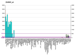
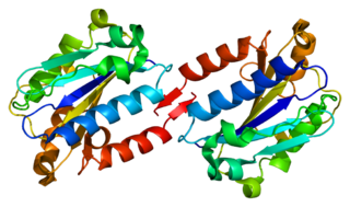
Selectin P ligand, also known as SELPLG or CD162, is a human gene.

<span class="mw-page-title-main">Selectin</span> Family of cell adhesion molecules

The selectins are a family of cell adhesion molecules. All selectins are single-chain transmembrane glycoproteins that share similar properties to C-type lectins due to a related amino terminus and calcium-dependent binding. Selectins bind to sugar moieties and so are considered to be a type of lectin, cell adhesion proteins that bind sugar polymers.

<span class="mw-page-title-main">P-selectin</span> Type-1 transmembrane protein

P-selectin is a type-1 transmembrane protein that in humans is encoded by the SELP gene.

<span class="mw-page-title-main">ICAM-1</span> Mammalian protein found in Homo sapiens

ICAM-1 also known as CD54 is a protein that in humans is encoded by the ICAM1 gene. This gene encodes a cell surface glycoprotein which is typically expressed on endothelial cells and cells of the immune system. It binds to integrins of type CD11a / CD18, or CD11b / CD18 and is also exploited by rhinovirus as a receptor for entry into respiratory epithelium.

<span class="mw-page-title-main">E-selectin</span>

E-selectin, also known as CD62 antigen-like family member E (CD62E), endothelial-leukocyte adhesion molecule 1 (ELAM-1), or leukocyte-endothelial cell adhesion molecule 2 (LECAM2), is a selectin cell adhesion molecule expressed only on endothelial cells activated by cytokines. Like other selectins, it plays an important part in inflammation. In humans, E-selectin is encoded by the SELE gene.

<span class="mw-page-title-main">Integrin alpha X</span> Mammalian protein found in Homo sapiens

CD11c, also known as Integrin, alpha X (ITGAX), is a gene that encodes for CD11c.

<span class="mw-page-title-main">Integrin alpha L</span> Mammalian protein found in Homo sapiens

Integrin, alpha L , also known as ITGAL, is a protein that in humans is encoded by the ITGAL gene. CD11a functions in the immune system. It is involved in cellular adhesion and costimulatory signaling. It is the target of the drug efalizumab.

High endothelial venules (HEV) are specialized post-capillary venules characterized by plump endothelial cells as opposed to the usual flatter endothelial cells found in regular venules. HEVs enable lymphocytes circulating in the blood to directly enter a lymph node.

Sialyl-Lewis <sup>X</sup> Chemical compound

Sialyl LewisX (sLeX), also known as cluster of differentiation 15s (CD15s) or stage-specific embryonic antigen 1 (SSEA-1), is a tetrasaccharide carbohydrate which is usually attached to O-glycans on the surface of cells. It is known to play a vital role in cell-to-cell recognition processes. It is also the means by which an egg attracts sperm; first, to stick to it, then bond with it and eventually form a zygote.

<span class="mw-page-title-main">Addressin</span> Protein-coding gene in the species Homo sapiens

Mucosal vascular addressin cell adhesion molecule 1 (MAdCAM-1) is a protein that in humans is encoded by the MADCAM1 gene. The protein encoded by this gene is an endothelial cell adhesion molecule that interacts preferentially with the leukocyte beta7 integrin LPAM-1, L-selectin, and VLA-4 on myeloid cells to direct leukocytes into mucosal and inflamed tissues. It is a member of the immunoglobulin superfamily and is similar to ICAM-1 and VCAM-1.

<span class="mw-page-title-main">CCL21</span> Mammalian protein found in Homo sapiens

Chemokine ligand 21 (CCL21) is a small cytokine belonging to the CC chemokine family. This chemokine is also known as 6Ckine, exodus-2, and secondary lymphoid-tissue chemokine (SLC). CCL21 elicits its effects by binding to a cell surface chemokine receptor known as CCR7. The main function of CCL21 is to guide CCR7 expressing leukocytes to the secondary lymphoid organs, such as lymph nodes and Peyer´s patches.

Glycosylation-dependent cell adhesion molecule-1 (GLYCAM1) is a proteoglycan ligand expressed on cells of the high endothelial venules in lymphoid tissues. It is the ligand for the receptor L-selectin allowing for naive lymphocytes to exit the bloodstream into lymphoid tissues. GLYCAM1 binds to L-selectin by presenting one or more O-linked carbohydrates to the lectin domain of the leukocyte cell surface selectin. Data suggests that GLYCAM1 is a hormone-regulated milk protein that is part of the milk mucin complex.

Lymphocyte homing receptors are cell adhesion molecules expressed on lymphocyte cell membranes that recognize addressins on target tissues. Lymphocyte homing refers to adhesion of the circulating lymphocytes in blood to specialized endothelial cells within lymphoid organs. These diverse tissue-specific adhesion molecules on lymphocytes and on endothelial cells contribute to the development of specialized immune responses.

<span class="mw-page-title-main">Leukocyte extravasation</span>

Leukocyte extravasation is the movement of leukocytes out of the circulatory system and towards the site of tissue damage or infection. This process forms part of the innate immune response, involving the recruitment of non-specific leukocytes. Monocytes also use this process in the absence of infection or tissue damage during their development into macrophages.

<span class="mw-page-title-main">FUT7</span> Protein-coding gene in the species Homo sapiens

Alpha-(1,3)-fucosyltransferase is an enzyme that in humans is encoded by the FUT7 gene.

Lymph node stromal cells are essential to the structure and function of the lymph node whose functions include: creating an internal tissue scaffold for the support of hematopoietic cells; the release of small molecule chemical messengers that facilitate interactions between hematopoietic cells; the facilitation of the migration of hematopoietic cells; the presentation of antigens to immune cells at the initiation of the adaptive immune system; and the homeostasis of lymphocyte numbers. Stromal cells originate from multipotent mesenchymal stem cells.

Gut-specific homing is the mechanism by which activated T cells and antibody-secreting cells (ASCs) are targeted to both inflamed and non-inflamed regions of the gut in order to provide an effective immune response. This process relies on the key interaction between the integrin α4β7 and the addressin MadCAM-1 on the surfaces of the appropriate cells. Additionally, this interaction is strengthened by the presence of CCR9, a chemokine receptor, which interacts with TECK. Vitamin A-derived retinoic acid regulates the expression of these cell surface proteins.

Integrin α4β7 is an integrin heterodimer composed of CD49d (alpha-4) subunit and beta-7 subunit noncovalently linked. LPAM-1 is expressed on the cell surface of leukocytes. This receptor is involved in lymphocyte trafficking pathway to site of inflammation in intestinal tissues.

References

  1. 1 2 3 GRCh38: Ensembl release 89: ENSG00000188404 - Ensembl, May 2017
  2. 1 2 3 GRCm38: Ensembl release 89: ENSMUSG00000026581 - Ensembl, May 2017
  3. "Human PubMed Reference:". National Center for Biotechnology Information, U.S. National Library of Medicine.
  4. "Mouse PubMed Reference:". National Center for Biotechnology Information, U.S. National Library of Medicine.
  5. 1 2 3 Ivetic A (March 2018). "A head-to-tail view of L-selectin and its impact on neutrophil behaviour". Cell and Tissue Research. 371 (3): 437–453. doi:10.1007/s00441-017-2774-x. PMC   5820395 . PMID   29353325.
  6. 1 2 3 4 5 6 7 8 9 10 11 12 Ivetic A, Hoskins Green HL, Hart SJ (2019-05-14). "L-selectin: A Major Regulator of Leukocyte Adhesion, Migration and Signaling". Frontiers in Immunology. 10: 1068. doi: 10.3389/fimmu.2019.01068 . PMC   6527602 . PMID   31139190.
  7. 1 2 3 Feng Y, Ma X, Deng L, Yao B, Xiong Y, Wu Y, et al. (May 2017). "Role of selectins and their ligands in human implantation stage". Glycobiology. 27 (5): 385–391. doi: 10.1093/glycob/cwx009 . PMID   28115423.
  8. Tvaroška I, Selvaraj C, Koča J (June 2020). "Selectins-The Two Dr. Jekyll and Mr. Hyde Faces of Adhesion Molecules-A Review". Molecules. 25 (12): 2835. doi: 10.3390/molecules25122835 . PMC   7355470 . PMID   32575485.
  9. Kerdiles YM, Beisner DR, Tinoco R, Dejean AS, Castrillon DH, DePinho RA, Hedrick SM (February 2009). "Foxo1 links homing and survival of naive T cells by regulating L-selectin, CCR7 and interleukin 7 receptor". Nature Immunology. 10 (2): 176–184. doi:10.1038/ni.1689. PMC   2856471 . PMID   19136962.
  10. Robbins SL, Cotran RS, Kumar V, Collins T (1998). Robbins Pathologic Basis of Disease. Philadelphia: W.B Saunders Company. ISBN   0-7216-7335-X.
  11. Kohn LA, Hao QL, Sasidharan R, Parekh C, Ge S, Zhu Y, et al. (October 2012). "Lymphoid priming in human bone marrow begins before expression of CD10 with upregulation of L-selectin". Nature Immunology. 13 (10): 963–971. doi:10.1038/ni.2405. PMC   3448017 . PMID   22941246.
  12. Björkman L, Christenson K, Davidsson L, Mårtensson J, Amirbeagi F, Welin A, et al. (June 2019). "Neutrophil recruitment to inflamed joints can occur without cellular priming". Journal of Leukocyte Biology. 105 (6): 1123–1130. doi:10.1002/JLB.3AB0918-369R. PMID   30570778. S2CID   58603580.
  13. James JL, Carter AM, Chamley LW (May 2012). "Human placentation from nidation to 5 weeks of gestation. Part I: What do we know about formative placental development following implantation?". Placenta. 33 (5): 327–334. doi:10.1016/j.placenta.2012.01.020. PMID   22374510.
  14. Segura J, He B, Ireland J, Zou Z, Shen T, Roth G, Sun PD (2021-09-29). "The Role of L-Selectin in HIV Infection". Frontiers in Microbiology. 12: 725741. doi: 10.3389/fmicb.2021.725741 . PMC   8511817 . PMID   34659153.
  15. Natoni A, Macauley MS, O'Dwyer ME (2016). "Targeting Selectins and Their Ligands in Cancer". Frontiers in Oncology. 6: 93. doi: 10.3389/fonc.2016.00093 . PMC   4834419 . PMID   27148485.

Further reading

This article incorporates text from the United States National Library of Medicine, which is in the public domain.