Mariannaea elegans

Last updated

Mariannaea elegans
Mariannaea elegans UAMH 4676.png
Scientific classification OOjs UI icon edit-ltr.svg
Domain: Eukaryota
Kingdom: Fungi
Division: Ascomycota
Class: Sordariomycetes
Order: Hypocreales
Family: Nectriaceae
Genus: Mariannaea
Species:
M. elegans
Binomial name
Mariannaea elegans
(Corda) Samson (1974)
Synonyms
  • Penicillium elegansCorda (1838)
  • Paecilomyces elegansCorda (1951)
  • Penicillium elegant Corda (1838)
  • Hormodendrum elegans (Corda) Bonord (1851)
  • Spicaria elegans (Corda) (1871)
  • Paecilomyces elegans (Corda) (1951)
  • Spicaria elegans var. sorghina Sacc.(1886)
  • Spicaria elegans var. microspora Jacz.(1922)

Mariannaea elegans an anamorphic fungus (i.e., it reproduces exclusively asexually). [1] It is mainly found on rotting wood and soil. [1] M. elegans is not pathogenic to humans, animals, or plants. [2]

Contents

Original line illustration of Penicillium elegans (= Mariannaea elegans), source: ACJ Corda. 1838. Icones fungorum hucusque cognitorum. 2:1-43. Penicillium elegans.png
Original line illustration of Penicillium elegans (= Mariannaea elegans), source: ACJ Corda. 1838. Icones fungorum hucusque cognitorum. 2:1-43.

History and taxonomy

Czech mycologist August Carl Joseph Corda named this species Penicillium elegans in 1838. This species was transferred to the genus Paecilomyces in 1951. [3] Later, in 1952, French mycologist, Gabriel Arnaud named the species Mariannaea elegans although he failed to provide a Latin description, which was a requirement for valid publication at the time. [4] [5] Arnaud noted that the genus Mariannaea shared many characteristics with genus Paecilomyces but was distinguished by the divergent nature of the conidial chains of Mariannaea. [4] [5] This conclusion was supported by Dutch mycologist, Robert Archibald Samson, who in 1974 validated the names of the genus and species by providing Latin diagnoses. [5] [6] The genus Mariannaea currently consists of eight species. [7] Two varieties of M. elegans exist: M. elegans var. elegans and M. elegans var. punicea. [7]

Growth and morphology

Mariannaea elegans is an anamorphic fungus (i.e., it reproduces asexually). [1] When M. elegans is grown on a Petri dish in 2% MEA (Malt Extract Agar; medium used in a Petri dish) and PDA (potato-dextrose agar) at 22 °C (72 °F) the growth and morphological characteristics listed below are observed. The colonies of M. elegans grows better in MEA than in PDA. [7] On average colonies of M. elegans can be observed with the naked eye having a diameter of 2.5–6 cm after 10 days of growth. [1] [8] [3] They appear thin and velvety (i.e. smooth) or floccose (i.e. woolly) or matted (i.e. powdery) and are odourless. [1] [8] Sometimes the colonies make a ring shaped pattern as might be seen on a jawbreaker candy. [1] Specimens isolated from wood possess brown colonies whose brown pigment fuses into the surrounding agar. [1] However, specimens isolated from soil possess reddish-purple colonies whose pigmentation does not diffuse in to the agar surrounding the colonies of M. elegans. [1] Colonies are white to cinnamon buff (peanut butter colour) and possess either smooth or rough walls. [1] The mycelium is either smooth-walled or rough-walled, white to olivaceous (the green colour of the mould on blue cheese) or pink. [1]

Conidiophores are arise from mycelia immersed in the agar. They are light brown, with a smooth or verrucose (i.e., bumpy) surface and grow to a maximum of 1000 μm long and reach a maximum of 10 μm in width at the base. [8] Conidiophores can also arise from aerial hyphae. [8] These conidiophores grow to 250 μm in length and 7 μm in width. [1] Additionally, they can grow to reach a maximum of 1400 μm in length and 23 μm in width at their base. [1] The tops narrow to 3-4 μm. [1] Alternatively, conidiophores can grow laterally from ascending mycelium (i.e., the mycelium is not immersed in the agar) or hyphal ropes. On average these conidiophores are smaller than the ones that grow from hypha immersed in agar. [1] They are smooth-walled or rough-walled, hyaline to pale yellowish. Bearing up to 4 diverging branches that are 9-22 μm long and 3-5 μm wide. [1] They may bear secondary branches. [1] Phialides are observed at the apex of the conidiophores and their branches. [1] They are hyaline in colour and possesses a smooth surface and ellipsoidal (i.e. shaped like a football) to fusiform (i.e. spindle) in shape. [1] They are 8-22 x 2.2-3.5 μm in width and they gradually become thinner reaching 0.8-1.4 μm in width at their apex. Growing obliquely form the apex of a phialide are long chains of conidia (singular conidium). Like phialides, they have a smooth surface and are ellipsoidal to fusiform in shape. [1] Their apex is sharply pointed and round at the base, on average 5.3 x 2.7 μm and reaches a maximum of 9.0 x 4.0 μm in width. [1]

The two varieties of Mariannaea elegans, M. elegans var. elegans and M. elegans var. punicea, are highly morphologically similar. [7] [1] Colonies of M. elegans var. elegans grow more slowly than those of M. elegans var. punicea. [1] Colonies of the former also tend to develop a brownish colour and ringed pattern whereas those of the latter tend to develop a reddish-purple pigmentation and lacking a ringed pattern. [1] M. elegans var. elegans is most commonly isolated from rotting wood whereas M. elegans var. punicea is only known from soil. [1] The genus Mariannaea is closely related to Clonastachys and Gliocladium . [1] They differ from Mariannaea because their conidiophores are clustered tightly together. [1] M. elegans is often misidentified as a member of the genus Paecilomyces because of its morphological similarity. [1] However, the phialides of Mariannaea are flask shaped (thick at the base gradually narrow towards the apex, in cross-section like a canoe paddle) whereas those of Paecilomyces tend to be shorter and stouter (in cross-section like a tennis racquet). [1]

Physiology

This fungus is not known to be a pathogen of humans, animals, or plants. [2] A study carried out in 2007 revealed anti-M. elegans cutaneous bacterial communities that live on the skin of amphibians. [9] The presence of these bacteria inhibits the growth of M. elegans on amphibians. The species grows optimally at a pH of 7.0; [7] however, it tolerates a pH range of 5.5-8.0. [3] The sporulation phase of M. elegans is sensitive to pH. It sporulates best at a pH range of 6.0-6.5 and below a pH of 5.0 sporulation is reduced. [3] The optimal temperature that it grows in is 30 °C (86 °F). [7] However, trances of M. elegans can be founds thriving at 50 °C (122 °F). [3] M. elegans produces amylase, beta-glucosidase, cellulase, and protease. [7] It is able to degrade cellulose. [3] When M. elegans is grown in vitro in the presence of different sugars (e.g., glucose, galactose, sucrose, mannose, fructose, and lactose) at 30 °C (86 °F) for 2 days in PDA distinct cell morphology is observed. [10] Growth in 2% of glucose, galactose, or sucrose leads to the formation of many small fungal cells. [10] Whereas the opposite is observed in 2% of mannose, fructose, or lactose, which lead to the formation of a few large fungal cells. [10] Sugar appears to be an important factor in the growth of M.elegans because in the absence of sugar it experiences a delay in growth. [10]

Habitat and ecology

Mariannaea elegans grows on decaying coniferous bark or wood, [3] [7] forest soil, [3] [7] house dust, [1] prairie and grassland soils, [3] calcareous soil, [3] running water that has little pollution, [3] waste stabilization ponds, [3] acid mine drainage stream, [3] fields treated with digested sewage sludge, [3] in arable soils, and on pine chips. [3]

Mariannaea elegans has been isolated from various regions in Canada: Quebec, Ontario, Manitoba, Saskatchewan, and British Columbia. [1] Both varieties have both been recorded in Germany, the Netherlands, the United States, and Canada. [3] However, only M. elegans var. elegans has also be found in France, the British Isles, Italy, Poland, and South Africa, whereas, M. elegans var. punicea has exclusively be found in Democratic Republic of the Congo. [3]

The ecological role of this species is not well known. It is involved in wood biodeterioration either through the formation of soft rot cavities or through cell wall erosion. [8] It may also influence the growth of other fungi. For example, at 25 °C (77 °F) it inhibits mycelial growth in the mushroom, Pholiota microspora ; but at12 °C (54 °F) it enhances mycelial growth of P. microspora. [3] It is capable of preventing sapstain (a blue to greyish-black colour) formation on wood treated with M. elegans. [11] This is important in the lumber industry because discoloured wood is less versatile and can not be used for some applications. [11] M. elegans has potential implication in humans. A study carried out in 2001 concluded that mariannaeaprone, a fungal metabolite made by M. elegans induces the aggregation of human platelet cells. [12] M. elegans may also be consumed by amoebae. [3]

Related Research Articles

<i>Trichoderma</i> Genus of fungi

Trichoderma is a genus of fungi in the family Hypocreaceae that is present in all soils, where they are the most prevalent culturable fungi. Many species in this genus can be characterized as opportunistic avirulent plant symbionts. This refers to the ability of several Trichoderma species to form mutualistic endophytic relationships with several plant species. The genomes of several Trichoderma specieshave been sequenced and are publicly available from the JGI.

<i>Acrophialophora fusispora</i> Species of ascomycete fungus found in soil, air and various plants

Acrophialophora fusispora is a poorly studied ascomycete fungus found in soil, air and various plants. A. fusispora is morphologically similar to the genera Paecilomyces and Masonia, but differ in the presence of pigmented conidiophores, verticillate phialides, and frequent sympodial proliferation. Moreover, A. fusispora is distinguished by its pigmented spindle-shaped conidia, covered with spiral bands. The fungus is naturally found in soils of tropical to temperate regions. The fungus has been identified as a plant and animal pathogen, and has recently been recognized as an emerging opportunistic human pathogen. A. fusispora infection in human is rare and has few documented clinical cases, but due to the rarity of the fungus and potential misidentification, the infections may be underdiagnosed. Clinical cases of A. fusispora include cases of keratitis, pulmonary colonization and infection, and cerebral infections. The fungus also has two documented cases of infection in dogs.

<i>Purpureocillium lilacinum</i> Species of fungus

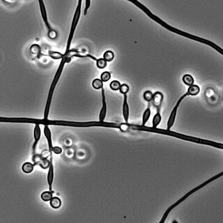
Purpureocillium lilacinum is a species of filamentous fungus in the family Ophiocordycipitaceae. It has been isolated from a wide range of habitats, including cultivated and uncultivated soils, forests, grassland, deserts, estuarine sediments and sewage sludge, and insects. It has also been found in nematode eggs, and occasionally from females of root-knot and cyst nematodes. In addition, it has frequently been detected in the rhizosphere of many crops. The species can grow at a wide range of temperatures – from 8 to 38 °C for a few isolates, with optimal growth in the range 26 to 30 °C. It also has a wide pH tolerance and can grow on a variety of substrates. P. lilacinum has shown promising results for use as a biocontrol agent to control the growth of destructive root-knot nematodes.

<i>Paecilomyces variotii</i> Species of fungus

Paecilomyces variotii, also known by the name Byssochlamys spectabilis for the sexual state, is a common environmental mold from the Phylum Ascomycota. It is widespread in the environment and can be found in composts, soils and wood, as well es a common environmental contaminant in indoor air and carpet dust. Ascospores of the sexual state of P. variotii are strongly heat-resistant. As such the fungus is a common contaminant of heat-treated foods and juices. Paecilomyces variotii has been associated with a number of infective diseases of humans and animals.

Aspergillus unguis is a species of fungus in the genus Aspergillus, and the asexual state (anamorph) of Emericella unguis. Aspergillus unguis is a filamentous soil-borne fungus found on decomposing plant matter and other moist substrates including with building materials and household dust. Aspergillus unguis occurs mainly in tropical and subtropical soils but has also been isolated from various marine and aquatic habitats. The species was first isolated in 1935 by Weill and L. Gaudin. Historically, A. unguis was assigned to the A. nidulans group, a common group of soil-borne fungi due to the resemblance of its ascospores and cleistothecia to those of Emericella nidulans. Aspergillus unguis is distinctive, however, in possessing spicular hyphae. A number of synonyms have been collapsed into this species, including Sterigmatocystis unguis, Aspergillus laokiashanensis and Aspergillus mellinus.

Penicillium verrucosum is a psychrophilic fungus which was discovered in Belgium and introduced by Dierckx in 1901. Six varieties of this species have been recognized based primarily on differences in colony colour: P. verrucosum var. album, P. verrucosum var. corymbiferum, P. verrucosum var. cyclopium, P. verrucosum var. ochraceum, P. verrucosum var. melanochlorum and P. verrucosum var. verrucosum. This fungus has important implications in food, specifically for grains and other cereal crops on which it grows. Its growth is carefully regulated in order to reduce food spoilage by this fungi and its toxic products. The genome of P. verrucosum has been sequenced and the gene clusters for the biosyntheses of its mycotoxins have been identified.

<i>Aspergillus clavatus</i> Species of fungus

Aspergillus clavatus is a species of fungus in the genus Aspergillus with conidia dimensions 3–4.5 x 2.5–4.5 μm. It is found in soil and animal manure. The fungus was first described scientifically in 1834 by the French mycologist John Baptiste Henri Joseph Desmazières.

Paecilomyces marquandii is a soil-borne filamentous fungus distributed throughout temperate to tropical latitudes worldwide including forest, grassland, sewage sludge and strongly metal polluted area characterized by high tolerance in heavy metals. Simultaneous toxic action of zinc and alachlor result an increase in uptake of metal in this fungus but disrupts the cell membrane. Paecilomyces marquandii is known to parasitize the mushroom, Cuphophyllus virgineus, in the family, Hygrophoraceae. Paecilomyces marquandii is categorised as a biosafety risk group 1 in Canada and is not thought to be a significant pathogen of humans or animals.

<i>Keratinophyton durum</i> Species of fungus

Keratinophyton durum is a keratinophilic fungus, that grows on keratin found in decomposing or shed animal hair and bird feathers. Various studies conducted in Canada, Japan, India, Spain, Poland, Ivory Coast and Iraq have isolated this fungus from decomposing animal hair and bird feathers using SDA and hair-bait technique. Presence of fungus in soil sediments and their ability to decompose hairs make them a potential human pathogen.

<i>Penicillium spinulosum</i> Species of fungus

Penicillium spinulosum is a non-branched, fast-growing fungus with a swelling at the terminal of the stipe (vesiculate) in the genus Penicillium. P. spinulosum is able to grow and reproduce in environment with low temperature and low water availability, and is known to be acidotolerant. P. spinulosum is ubiquitously distributed, and can often be isolated from soil. Each individual strain of P. spinulosum differs from others in their colony morphology, including colony texture, amount of sporulation and roughness of conidia and conidiophores.

Aspergillus wentii is an asexual, filamentous, endosymbiotic fungus belonging to the mold genus, Aspergillus. It is a common soil fungus with a cosmopolitan distribution, although it is primarily found in subtropical regions. Found on a variety of organic materials, A. wentii is known to colonize corn, cereals, moist grains, peanuts and other ground nut crops. It is also used in the manufacture of biodiesel from lipids and is known for its ability to produce enzymes used in the food industry.

<i>Metarhizium granulomatis</i> Species of fungus

Metarhizium granulomatis is a fungus in the family Clavicipitaceae associated with systemic mycosis in veiled chameleons. The genus Metarhizium is known to infect arthropods, and collectively are referred to green-spored asexual pathogenic fungi. This species grows near the roots of plants and has been reported as an agent of disease in captive veiled chameleons. The etymology of the species epithet, "granulomatis" refers to the ability of the fungus to cause granulomatous disease in susceptible reptiles.

Sarocladium kiliense is a saprobic fungus that is occasionally encountered as a opportunistic pathogen of humans, particularly immunocompromised and individuals. The fungus is frequently found in soil and has been linked with skin and systemic infections. This species is also known to cause disease in the green alga, Cladophora glomerata as well as various fruit and vegetable crops grown in warmer climates.

<i>Phialophora fastigiata</i> Species of fungus

Phialophora fastigiata is a mitosporic, saprophytic fungus commonly found in soil, and on wood, and wood-pulp. This species was initially placed in the genus Cadophora but was later transferred to the genus Phialophora based on morphological and growth characteristics. In culture, P. fastigiata produces olive-brown, velvety colonies. The fungus is recognizable microscopically due to the presence of distinctive, funnel-shaped cuffs (collarettes) encircling the tips of phialides that bear slimy conidia. The fungus is often implicated in soft-rot wood decay due to its ability to degrade lignin, cellulose and pectin. It has also been reported to cause blue staining of wood and wood pulp.

<i>Torula herbarum</i> Species of fungus

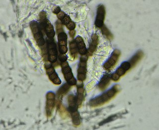
Torula herbarum is a darkly-pigmented filamentous fungus in the phylum Ascomycota. It is often included in the unrelated but morphologically similar group of fungi known as sooty molds. It was first described by Persoon in the genus Monilinia based on similarity to the agent of brown rot of stone fruit but later transferred to the genus Torula by Link. Conidia of T. herbarum are dark brown or olivaceous colour and have a distinctive shape and number of cells. T. herbarum produces secondary metabolites with cytotoxic activity towards bacteria and human cancer cells.

<i>Trichoderma koningii</i> Species of fungus

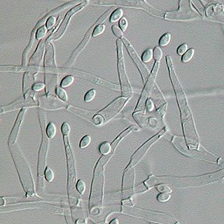
Trichoderma koningii is a very common soil dwelling saprotroph with a worldwide distribution. It has been heavily exploited for agricultural use as an effective biopesticide, having been frequently cited as an alternative biological control agent in the regulation of fungi-induced plant diseases. They are endosymbionts associated with plant root tissues, exhibiting mycoparasitism and promoting plant growth due to their capacity to produce different secondary metabolites.

Myriodontium keratinophilum is a fungus widespread in nature, most abundantly found in keratin-rich environments such as feathers, nails and hair. Despite its ability to colonize keratinous surfaces of human body, the species has been known to be non-pathogenic in man and is phylogentically distant to other human pathogenic species, such as anthropophilic dermatophytes. However, its occasional isolation from clinical specimens along with its keratinolytic properties suggest the possibility it may contribute to disease.

Harzia acremonioides is a species of seed-borne fungus that occurs in the soil. It has been categorized in the Ceratostomataceae family and under the genus Harzia. The genus Harzia contained up to three accepted species: H. acremonioides, H. verrucose, and H. velatea in 1974. Within the genus Harzia, H. acremonioides is one of the most common species that can be found in all climate regions around the world.

<i>Mariannaea</i> Genus of fungi

Mariannaea is a genus of fungi belonging to the family Nectriaceae.

<i>Xenodevriesia</i> Species of ascomycete fungus

Xenodevriesia strelitziicola is a pathogenic ascomycete fungus in the class Dothideomycetes that infects the South African plant Strelitzia. It is the only species of the monotypic genus Xenodevriesia and family Xenodevriesiaceae.

References

  1. 1 2 3 4 5 6 7 8 9 10 11 12 13 14 15 16 17 18 19 20 21 22 23 24 25 26 27 28 29 30 Bisette, John (1979). "Mariannaea elegans". Fungi Canadensis.
  2. 1 2 "UAMH Centre for Global Microfungal Biodiversity". www.uamh.ca.
  3. 1 2 3 4 5 6 7 8 9 10 11 12 13 14 15 16 17 18 19 Domsch, Klaus Heinz; Grams, Walter; Anderson, Traute- Heidi (1980). Compendium of soil fungi. London: Academic Press. pp.  409–410. ISBN   978-0122204012.
  4. 1 2 R, Arnaud (1886). "Bulletin trimestriel". La Société. 5: 196.
  5. 1 2 3 Samson, R.A. (10 June 1974). "Paecilomyces and some allied hyphomycetes". Studies in Mycology. 6: 1–119.
  6. "Wiki Species - Mycological authors: R.A. Samson". 2017.
  7. 1 2 3 4 5 6 7 8 9 Tang, Longqing; Hyun, Min Woo; Yun, Yeo Hong; Suh, Dong Yeon; Kim, Seong Hwan; Sung, Gi Ho (March 16, 2012). "New record of Mariannaea elegans var. elegans in Korea". Mycobiology. 40 (1): 14–19. doi:10.5941/myco.2012.40.1.014. PMC   3385148 . PMID   22783129.
  8. 1 2 3 4 5 C.J.K., Wang; R.A., Zabel (1990). Identification manual for fungi from utility poles in the eastern united states. American Type Culture Collection. pp.  248–249. ISBN   978-0930009311.
  9. Lauer, Antje; Simon, Mary Alice; Banning, Jenifer L.; Lam, Brianne A.; Harris, Reid N. (December 13, 2007). "Diversity of cutaneous bacteria with antibacterial activity isolated from female four-toed salamanders". The ISME Journal. 2 (2): 145–157. doi: 10.1038/ismej.2007.110 . PMID   18079731.
  10. 1 2 3 4 Tani, Yasushi; Yamashita, Yasunori; Saito, Shigeki; Mihara, Hisaaki (2014). "Effects of Sugars and Salt on the Production of Glycophingolipids in Mariannaea elegans". Trace Nutrients Research. 31: 27–44.
  11. 1 2 Seifert, Nepean; Colette Breuil, Vanier; Mes-Hartree, Mary. "Sapstain Control Method Using Mariannaea Elegans" (PDF). FPO Driving IP Forward. Retrieved November 27, 2018.
  12. Fabian, K; Anke, T; Sterner, O (2001). "Mariannaepyrone--a new inhibitor of thrombroxane A2 induced platelet aggregation". Journal of Biosciences (56): 106–110.