
Trematoda is a class of flatworms known as flukes or trematodes. They are obligate internal parasites with a complex life cycle requiring at least two hosts. The intermediate host, in which asexual reproduction occurs, is usually a snail. The definitive host, where the flukes sexually reproduce, is a vertebrate. Infection by trematodes can cause disease in all five traditional vertebrate classes: mammals, birds, amphibians, reptiles, and fish.

Clonorchis sinensis, the Chinese liver fluke, is a liver fluke belonging to the class Trematoda, phylum Platyhelminthes. It infects fish-eating mammals, including humans. In humans, it infects the common bile duct and gall bladder, feeding on bile. It was discovered by British physician James McConnell at the Medical College Hospital in Calcutta (Kolkata) in 1874. The first description was given by Thomas Spencer Cobbold, who named it Distoma sinense. The fluke passes its lifecycle in three different hosts, namely freshwater snail as first intermediate hosts, freshwater fish as second intermediate host, and mammals as definitive hosts.

Fasciola hepatica, also known as the common liver fluke or sheep liver fluke, is a parasitic trematode of the class Trematoda, phylum Platyhelminthes. It infects the livers of various mammals, including humans, and is transmitted by sheep and cattle to humans the world over. The disease caused by the fluke is called fasciolosis or fascioliasis, which is a type of helminthiasis and has been classified as a neglected tropical disease. Fasciolosis is currently classified as a plant/food-borne trematode infection, often acquired through eating the parasite's metacercariae encysted on plants. F. hepatica, which is distributed worldwide, has been known as an important parasite of sheep and cattle for decades and causes significant economic losses in these livestock species, up to £23 million in the UK alone. Because of its relatively large size and economic importance, it has been the subject of many scientific investigations and may be the best-known of any trematode species. F. hepatica's closest relative is Fasciola gigantica. These two flukes are sister species; they share many morphological features and can mate with each other.

The Aspidogastrea is a small group of flukes comprising about 80 species. It is a subclass of the trematoda, and sister group to the Digenea. Species range in length from approximately one millimeter to several centimeters. They are parasites of freshwater and marine molluscs and vertebrates. Maturation may occur in the mollusc or vertebrate host. None of the species has any economic importance, but the group is of very great interest to biologists because it has several characters which appear to be archaic.

Trematodes are parasitic flatworms of the class Trematoda, specifically parasitic flukes with two suckers: one ventral and the other oral. Trematodes are covered by a tegument, that protects the organism from the environment by providing secretory and absorptive functions.

Schistosoma haematobium is a species of digenetic trematode, belonging to a group (genus) of blood flukes (Schistosoma). It is found in Africa and the Middle East. It is the major agent of schistosomiasis, the most prevalent parasitic infection in humans. It is the only blood fluke that infects the urinary tract, causing urinary schistosomiasis, and is the leading cause of bladder cancer. The diseases are caused by the eggs.

Echinostoma is a genus of trematodes (flukes), which can infect both humans and other animals. These intestinal flukes have a three-host life cycle with snails or other aquatic organisms as intermediate hosts, and a variety of animals, including humans, as their definitive hosts.

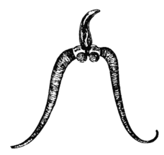
Leucochloridium paradoxum, the green-banded broodsac, is a parasitic flatworm. Its intermediate hosts are land snails, usually of the genus Succinea. The pulsating, green broodsacs fill the eye stalks of the snail, thereby attracting predation by birds, the primary host. These broodsacs visually imitate caterpillars, a prey of birds. The adult parasite lives in the bird's cloaca, releasing its eggs into the faeces.
Metagonimoides oregonensis is a trematode, or fluke worm, in the family Heterophyidae. This North American parasite is found primarily in the intestines of raccoons, American minks, frogs in the genus Rana, and freshwater snails in the genus Goniobasis. It was first described in 1931 by E. W. Price. The parasite has a large distribution, from Oregon to North Carolina. Adult flukes vary in host range and morphology dependent on the geographical location. This results in different life cycles, as well as intermediate hosts, across the United States. On the west coast, the intermediate host is freshwater snails (Goniobasis), while on the east coast the intermediate host is salamanders (Desmognathus). The parasites on the west coast are generally much larger than on the east coast. For example, the pharynx as well as the body of the parasite are distinctly larger in Oregon than in North Carolina. The reverse pattern is observed on the east coast for uterine eggs, which are larger on the west coast. In snails, there is also a higher rate of infection in female snails than in males. Research on the life history traits of the parasites have been performed with hamsters and frogs as model species.

Leucochloridium variae, the brown-banded broodsac, is a species of trematode whose life cycle involves the alternate parasitic invasion of certain species of snail and bird. While there is no external evidence of the worm's existence within the bird host, the invasion of the snail host involves the grotesque swelling of one or both of the snail's eye stalks. This invasion does not cause the snail's death.
Bucephalus polymorphus is a type of flatworm. This species is within the Bucephalidae family of Digenea, which in turn is a subclass of Trematodes within the phylum Platyhelminthes. It is characterized by having a mouth near the middle of its body, along with a sac-like gut. The mouth opening is located in the centre of the ventral surface. This is a specific body type of cecaria known as a gastrostome.
Echinostoma revolutum is a trematode parasites, of which the adults can infect birds and mammals, including humans. In humans, it causes echinostomiasis.
Echinostoma hortense is an intestinal fluke of the class Trematoda, which has been found to infect humans in East Asian countries such as Korea, China, and Japan. This parasite resides in the intestines of birds, rats and other mammals such as humans. While human infections are very rare in other regions of the world, East Asian countries have reported human infections up to about 24% of the population in some endemic sub-regions. E. hortense infections are zoonotic infections, which occurs from eating raw or undercooked freshwater fish. The primary disease associated with an E. hortense infection is called echinostomiasis, which is a general name given to diseases caused by Trematodes of the genus Echinostoma.
Echinostoma cinetorchis is a species of human intestinal fluke, a trematode in the family Echinostomatidae.

Clinostomum marginatum is a species of parasitic fluke. It is commonly called the "yellow grub". It is found in many freshwater fish in North America, and no fish so far is immune to this parasite. It is also found in frogs. Clinostomum marginatum can also be found in the mouth of aquatic birds such as herons and egrets. They are commonly present in the esophagus of fish-eating birds and reptiles. Eggs of these trematodes are shed in the feces of aquatic birds and released into water. Aquatic birds become hosts of this parasite by ingesting infected freshwater fish. The metacercariae are found right beneath the skin or in the muscles of host fish.
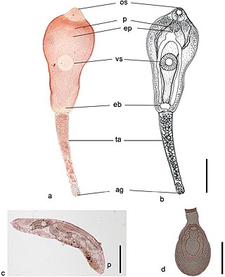
Philophthalmus gralli, commonly known as the Oriental avian eye fluke, parasitises the conjunctival sac of the eyes of many species of birds, including birds of the orders Galliformes and Anseriformes. In Brazil this parasite was reported in native Anseriformes species. It was first discovered by Mathis and Leger in 1910 in domestic chickens from Hanoi, Vietnam. Birds are definitive hosts and freshwater snail species are intermediate hosts. Human cases of philophthalmosis are rare, but have been previously reported in Europe, Asia, and America.
Paramphistomum is a genus of parasitic flatworms belonging to the digenetic trematodes. It includes flukes which are mostly parasitising livestock ruminants, as well as some wild mammals. They are responsible for the serious disease called paramphistomiasis, also known as amphistomosis, especially in cattle and sheep. Its symptoms include profuse diarrhoea, anaemia, lethargy, and often result in death if untreated. They are found throughout the world, and most abundantly in livestock farming regions such as Australia, Asia, Africa, Eastern Europe, and Russia.
Echinostoma caproni is a species of 37-spined Egyptian echinostome. It is naturally found in Cameroon, Congo, Egypt, Madagascar, and Togo.

Metagonimus yokogawai, or the Yokogawa fluke, is a species of a trematode, or fluke worm, in the family Heterophyidae.
Echinostoma bolschewense is a species of echinostome from the Czech Republic, Russia, and the Slovak Republic.