Monocercomonoides

Last updated

Contents

Monocercomonoides
Monocermonoides melolanthae.jpg
Monocercomonoides melolanthae
Scientific classification
Domain:
(unranked):
Phylum:
Class:
Order:
Family:
Genus:
Monocercomonoides

Travis, 1932
Species

Monocercomonoides is a genus of flagellate Excavata belonging to the order Oxymonadida. It was established by Bernard V. Travis and was first described as those with "polymastiginid flagellates having three anterior flagella and a trailing one originating at a single basal granule located in front of the anteriorly positioned nucleus, and a more or less well-defined axostyle". [14] It is the first eukaryotic genus to be found to completely lack mitochondria, and all hallmark proteins responsible for mitochondrial function. The genus also lacks any other mitochondria related organelles (MROs) such as hydrogenosomes or mitosomes. [15] Data suggests that the absence of mitochondria is not an ancestral feature, but rather due to secondary loss. Monocercomonoides sp. was found to obtain energy through an enzymatic action of nutrients absorbed from the environment. [15] The genus has replaced the iron-sulfur cluster assembly pathway with a cytosolic sulfur mobilization system, likely acquired by horizontal gene transfer from a eubacterium of a common ancestor of oxymonads. [16] These organisms are significant because they undermine assumptions that eukaryotes must have mitochondria to properly function. The genome of Monocercomonoides exilis has approximately 82 million base pairs (82 Mbp), with 18 152 predicted protein-coding genes. [17]

Habitat and ecology

Most Monocercomonoides species are obligate animal symbionts that live in the digestive tracts of insects, amphibians, reptiles, and mammals. [18] Monocercomonoides are common in insect orders Orthoptera and Coleoptera. The species Monocercomonoides qadrii are found in the rectum of the larva of the dung-beetle ( Oryctes rhinoceros ). [19] M. caviae, M. wenrichi, M. quadrifunilis, and M. exilis are found in the caecum of guinea pigs, and M. caprae has been found in the rumen of goats. [20] Interestingly, some Monocercomonoides species were isolated from cesspits, [21] suggesting that they might be able to survive outside of the host in certain environmental conditions. The organism uses enzymes found in its cytoplasm to break down food and furnish energy since there is no mitochondria or oxygen presence. [22] It has been noted that Monocercomonoides ingest bacteria or wood and feed by pinocytosis, however, limited studies have been done on feeding style.

Morphology

Monocercomonoides are small free-swimming, single-cell organisms ranging from 5-12μm in length, and 4.5-14.5μm in width. [19] The body may be ovoidal, pyriform, spherical or subspherical; however, they lack holdfasts and have small axostyles. [23] The axostyle is a single, contractible appendage made of microtubules that originates from the posterior end of the preaxostyle, and is situated near the posterior pair of the basal bodies (known as blepharoplast in older cytological literature). [23] The cytoskeleton is based around four basal bodies, an anterior pair and a posterior pair. [24] The preaxostyle runs between the two pairs of basal bodies and is composed of a broad, curved sheet of microtubules. [24] The inner face of the microtubule sheet adheres to a paracrystalline fibre (about 50 nm thick) which is a common characteristic of all oxymonads. [24] Monocercomonoides sp. has four flagella that originate in two pairs and arise from each basal body found in the anterior end. [23] Three of the four flagella are roughly equal in length (9.5-18μm) and the fourth trailing flagellum is slightly longer, measuring between 9.0 and 24.5μm. [19] The flagella have a beating action and are used for rapid movement. The proximal part of the long flagellum may adhere to the pellicle, which causes it to trail posteriorly. [23] The trailing flagellum is always directed backwards and is attached to the body for a considerable distance (6-9μm) by an accessory filament called a funis. [19] There are one to four filaments (rib-like strictures) extending backwards beneath the body surface. [19] In some parasites, the flagella end in acronemes. The nucleus is generally situated near the anterior end of the body and contains a central endosome surrounded by chromatin granules, some species have pelta-like structures below the nucleus. [23] The cytoplasm is granular with or without vacuoles. [23] Electron microscopic imaging of Monocercomonoides has found that the intracellular morphology lacks any Golgi apparatus, mitochondria, or potential homologs of the two; Golgi-associated proteins have been detected, but mitochondrial ones have not. [15]

Metabolic processes

Monocercomonoides sp. strain PA203 (later described as M. exilis [21] ) is the first eukaryote discovered to lack any trace of mitochondria. In all other eukaryotes that seemingly lack mitochondria, there is nuclear DNA that contains some of the genes required to assemble mitochondria, but no such genes are present in Monocercomonoides. [15] It also lacks any genes ordinarily found in mitochondrial DNA, and genes used to make the energy-extracting enzymes present in mitochondria. Monocercomonoides are able to get some energy from glucose using anaerobic metabolic pathways that operate in the cytoplasm, however, most of its energy is obtained using enzymes that break down the amino acid arginine. [24]

Glycolytic pathway

Each molecule of glucose catabolized in Monocercomonoides yields less ATP compared to mitochondrial eukaryotes that use the tricarboxylic acid cycle and electron transport chain. [25] To aid in energy conservation, Monocercomonoides has adapted alternative glycolytic enzymes. Four alternative glycolytic enzymes include pyrophosphate-fructose-6-phosphate phosphotransferase (PFP), fructose-bisphosphate aldolase class II (FBA class II), 2,3-bisphosphoglycerate independent phosphoglycerate mutase (iPGM), and pyruvate phosphate dikinase (PPDK). [25] Glucose-6-phosphate isomerase (GPI) is predicted to be in Monocercomonoides  since it is universally distributed among Eukaryotes, Bacteria, and some Archaea and essential in catabolic glycolysis, but has not yet been found. [25] Most of the glycolytic enzymes are the standard eukaryotic versions, making Monocercomonoides' metabolic pathway a mosaic similar to that of other anaerobes. [25]

The addition of PPDK to the conversion of phosphoenolpyruvate to pyruvate (typically catalyzed solely by pyruvate kinase) has a strong effect on ATP conservation. [25] Both PFP and PPDK rely on inorganic phosphate (PPi) as the phosphate donor;  therefore rather than hydrolyzing ATP, the ATP yield is increased by using a by-product of the cell's anabolic processes as an energy source. [25] These reactions are able to allow for greater ATP conservation and regulation of glycolysis due to the PPDK's reversible nature and use of inorganic phosphate where pyruvate kinase only catalyzes the forward reaction. [25]

Arginine deiminase pathway

In addition to the adjusted glycolysis, Monocercomonoides contain enzymes needed in the arginine deiminase (degradation) pathway. [15] The arginine deiminase pathway may be used for ATP production, as in Giardia intestinalis and Trichomonas vaginalis . [15] In G. intestinalis (an anaerobic unicellular eukaryote) this pathway produces eight times more ATP than sugar metabolism, and a similar output is expected in Monocercomonoides, but has yet to be confirmed. [15] All 3 enzymes of the arginine deiminase pathway are localized in the cytosol of Monocercomonoides exilis which may reflect an ancestral state in Metamonada. [26]

Iron-sulfur cluster

Iron-sulfur clusters are important protein components that are synthesized by mitochondria. [16] The main function of these small inorganic prosthetic groups is mediating electron transport, which makes them a key part of photosynthesis, respiration, DNA replication/repair, and regulation of gene expression. [16] In eukaryotic cells, the common pathway for Fe-S cluster synthesis is ISC (iron-sulfur cluster). In the cytosol, a cytosolic iron-sulfur cluster assembly (CIA) forms Fe-S cluster-containing proteins that are responsible for the maturation of nuclear Fe-S proteins. CIA is unique to eukaryotes and does not have prokaryotic homologs. [16] The mitochondrial ISC pathway is believed to be necessary for the function of CIA since it synthesizes and transports uncharacterized sulfur-containing precursor to the cytosol, and is a major reason for retention of mitochondrial-related organelles in anaerobic eukaryotes. [16] The genus Monocercomonoides contains the CIA pathways but completely lacks the ISC pathway, along with any mitochondrial proteins. [16] The genus contains a reduced version of the SUF (sulfur utilization factor) pathway, having only three proteins - SufB, SufC, and SufU. [16] The SUF pathway is a known pathway of prokaryotes, and it is believed that the genes used to build Monocercomonoides' SUF system had to have come from prokaryotes. [16] However, Monocercomonoides' SUF proteins were found to not be related to plastid homologues, or any other microbial eukaryotes. [16] It was proposed that the pathway was acquired from a eubacterium by horizontal gene transfer (HGT) in the common ancestor of Monocercomonoides and Paratrismastrix (a sister taxon of oxymonads). [16] The genetic acquisition has not been demonstrated despite the assumption that it must have occurred.

Mitochondrial acquisition and loss

Monocercomonoides contain no detectable sign that mitochondria were ever part of the organism. [15] However, since it is widely accepted that all eukaryotes have a common ancestor that evolved mitochondria, it is believed that mitochondria must have once been present in the ancestors to oxymonads and then secondarily lost. The amitchondrial genus demonstrates that mitochondria are not absolutely essential for life of a eukaryotic cell.

Genomic structure

The lack of mitochondria or any mitochondria-related organelles in Monocercomonoides exilis is confirmed by its genome sequence. A complete genome sequence analysis of Monocercomonoidesexilis strain PA203 from Chinchilla lanigera was conducted. [15] The estimated size of the genome is ~75Mb and the number of predicted protein-coding genes is 16,629. [15] A more recent re-sequencing of the genome using Oxford nanopore showed that the genome is ~82 Mbp in size. [17] Homology searches reveal a lack of genes that encode mitochondrial import machinery, metabolite transport proteins, and iron-sulfur clusters. [15] [17] Additionally, an absence of targeted important genes and genes coding for mitochondrial membrane proteins were revealed when a search for specific N-terminal and C-terminal sequences was conducted. [15] [17] Genes that are typically encoded on mitochondrial genomes (mtDNA) were not found among the assembled scaffold, suggesting Monocercomonoides lacks mtDNA. [15] 18S RNA genes were sequenced and found to be 2,927 nt long, and is among the longest known. [15] [21] Some expansions were specific to Monocercomonoides, but many were similar to those in other oxymonad genera but substantially longer. [15] Comparisons of genes coding for 𝛼-tubulin, 𝛽-tubulin, 𝛾-tubulin, EF-1𝛼, EF-2, cytHSP70, ubiquitin, 18S rRNA, and HSP90 allow the placement of oxymonads near diplomonads and trichomonads, with Monocercomonoides and Streblomastix making up the oxymonad branch. [15]

Related Research Articles

<span class="mw-page-title-main">Mitochondrion</span> Organelle in eukaryotic cells responsible for respiration

A mitochondrion is an organelle found in the cells of most eukaryotes, such as animals, plants and fungi. Mitochondria have a double membrane structure and use aerobic respiration to generate adenosine triphosphate (ATP), which is used throughout the cell as a source of chemical energy. They were discovered by Albert von Kölliker in 1857 in the voluntary muscles of insects. The term mitochondrion was coined by Carl Benda in 1898. The mitochondrion is popularly nicknamed the "powerhouse of the cell", a phrase coined by Philip Siekevitz in a 1957 article of the same name.

<span class="mw-page-title-main">Oxidative phosphorylation</span> Metabolic pathway

Oxidative phosphorylation or electron transport-linked phosphorylation or terminal oxidation is the metabolic pathway in which cells use enzymes to oxidize nutrients, thereby releasing chemical energy in order to produce adenosine triphosphate (ATP). In eukaryotes, this takes place inside mitochondria. Almost all aerobic organisms carry out oxidative phosphorylation. This pathway is so pervasive because it releases more energy than alternative fermentation processes such as anaerobic glycolysis.

<span class="mw-page-title-main">Symbiogenesis</span> Evolutionary theory holding that eukaryotic organelles evolved through symbiosis with prokaryotes

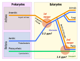
Symbiogenesis is the leading evolutionary theory of the origin of eukaryotic cells from prokaryotic organisms. The theory holds that mitochondria, plastids such as chloroplasts, and possibly other organelles of eukaryotic cells are descended from formerly free-living prokaryotes taken one inside the other in endosymbiosis. Mitochondria appear to be phylogenetically related to Rickettsiales bacteria, while chloroplasts are thought to be related to cyanobacteria.

<span class="mw-page-title-main">Cellular respiration</span> Process to convert glucose to ATP in cells

Cellular respiration is the process by which biological fuels are oxidized in the presence of an inorganic electron acceptor, such as oxygen, to drive the bulk production of adenosine triphosphate (ATP), which contains energy. Cellular respiration may be described as a set of metabolic reactions and processes that take place in the cells of organisms to convert chemical energy from nutrients into ATP, and then release waste products.

<span class="mw-page-title-main">Excavata</span> Supergroup of unicellular organisms belonging to the domain Eukaryota

Excavata is an extensive and diverse but paraphyletic group of unicellular Eukaryota. The group was first suggested by Simpson and Patterson in 1999 and the name latinized and assigned a rank by Thomas Cavalier-Smith in 2002. It contains a variety of free-living and symbiotic protists, and includes some important parasites of humans such as Giardia and Trichomonas. Excavates were formerly considered to be included in the now obsolete Protista kingdom. They were distinguished from other lineages based on electron-microscopic information about how the cells are arranged. They are considered to be a basal flagellate lineage.

<span class="mw-page-title-main">Metamonad</span> Phylum of excavate protists

The metamonads are a large group of flagellate amitochondriate microscopic eukaryotes. Their composition is not entirely settled, but they include the retortamonads, diplomonads, and possibly the parabasalids and oxymonads as well. These four groups are all anaerobic, occurring mostly as symbiotes or parasites of animals, as is the case with Giardia lamblia which causes diarrhea in mammals.

The Oxymonads are a group of flagellated protists found exclusively in the intestines of animals, mostly termites and other wood-eating insects. Along with the similar parabasalid flagellates, they harbor the symbiotic bacteria that are responsible for breaking down cellulose. There is no evidence for presence of mitochondria in oxymonads and 3 species have been shown to completely lack any molecular markers of mitochondria.

A mitosome is a mitochondrion-related organelle (MRO) found in a variety of parasitic unicellular eukaryotes, such as members of the supergroup Excavata. The mitosome was first discovered in 1999 in Entamoeba histolytica, an intestinal parasite of humans, and mitosomes have also been identified in several species of Microsporidia and in Giardia intestinalis.

<span class="mw-page-title-main">Malate–aspartate shuttle</span> Biochemical system for transporting electrons produced during glycolysis

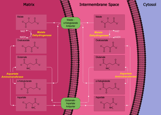
The malate–aspartate shuttle is a biochemical system for translocating electrons produced during glycolysis across the semipermeable inner membrane of the mitochondrion for oxidative phosphorylation in eukaryotes. These electrons enter the electron transport chain of the mitochondria via reduction equivalents to generate ATP. The shuttle system is required because the mitochondrial inner membrane is impermeable to NADH, the primary reducing equivalent of the electron transport chain. To circumvent this, malate carries the reducing equivalents across the membrane.

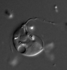
A symbiotic eukaryote that lives in the hindgut of termites, Streblomastix is a protist associated with a community of ectosymbiotic bacteria.

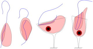
Trimastix is a genus of excavate protists, the sole occupant of the order Trimastigida. Trimastix are bacterivorous, free living and anaerobic. It was first observed in 1881 by William Kent. There are few known species, and the genus's role in the ecosystem is largely unknown. However, it is known that they generally live in marine environments within the tissues of decaying organisms to maintain an anoxic environment. Much interest in this group is related to its close association with other members of Preaxostyla. These organisms do not have classical mitochondria, and as such, much of the research involving these microbes is aimed at investigating the evolution of mitochondria.

Anaeromonadea, also known as Preaxostyla, is a class of excavate protists, comprising the oxymonads, Trimastix, and Paratrimastix. This group is studied as a model system for reductive evolution of mitochondria, because it includes both organisms with anaerobic mitochondrion-like organelles, and those that have completely lost their mitochondria.

<span class="mw-page-title-main">Jakobid</span>

Jakobids are an order of free-living, heterotrophic, flagellar eukaryotes in the supergroup Excavata. They are small, and can be found in aerobic and anaerobic environments. The order Jakobida, believed to be monophyletic, consists of only twenty species at present, and was classified as a group in 1993. There is ongoing research into the mitochondrial genomes of jakobids, which are unusually large and bacteria-like, evidence that jakobids may be important to the evolutionary history of eukaryotes.

<i>Proteromonas</i> Genus of single-celled organisms

Proteromonas is a genus of single-celled biflagellated microbial eukaryotes belonging to the Superphylum Stramenopiles which are characterized by the presence of tripartite, hair-like structures on the anteriorly-directed larger of the two flagella. Proteromonas on the other hand are notable by having tripartite hairs called somatonemes not on the flagella but on the posterior of the cell. Proteromonas are closely related to Karotomorpha and Blastocystis, which belong to the Opalines group.

<i>Jakoba</i> Genus of Eukaryotic Organisms

Jakoba is a genus in the taxon Excavata, and currently has a single described species, Jakoba libera described by Patterson in 1990, and named in honour of Dutch botanist Jakoba Ruinen.

<span class="mw-page-title-main">Arginine repressor ArgR</span>

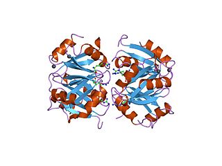
In molecular biology, the arginine repressor (ArgR) is a repressor of prokaryotic arginine deiminase pathways.

<span class="mw-page-title-main">Diplonemidae</span> Family of protozoans

Diplonemidae is a family of biflagellated unicellular protists that may be among the more diverse and common groups of planktonic organisms in the ocean. Although this family is currently made up of three named genera; Diplonema, Rhynchopus, and Hemistasia, there likely exist thousands of still unnamed genera. Organisms are generally colourless and oblong in shape, with two flagella emerging from a subapical pocket. They possess a large mitochondrial genome composed of fragmented linear DNA. These non-coding sequences must be massively trans-spliced, making it one of the most complicated post-transcriptional editing process known to eukaryotes.

<span class="mw-page-title-main">Andrew J. Roger</span> Canadian-Australian molecular biologist

Andrew J. Roger is a Canadian-Australian molecular biologist and evolutionary bioinformatician. He is currently a professor in the Department of Biochemistry and Molecular Biology at Dalhousie University and was the founding director of the inter-departmental Centre for Comparative Genomics and Evolutionary Bioinformatics (CGEB).

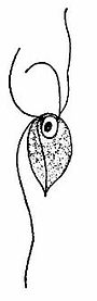
Rhodelphis is a single-celled archaeplastid that lives in aquatic environments and is the sister group to red algae and possibly Picozoa. While red algae have no flagellated stages and are generally photoautotrophic, Rhodelphis is a flagellated predator containing a non-photosynthetic plastid. This group is important to the understanding of plastid evolution because they provide insight into the morphology and biochemistry of early archaeplastids. Rhodelphis contains a remnant plastid that is not capable of photosynthesis, but may play a role in biochemical pathways in the cell like heme synthesis and iron-sulfur clustering. The plastid does not have a genome, but genes are targeted to it from the nucleus. Rhodelphis is ovoid with a tapered anterior end bearing two perpendicularly-oriented flagella.

<i>Paratrimastix pyriformis</i> Species of protists

Paratrimastix pyriformis is a species of free-living anaerobic freshwater bacteriovorous flagellated protists formerly known as Trimastix pyriformis and Tetramitus pyriformis.

References

  1. 1 2 Mali, M.; Kulkarni, S.; Mali, S. (2001). "Two species of flagellates of the genus Monocercomonoides Travis, 1932 from the gut of dung beetle larva (Oryctes rhinoceros) in India". Geobios (Jodhpur). 28 (4): 201–204.
  2. Mali, M.; Patil, D. (2003). "The morphology of Monocercomonoides aurangabadae n. sp. a flagellata from the gut of Blatta germanica". Uttar Pradesh Journal of Zoology. 23 (2): 117–119.
  3. Jensen, E.A.; Hammond, D.M. (1964). "A morphological study of trichomonads and related flagellates from the bovine digestive tract". Journal of Protozoology. 11 (3): 386–394. doi:10.1111/j.1550-7408.1964.tb01768.x. PMID   14207121.
  4. Krishnamurthy, R.; Madre, V.E. (1979). "Studies on two flagellates of the genus Monocercomonoides Travis, 1932 (Mastigophora: Polymastigina) from amphibians and reptiles in India". Acta Protozoologica. 18 (2): 251–257.
  5. 1 2 3 Rao, T.B. (1969). "The morphology and incidence of the genus Monocercomonoides (Grassi, 1879) Travis, 1932, of insects found in Andhra Pradesh, India". Proceedings of the Indian Academy of Sciences, Section B. 70 (5): 208–214. doi:10.1007/BF03052226. S2CID   127320109 . Retrieved 11 February 2018.
  6. Sultana, T.; Krishnamurthy, R. (1978). "Monocercomonoides gryllusae n. sp. (Mastigophora: Oxymonadida) from Gryllus bimaculatus". Geobios (Jodhpur). 5 (3): 114–115.
  7. Radek, R. (1994). "Monocercomonoides termitis n. sp., an oxymonad from the lower termite Kalotermes sinaicus". Archiv für Protistenkunde. 144 (4): 373–382. doi:10.1016/S0003-9365(11)80240-X.
  8. Radek, R. (1997). "Monocercomonoides hausmanni nom. nov, a New Species Name for M. termitis Radek, 1994". Archiv für Protistenkunde. 147 (3–4): 411. doi:10.1016/S0003-9365(97)80068-1.
  9. Mali, M.; Mali, S. (2004). "The Monocercomonoides khultabadae n.sp., a new flagellate from the gut of Pycnoscelus surinamensis". Uttar Pradesh Journal of Zoology. 24 (1): 55–58.
  10. 1 2 Krishnamurthy, R.; Sultana, T. (1976). "The morphology of two new flagellates of the genus Monocercomonoides Travis, 1932 from insects in India". Proceedings of the Indian Academy of Sciences, Section B. 84 (3): 109–115. doi:10.1007/BF03045588. S2CID   104326570 . Retrieved 11 February 2018.
  11. 1 2 Krishnamurthy, R. (1967). "Two new species of the genus Monocercomonoides Travis, 1932 (protozoa: Mastigophora) from reptiles". Proceedings of the Indian Academy of Sciences, Section B. 66 (5): 184–191. doi:10.1007/BF03052183. S2CID   83345325 . Retrieved 11 February 2018.
  12. Abraham, R. (961). "A description of Monocercomonoides sayeedi n. sp., from the rumen of an Indian goat". Zeitschrift für Parasitenkunde. 20 (6): 558–562. doi:10.1007/BF00333238. S2CID   26344904.
  13. Krishnamurthy, R.; Sultana, T. (1979). "A new flagellate of the genus Monocercomonoides Travis, 1932 from a termite". Proceedings of the National Academy of Sciences, India Section B. 49 (2): 85–87.
  14. Travis, B. V. 1932. A Discussion of Synonymy in the Nomenclature of Certain Insect Flagellates, with the Description of a New Flagellate from the Larvae of Ligyrodes relictus Say (Coleoptera-Scarabaeidae). Iowa State Coll J. Sci., 6, 317–323.
  15. 1 2 3 4 5 6 7 8 9 10 11 12 13 14 15 16 Karnkowska, Anna; Vacek, Vojtěch; Zubáčová, Zuzana; Treitli, Sebastian C.; Petrželková, Romana; Eme, Laura; Novák, Lukáš; Žárský, Vojtěch; Barlow, Lael D.; Herman, Emily K.; Soukal, Petr (2016). "A Eukaryote without a Mitochondrial Organelle". Current Biology. 26 (10): 1274–1284. Bibcode:2016CBio...26.1274K. doi: 10.1016/j.cub.2016.03.053 . PMID   27185558. S2CID   3933236.
  16. 1 2 3 4 5 6 7 8 9 10 Vacek, V., Novak, L. V. F., Treitli, S. C., et al. 2018. Fe–S Cluster Assembly in Oxymonads and Related Protists. Molecular Biology and Evolution. 35(11): 2712-2718.
  17. 1 2 3 4 Treitli, Sebastian Cristian; Peña-Diaz, Priscila; Hałakuc, Paweł; Karnkowska, Anna; Hampl, VladimírYR 2021 (2021). "High quality genome assembly of the amitochondriate eukaryote Monocercomonoides exilis". Microbial Genomics. 7 (12): 000745. doi: 10.1099/mgen.0.000745 . ISSN   2057-5858. PMC   8767320 . PMID   34951395.{{cite journal}}: CS1 maint: numeric names: authors list (link)
  18. Hampl, Vladimir (2017), Archibald, John M.; Simpson, Alastair G.B.; Slamovits, Claudio H. (eds.), "Preaxostyla", Handbook of the Protists, Cham: Springer International Publishing, pp. 1139–1174, doi:10.1007/978-3-319-28149-0_8, ISBN   978-3-319-28147-6 , retrieved 2022-08-01
  19. 1 2 3 4 5 Bhaskar Roa, T. 1969. The morphology and Incidence of the genus Monocercomonoides of insects found in Andhra Pradesh, India. Proceedings of the Indian Academy of Sciences. 70(5): 208-214.
  20. Flynn, F. J. 1923. Parasites of Laboratory Animals. Iowa: Blackwell Publishing.
  21. 1 2 3 Treitli, Sebastian C.; Kotyk, Michael; Yubuki, Naoji; Jirounková, Eliška; Vlasáková, Jitka; Smejkalová, Pavla; Šípek, Petr; Čepička, Ivan; Hampl, Vladimír (2018-11-01). "Molecular and Morphological Diversity of the Oxymonad Genera Monocercomonoides and Blattamonas gen. nov". Protist. 169 (5): 744–783. doi:10.1016/j.protis.2018.06.005. ISSN   1434-4610. PMID   30138782. S2CID   52077969.
  22. Leslie, M. 2016. First eukaryotes found without a normal cellular power supply. Science Mag.
  23. 1 2 3 4 5 6 Laird, M. 1955. Intestinal Flagellates from Some New Zealand Insects. Transactions and Proceedings of the Royal Society of New Zealand. 84: 297-307.
  24. 1 2 3 4 Simpson, A. G. B., Radek, R., Dacks, J. B., and O'Kelly, C. J. 2002. How oxymonads lost their groove: An ultrastructural comparison of Monocercomonoides and excavate taxa. Journal of Eukaryotic Microbiology 49: 239-248.
  25. 1 2 3 4 5 6 7 Liapounova, N. A., Hampl, V., Gordon, P. M., Sensen, C.W., Gedamu, L., and Dacks, J.B. 2006. Reconstructing the mosaic glycolytic pathway of the anaerobic eukaryote Monocercomonoides. Eukaryotic Cell 5(12): 2138-2146.
  26. Novák, Lukáš; Zubáčová, Zuzana; Karnkowska, Anna; Kolisko, Martin; Hroudová, Miluše; Stairs, Courtney W.; Simpson, Alastair G. B.; Keeling, Patrick J.; Roger, Andrew J.; Čepička, Ivan; Hampl, Vladimír (2016-10-06). "Arginine deiminase pathway enzymes: evolutionary history in metamonads and other eukaryotes". BMC Evolutionary Biology. 16 (1): 197. Bibcode:2016BMCEE..16..197N. doi: 10.1186/s12862-016-0771-4 . ISSN   1471-2148. PMC   5052871 . PMID   27716026.