Nesfatin-1

Last updated

Nesfatin-1 is a neuropeptide produced in the hypothalamus of mammals. It participates in the regulation of hunger and fat storage. [1] Increased nesfatin-1 in the hypothalamus contributes to diminished hunger, a 'sense of fullness', and a potential loss of body fat and weight.

Contents

A study of metabolic effects of nesfatin-1 in rats was done in which subjects administered nesfatin-1 ate less, used more stored fat and became more active. Nesfatin-1-induced inhibition of feeding may be mediated through the inhibition of orexigenic neurons. [2] In addition, the protein stimulated insulin secretion from the pancreatic beta cells of both rats and mice. [3]

Biochemistry

Nesfatin-1 is a polypeptide encoded in the N-terminal region of the protein precursor, Nucleobindin-2 (NUCB2). Recombinant human Nesfatin-1 is a 9.7 kDa protein containing 82 amino acid residues. [4] Nesfatin-1 is expressed in the hypothalamus, in other areas of the brain, and in pancreatic islets, gastric endocrine cells and adipocytes.

Satiety

Nesfatin/NUCB2 is expressed in the appetite-control hypothalamic nuclei such as paraventricular nucleus (PVN), arcuate nucleus (ARC), supraoptic nucleus (SON) of hypothalamus, lateral hypothalamic area (LHA), and zona incerta in rats. Nesfatin-1 immunoreactivity was also found in the brainstem nuclei such as nucleus of the solitary tract (NTS) and Dorsal nucleus of vagus nerve.

Brain

Nesfatin-1 can cross the blood–brain barrier without saturation. [5]

The receptors within the brain are in the hypothalamus and the solitary nucleus, where nesfatin-1 is believed to be produced via peroxisome proliferator-activated receptors (PPARs). It appears there is a relationship between nesfatin-1 and cannabinoid receptors. Nesfatin-1-induced inhibition of feeding may be mediated through the inhibition of orexigenic NPY neurons.

Nesfatin/NUCB2 expression has been reported to be modulated by starvation and re-feeding in the Paraventricular nucleus (PVN) and supraoptic nucleus (SON) of the brain. Nesfatin-1 influences the excitability of a large proportion of different subpopulations of neurons located in the PVN. It is also reported that magnocellular oxytocin neurons are activated during feeding, and ICV infusion of oxytocin antagonist increases food intake, indicating a possible role of oxytocin in the regulation of feeding behavior. In addition, it is proposed that feeding-activated nesfatin-1 neurons in the PVN and SON could play an important role in the postprandial regulation of feeding behavior and energy homeostasis. [6] [7]

Nesfatin-1 immunopositive neurons are also located in the arcuate nucleus (ARC). Nesfatin-1 immunoreactive neurons in the ARC are activated by simultaneous injection of ghrelin and desacyl ghrelin, nesfatin-1 may be involved in the desacyl ghrelin-induced inhibition of the orexigenic effect of peripherally administered ghrelin in freely fed rat.

Nesfatin-1 was co-expressed with melanin concentrating hormone (MCH) in tuberal hypothalamic neurons. Nesfatin-1 co-expressed in MCH neurons may play a complex role not only in the regulation of food intake, but also in other essential integrative brain functions involving MCH signaling, ranging from autonomic regulation, stress, mood, cognition to sleep. [8]

Metabolism

There is growing evidence that nesfatin-1 may play an important role in the regulation of food intake and glucose homeostasis. [9] For instance, continuous infusion of nesfatin-1 into the third brain ventricle significantly decreased food intake and body weight gain in rats. In previous studies, it was demonstrated that plasma nesfatin-1 levels were elevated in patients with type 2 diabetes mellitus (T2DM) and associated with BMI, plasma insulin, and the homeostasis model assessment of insulin resistance. [10] [11]

It was found that central nesfatin-1 resulted in a marked suppression of hepatic PEPCK mRNA and protein levels in both standard diet (SD) and high fat diet (HFD) rats but failed to alter glucose 6-phosphatase (G-6-Pase) activity and protein expression. Central nesfatin-1 appeared to antagonize the effect of HFD on increasing PEPCK gene expression in vivo. In agreement with decreasing PEPCK gene expression, central nesfatin-1 also resulted in a reduced PEPCK enzyme activity, further confirming that it affected PEPCK rather than G-6-Pase. [11]

The part of the glucose entering the liver is phosphorylated by glucokinase and then dephosphorylated by G-6-Pase. This futile cycle between glucokinase and G-6-Pase is named glucose cycling, and it accounts for the difference between the total flux through G-6-Pase and glucose production. G-6-Pase catalyzes the last step in both gluconeogenesis and glycogenolysis, and PEPCK is responsible only for gluconeogenesis. In this study, central nesfatin-1 led to a marked suppression of hepatic PEPCK protein and activity, but failed to alter hepatic G-6-Pase activity, suggesting that PEPCK may be more sensitive to short-term central nesfatin-1 exposure than G-6-Pase. In addition, suppression of HGP by central nesfatin-1 was dependent on an inhibition of the substrate flux through G-6-Pase and not on a decrease in the amount of G-6-Pase enzyme. Thus, in SD and HFD rats, central nesfatin-1 may have decreased glucose production mainly via decreasing gluconeogenesis and PEPCK activity. [11]

Recently, it has been reported that ICV nesfatin-1 produced a dose-dependent delay of gastric emptying. [11] [12]

To further delineate the mechanism by which central nesfatin-1 modulates glucose homeostasis, we assessed the effects of central nesfatin-1 on the phosphorylation of several proteins in the INSRIRS-1AMPKAkt signaling cascade in the liver. We found that central nesfatin-1 significantly augmented InsR and IRS-1 tyrosine phosphorylation. These results demonstrated that central nesfatin-1 in both SD and HFD rats resulted in a stimulation of liver insulin signaling that could account for the increased insulin sensitivity and improving glucose metabolism. [11]

AMPK is a key regulator of both lipid and glucose metabolism. It has been referred to as a metabolic master switch, because its activity is regulated by the energy status of the cell. In this study, we demonstrate that central nesfatin-1 resulted in increased phosphorylation of AMPK accompanied by a marked suppression of hepatic PEPCK activity, mRNA, and protein levels in both SD and HFD rats. Notably, central nesfatin-1 appears to prevent the obesity-driven decrease in phospho-AMPK levels in HFD-fed rats. Because hepatic AMPK controls glucose homeostasis mainly through the inhibition of gluconeogenic gene expression and glucose production, the suppressive effect of central nesfatin-1 on the HGP (Hepatic Glucose Production) can be attributed partly to its ability to suppress the expression of PEPCK mRNA and protein through AMPK activation. Furthermore, the activation of AMPK has been shown to enhance glucose uptake in skeletal muscle. Therefore, increased AMPK phosphorylation by central nesfatin-1 may also have been responsible for the improved glucose uptake in muscle. [11]

Akt is a key effector of insulin-induced inhibition of HGP and stimulation of muscle glucose uptake. We therefore examined the effects of central nesfatin-1 on Akt phosphorylation in vivo. We found that central nesfatin-1 produced a pronounced increase in insulin-mediated phosphorylation of Akt in the liver of HFD-fed rats. This increase was paralleled by an increase in muscle glucose uptake and inhibition of HGP. This provided correlative evidence that Akt activation may be involved in nesfatin-1 signaling and its effects on glucose homeostasis and insulin sensitivity. [11]

The mTOR pathway has emerged as a molecular mediator of insulin resistance, which can be activated by both insulin and nutrients. It is needed to fully activate AKT and consists of two discrete protein complexes, TORC1 and TORC2, only one of which, TORC1, binds rapamycin. In addition to mTOR, the TORC2 complex contains RICTOR, mLST8, and SIN1 and regulates insulin action and Akt phosphorylation. Thus, mTOR sits at a critical juncture between insulin and nutrient signaling, making it important both for insulin signaling downstream from Akt and for nutrient sensing. Until now, it has not been known whether nesfatin-1 affects activation of mTOR. To gain further insight into the mechanism underlying the insulin-sensitizing effects of ICV nesfatin-1, we assessed mTOR and TORC2 phosphorylation in liver samples of SD- and HFD-fed animals. Both mTOR and TORC2 phosphorylations were increased in livers from these rats, demonstrating activation of mTOR and TORC2 by central nesfatin-1 in vivo. As mTOR kinase activity is required for Akt phosphorylation, the observed increased Akt phosphorylation may have been caused by the concomitant activation of the mTOR/TORC2. Thus, it's postulated that the mTOR/TORC2 plays a role as a negative-feedback mechanism in the regulation of metabolism and insulin sensitivity mediated by central nesfatin-1. [11]

See also

Related Research Articles

<span class="mw-page-title-main">Hypothalamus</span> Area of the brain below the thalamus

The hypothalamus is a part of the brain that contains a number of small nuclei with a variety of functions. One of the most important functions is to link the nervous system to the endocrine system via the pituitary gland. The hypothalamus is located below the thalamus and is part of the limbic system. In the terminology of neuroanatomy, it forms the ventral part of the diencephalon. All vertebrate brains contain a hypothalamus. In humans, it is the size of an almond.

<span class="mw-page-title-main">AMP-activated protein kinase</span> Class of enzymes

5' AMP-activated protein kinase or AMPK or 5' adenosine monophosphate-activated protein kinase is an enzyme that plays a role in cellular energy homeostasis, largely to activate glucose and fatty acid uptake and oxidation when cellular energy is low. It belongs to a highly conserved eukaryotic protein family and its orthologues are SNF1 in yeast, and SnRK1 in plants. It consists of three proteins (subunits) that together make a functional enzyme, conserved from yeast to humans. It is expressed in a number of tissues, including the liver, brain, and skeletal muscle. In response to binding AMP and ADP, the net effect of AMPK activation is stimulation of hepatic fatty acid oxidation, ketogenesis, stimulation of skeletal muscle fatty acid oxidation and glucose uptake, inhibition of cholesterol synthesis, lipogenesis, and triglyceride synthesis, inhibition of adipocyte lipogenesis, inhibition of adipocyte lipolysis, and modulation of insulin secretion by pancreatic β-cells.

<span class="mw-page-title-main">Glucokinase</span> Enzyme participating to the regulation of carbohydrate metabolism

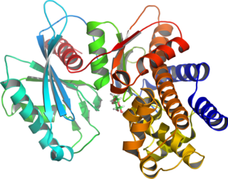
Glucokinase is an enzyme that facilitates phosphorylation of glucose to glucose-6-phosphate. Glucokinase occurs in cells in the liver and pancreas of humans and most other vertebrates. In each of these organs it plays an important role in the regulation of carbohydrate metabolism by acting as a glucose sensor, triggering shifts in metabolism or cell function in response to rising or falling levels of glucose, such as occur after a meal or when fasting. Mutations of the gene for this enzyme can cause unusual forms of diabetes or hypoglycemia.

<span class="mw-page-title-main">Arcuate nucleus</span>

The arcuate nucleus of the hypothalamus is an aggregation of neurons in the mediobasal hypothalamus, adjacent to the third ventricle and the median eminence. The arcuate nucleus includes several important and diverse populations of neurons that help mediate different neuroendocrine and physiological functions, including neuroendocrine neurons, centrally projecting neurons, and astrocytes. The populations of neurons found in the arcuate nucleus are based on the hormones they secrete or interact with and are responsible for hypothalamic function, such as regulating hormones released from the pituitary gland or secreting their own hormones. Neurons in this region are also responsible for integrating information and providing inputs to other nuclei in the hypothalamus or inputs to areas outside this region of the brain. These neurons, generated from the ventral part of the periventricular epithelium during embryonic development, locate dorsally in the hypothalamus, becoming part of the ventromedial hypothalamic region. The function of the arcuate nucleus relies on its diversity of neurons, but its central role is involved in homeostasis. The arcuate nucleus provides many physiological roles involved in feeding, metabolism, fertility, and cardiovascular regulation.

<span class="mw-page-title-main">Ghrelin</span> Peptide hormone involved in appetite regulation

Ghrelin is a hormone produced by enteroendocrine cells of the gastrointestinal tract, especially the stomach, and is often called a "hunger hormone" because it increases the drive to eat. Blood levels of ghrelin are highest before meals when hungry, returning to lower levels after mealtimes. Ghrelin may help prepare for food intake by increasing gastric motility and stimulating the secretion of gastric acid.

<span class="mw-page-title-main">Protein kinase B</span> Set of three serine/threonine-specific protein kinases

Protein kinase B (PKB), also known as Akt, is the collective name of a set of three serine/threonine-specific protein kinases that play key roles in multiple cellular processes such as glucose metabolism, apoptosis, cell proliferation, transcription, and cell migration.

mTOR Mammalian protein found in humans

The mammalian target of rapamycin (mTOR), also referred to as the mechanistic target of rapamycin, and sometimes called FK506-binding protein 12-rapamycin-associated protein 1 (FRAP1), is a kinase that in humans is encoded by the MTOR gene. mTOR is a member of the phosphatidylinositol 3-kinase-related kinase family of protein kinases.

<span class="mw-page-title-main">Branched-chain amino acid</span> Amino acid with a branched carbon chain

A branched-chain amino acid (BCAA) is an amino acid having an aliphatic side-chain with a branch. Among the proteinogenic amino acids, there are three BCAAs: leucine, isoleucine, and valine. Non-proteinogenic BCAAs include 2-aminoisobutyric acid and alloisoleucine.

<span class="mw-page-title-main">Lateral hypothalamus</span>

The lateral hypothalamus (LH), also called the lateral hypothalamic area (LHA), contains the primary orexinergic nucleus within the hypothalamus that widely projects throughout the nervous system; this system of neurons mediates an array of cognitive and physical processes, such as promoting feeding behavior and arousal, reducing pain perception, and regulating body temperature, digestive functions, and blood pressure, among many others. Clinically significant disorders that involve dysfunctions of the orexinergic projection system include narcolepsy, motility disorders or functional gastrointestinal disorders involving visceral hypersensitivity, and eating disorders.

The PHLPP isoforms are a pair of protein phosphatases, PHLPP1 and PHLPP2, that are important regulators of Akt serine-threonine kinases and conventional/novel protein kinase C (PKC) isoforms. PHLPP may act as a tumor suppressor in several types of cancer due to its ability to block growth factor-induced signaling in cancer cells.

<span class="mw-page-title-main">RPTOR</span> Protein-coding gene in the species Homo sapiens

Regulatory-associated protein of mTOR also known as raptor or KIAA1303 is an adapter protein that is encoded in humans by the RPTOR gene. Two mRNAs from the gene have been identified that encode proteins of 1335 and 1177 amino acids long.

<span class="mw-page-title-main">DEPTOR</span> Protein-coding gene in the species Homo sapiens

DEP domain-containing mTOR-interacting protein (DEPTOR) also known as DEP domain-containing protein 6 (DEPDC6) is a protein that in humans is encoded by the DEPTOR gene.

The Akt signaling pathway or PI3K-Akt signaling pathway is a signal transduction pathway that promotes survival and growth in response to extracellular signals. Key proteins involved are PI3K and Akt.

<span class="mw-page-title-main">Forkhead box protein O1</span> Protein

Forkhead box protein O1 (FOXO1), also known as forkhead in rhabdomyosarcoma (FKHR), is a protein that in humans is encoded by the FOXO1 gene. FOXO1 is a transcription factor that plays important roles in regulation of gluconeogenesis and glycogenolysis by insulin signaling, and is also central to the decision for a preadipocyte to commit to adipogenesis. It is primarily regulated through phosphorylation on multiple residues; its transcriptional activity is dependent on its phosphorylation state.

<span class="mw-page-title-main">PI3K/AKT/mTOR pathway</span> Cell cycle regulation pathway

The PI3K/AKT/mTOR pathway is an intracellular signaling pathway important in regulating the cell cycle. Therefore, it is directly related to cellular quiescence, proliferation, cancer, and longevity. PI3K activation phosphorylates and activates AKT, localizing it in the plasma membrane. AKT can have a number of downstream effects such as activating CREB, inhibiting p27, localizing FOXO in the cytoplasm, activating PtdIns-3ps, and activating mTOR which can affect transcription of p70 or 4EBP1. There are many known factors that enhance the PI3K/AKT pathway including EGF, shh, IGF-1, insulin, and CaM. Both leptin and insulin recruit PI3K signalling for metabolic regulation. The pathway is antagonized by various factors including PTEN, GSK3B, and HB9.

mTOR inhibitors Class of pharmaceutical drugs

mTOR inhibitors are a class of drugs that inhibit the mechanistic target of rapamycin (mTOR), which is a serine/threonine-specific protein kinase that belongs to the family of phosphatidylinositol-3 kinase (PI3K) related kinases (PIKKs). mTOR regulates cellular metabolism, growth, and proliferation by forming and signaling through two protein complexes, mTORC1 and mTORC2. The most established mTOR inhibitors are so-called rapalogs, which have shown tumor responses in clinical trials against various tumor types.

mTORC1 Protein complex

mTORC1, also known as mammalian target of rapamycin complex 1 or mechanistic target of rapamycin complex 1, is a protein complex that functions as a nutrient/energy/redox sensor and controls protein synthesis.

mTOR Complex 2 (mTORC2) is an acutely rapamycin-insensitive protein complex formed by serine/threonine kinase mTOR that regulates cell proliferation and survival, cell migration and cytoskeletal remodeling. The complex itself is rather large, consisting of seven protein subunits. The catalytic mTOR subunit, DEP domain containing mTOR-interacting protein (DEPTOR), mammalian lethal with sec-13 protein 8, and TTI1/TEL2 complex are shared by both mTORC2 and mTORC1. Rapamycin-insensitive companion of mTOR (RICTOR), mammalian stress-activated protein kinase interacting protein 1 (mSIN1), and protein observed with rictor 1 and 2 (Protor1/2) can only be found in mTORC2. Rictor has been shown to be the scaffold protein for substrate binding to mTORC2.

Immunometabolism is a branch of biology that studies the interplay between metabolism and immunology in all organisms. In particular, immunometabolism is the study of the molecular and biochemical underpinninngs for i) the metabolic regulation of immune function, and ii) the regulation of metabolism by molecules and cells of the immune system. Further categorization includes i) systemic immunometabolism and ii) cellular immunometabolism.

The food-entrainable oscillator (FEO) is a circadian clock that can be entrained by varying the time of food presentation. It was discovered when a rhythm was found in rat activity. This was called food anticipatory activity (FAA), and this is when the wheel-running activity of mice decreases after feeding, and then rapidly increases in the hours leading up to feeding. FAA appears to be present in non-mammals (pigeons/fish), but research heavily focuses on its presence in mammals. This rhythmic activity does not require the suprachiasmatic nucleus (SCN), the central circadian oscillator in mammals, implying the existence of an oscillator, the FEO, outside of the SCN, but the mechanism and location of the FEO is not yet known. There is ongoing research to investigate if the FEO is the only non-light entrainable oscillator in the body.

References

  1. Oh-i, Shinsuke; Shimizu, Hiroyuki; Satoh, Tetsurou; Okada, Shuichi; Adachi, Sachika; Inoue, Kinji; Eguchi, Hiroshi; Yamamoto, Masanori; et al. (2006). "Identification of nesfatin-1 as a satiety molecule in the hypothalamus". Nature. 443 (7112): 709–12. Bibcode:2006Natur.443..709O. doi:10.1038/nature05162. PMID   17036007. S2CID   4366701.
  2. Price, Christopher J.; Samson, Willis K.; Ferguson, Alastair V. (2008). "Nesfatin-1 inhibits NPY neurons in the arcuate nucleus". Brain Research. 1230: 99–106. doi:10.1016/j.brainres.2008.06.084. PMC   2590930 . PMID   18625211.
  3. Gonzalez, R.; Reingold, B. K.; Gao, X.; Gaidhu, M. P.; Tsushima, R.; Unniappan, S. (2011). "Nesfatin-1 Exerts a Direct, Glucose-Dependent Insulinotropic Action on Mouse Islet Beta and MIN6 Cells". Journal of Endocrinology. 208 (3): R9–R16. doi: 10.1530/JOE-10-0492 . PMID   21224288.
  4. ProSci inc. "Nesfatin-1 Recombinant Protein". Archived from the original on 11 April 2013. Retrieved 21 March 2013.
  5. Pan, Weihong; Hsuchou, Hung; Kastin, Abba J. (2007). "Nesfatin-1 crosses the blood–brain barrier without saturation". Peptides. 28 (11): 2223–8. doi:10.1016/j.peptides.2007.09.005. PMID   17950952. S2CID   17973594.
  6. Stengel, A.; Tache, Y. (2011). "Minireview: Nesfatin-1--An Emerging New Player in the Brain-Gut, Endocrine, and Metabolic Axis". Endocrinology. 152 (11): 4033–8. doi:10.1210/en.2011-1500. PMC   3199002 . PMID   21862618.
  7. Maejima, Yuko; Sedbazar, Udval; Suyama, Shigetomo; Kohno, Daisuke; Onaka, Tatsushi; Takano, Eisuke; Yoshida, Natsu; Koike, Masato; et al. (2009). "Nesfatin-1-Regulated Oxytocinergic Signaling in the Paraventricular Nucleus Causes Anorexia through a Leptin-Independent Melanocortin Pathway". Cell Metabolism. 10 (5): 355–65. doi: 10.1016/j.cmet.2009.09.002 . PMID   19883614.
  8. Shimizu, H; Ohsaki, A; Oh-I, S; Okada, S; Mori, M (May 2009). "A new anorexigenic protein, nesfatin-1". Peptides. 30 (5): 995–8. doi:10.1016/j.peptides.2009.01.002. PMID   19452636. S2CID   3837217.
  9. Shimizu, H.; Oh-i, S.; Hashimoto, K.; Nakata, M.; Yamamoto, S.; Yoshida, N.; Eguchi, H.; Kato, I.; et al. (2008). "Peripheral Administration of Nesfatin-1 Reduces Food Intake in Mice: The Leptin-Independent Mechanism". Endocrinology. 150 (2): 662–71. doi: 10.1210/en.2008-0598 . PMID   19176321.
  10. Zhang, Z.; Li, L.; Yang, M.; Liu, H.; Boden, G.; Yang, G. (2011). "Increased Plasma Levels of Nesfatin-1 in Patients with Newly Diagnosed Type 2 Diabetes Mellitus". Experimental and Clinical Endocrinology & Diabetes. 120 (2): 91–95. doi: 10.1055/s-0031-1286339 . PMID   22020667.
  11. 1 2 3 4 5 6 7 8 Yang, M.; Zhang, Z.; Wang, C.; Li, K.; Li, S.; Boden, G.; Li, L.; Yang, G. (2012). "Nesfatin-1 Action in the Brain Increases Insulin Sensitivity Through Akt/AMPK/TORC2 Pathway in Diet-Induced Insulin Resistance". Diabetes. 61 (8): 1959–68. doi:10.2337/db11-1755. PMC   3402309 . PMID   22688332.
  12. Stengel, A.; Goebel, M.; Wang, L.; Rivier, J.; Kobelt, P.; Monnikes, H.; Lambrecht, N. W. G.; Tache, Y. (2009). "Central Nesfatin-1 Reduces Dark-Phase Food Intake and Gastric Emptying in Rats: Differential Role of Corticotropin-Releasing Factor2 Receptor". Endocrinology. 150 (11): 4911–9. doi:10.1210/en.2009-0578. PMC   2775975 . PMID   19797401.