
The cell is the basic structural and functional unit of all forms of life. Every cell consists of cytoplasm enclosed within a membrane; many cells contain organelles, each with a specific function. The term comes from the Latin word cellula meaning 'small room'. Most cells are only visible under a microscope. Cells emerged on Earth about 4 billion years ago. All cells are capable of replication, protein synthesis, and motility.
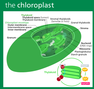
A chloroplast is a type of membrane-bound organelle known as a plastid that conducts photosynthesis mostly in plant and algal cells. The photosynthetic pigment chlorophyll captures the energy from sunlight, converts it, and stores it in the energy-storage molecules ATP and NADPH while freeing oxygen from water in the cells. The ATP and NADPH is then used to make organic molecules from carbon dioxide in a process known as the Calvin cycle. Chloroplasts carry out a number of other functions, including fatty acid synthesis, amino acid synthesis, and the immune response in plants. The number of chloroplasts per cell varies from one, in unicellular algae, up to 100 in plants like Arabidopsis and wheat.
In cell biology, an organelle is a specialized subunit, usually within a cell, that has a specific function. The name organelle comes from the idea that these structures are parts of cells, as organs are to the body, hence organelle, the suffix -elle being a diminutive. Organelles are either separately enclosed within their own lipid bilayers or are spatially distinct functional units without a surrounding lipid bilayer. Although most organelles are functional units within cells, some function units that extend outside of cells are often termed organelles, such as cilia, the flagellum and archaellum, and the trichocyst.
A plastid, pl. plastids, is a membrane-bound organelle found in the cells of plants, algae, and some other eukaryotic organisms. They are considered to be intracellular endosymbiotic cyanobacteria.

Chromista is a proposed but polyphyletic biological kingdom, refined from the Chromalveolata, consisting of single-celled and multicellular eukaryotic species that share similar features in their photosynthetic organelles (plastids). It includes all eukaryotes whose plastids contain chlorophyll c and are surrounded by four membranes. If the ancestor already possessed chloroplasts derived by endosymbiosis from red algae, all non-photosynthetic Chromista have secondarily lost the ability to photosynthesise. Its members might have arisen independently as separate evolutionary groups from the last eukaryotic common ancestor.

A unicellular organism, also known as a single-celled organism, is an organism that consists of a single cell, unlike a multicellular organism that consists of multiple cells. Organisms fall into two general categories: prokaryotic organisms and eukaryotic organisms. Most prokaryotes are unicellular and are classified into bacteria and archaea. Many eukaryotes are multicellular, but some are unicellular such as protozoa, unicellular algae, and unicellular fungi. Unicellular organisms are thought to be the oldest form of life, with early protocells possibly emerging 3.8–4.8 billion years ago.
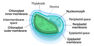
Nucleomorphs are small, vestigial eukaryotic nuclei found between the inner and outer pairs of membranes in certain plastids. They are thought to be vestiges of primitive red and green algal nuclei that were engulfed by a larger eukaryote. Because the nucleomorph lies between two sets of membranes, nucleomorphs support the endosymbiotic theory and are evidence that the plastids containing them are complex plastids. Having two sets of membranes indicate that the plastid, a prokaryote, was engulfed by a eukaryote, an alga, which was then engulfed by another eukaryote, the host cell, making the plastid an example of secondary endosymbiosis.

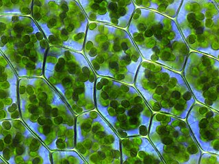
The green algae are a group of chlorophyll-containing autotrophic eukaryotes consisting of the phylum Prasinodermophyta and its unnamed sister group that contains the Chlorophyta and Charophyta/Streptophyta. The land plants (Embryophytes) have emerged deep in the Charophyte alga as a sister of the Zygnematophyceae. Since the realization that the Embryophytes emerged within the green algae, some authors are starting to include them. The completed clade that includes both green algae and embryophytes is monophyletic and is referred to as the clade Viridiplantae and as the kingdom Plantae. The green algae include unicellular and colonial flagellates, most with two flagella per cell, as well as various colonial, coccoid (spherical), and filamentous forms, and macroscopic, multicellular seaweeds. There are about 22,000 species of green algae, many of which live most of their lives as single cells, while other species form coenobia (colonies), long filaments, or highly differentiated macroscopic seaweeds.
Euglena gracilis is a freshwater species of single-celled alga in the genus Euglena. It has secondary chloroplasts, and is a mixotroph able to feed by photosynthesis or phagocytosis. It has a highly flexible cell surface, allowing it to change shape from a thin cell up to 100 μm long to a sphere of approximately 20 μm. Each cell has two flagella, only one of which emerges from the flagellar pocket (reservoir) in the anterior of the cell, and can move by swimming, or by so-called "euglenoid" movement across surfaces. E. gracilis has been used extensively in the laboratory as a model organism, particularly for studying cell biology and biochemistry.
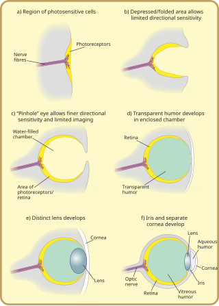
Many scientists have found the evolution of the eye attractive to study because the eye distinctively exemplifies an analogous organ found in many animal forms. Simple light detection is found in bacteria, single-celled organisms, plants and animals. Complex, image-forming eyes have evolved independently several times.

The eyespot apparatus is a photoreceptive organelle found in the flagellate or (motile) cells of green algae and other unicellular photosynthetic organisms such as euglenids. It allows the cells to sense light direction and intensity and respond to it, prompting the organism to either swim towards the light, or away from it. A related response occurs when cells are briefly exposed to high light intensity, causing the cell to stop, briefly swim backwards, then change swimming direction. Eyespot-mediated light perception helps the cells in finding an environment with optimal light conditions for photosynthesis. Eyespots are the simplest and most common "eyes" found in nature, composed of photoreceptors and areas of bright orange-red red pigment granules. Signals relayed from the eyespot photoreceptors result in alteration of the beating pattern of the flagella, generating a phototactic response.
The following outline is provided as an overview of and topical guide to cell biology:

The Gymnodiniales are an order of dinoflagellates, of the class Dinophyceae. Members of the order are known as gymnodinioid or gymnodinoid. They are athecate, or lacking an armored exterior, and as a result are relatively difficult to study because specimens are easily damaged. Many species are part of the marine plankton and are of interest primarily due to being found in algal blooms. As a group the gymnodinioids have been described as "likely one of the least known groups of the open ocean phytoplankton."

The Warnowiaceae are a family of athecate dinoflagellates. Members of the family are known as warnowiids. The family is best known for a light-sensitive subcellular structure known as the ocelloid, a highly complex arrangement of organelles with a structure directly analogous to the eyes of multicellular organisms. The ocelloid has been shown to be composed of multiple types of endosymbionts, namely mitochondria and at least one type of plastid.

A nematocyst is a subcellular structure or organelle containing extrusive filaments found in two families of athecate dinoflagellates, the Warnowiaceae and Polykrikaceae. It is distinct from the similar subcellular structures found in the cnidocyte cells of cnidarians, a group of multicellular organisms including jellyfish and corals; such structures are also often called nematocysts, and cnidocytes are sometimes referred to as nematocytes. It is unclear whether the relationship between dinoflagellate and cnidarian nematocysts is a case of convergent evolution or common descent, although molecular evidence has been interpreted as supporting an endosymbiotic origin for cnidarian nematocysts.

The Polykrikaceae are a family of athecate dinoflagellates of the order Gymnodiniales. Members of the family are known as polykrikoids. The family contains two genera: Polykrikos and Pheopolykrikos.

Erythropsidinium is a genus of dinoflagellates of the family Warnowiaceae.
Durinskia is a genus of dinoflagellates that can be found in freshwater and marine environments. This genus was created to accommodate its type species, Durinskia baltica, after major classification discrepancies were found. While Durinskia species appear to be typical dinoflagellates that are armored with cellulose plates called theca, the presence of a pennate diatom-derived tertiary endosymbiont is their most defining characteristic. This genus is significant to the study of endosymbiotic events and organelle integration since structures and organelle genomes in the tertiary plastids are not reduced. Like some dinoflagellates, species in Durinskia may cause blooms.
A plastid is a membrane-bound organelle found in plants, algae and other eukaryotic organisms that contribute to the production of pigment molecules. Most plastids are photosynthetic, thus leading to color production and energy storage or production. There are many types of plastids in plants alone, but all plastids can be separated based on the number of times they have undergone endosymbiotic events. Currently there are three types of plastids; primary, secondary and tertiary. Endosymbiosis is reputed to have led to the evolution of eukaryotic organisms today, although the timeline is highly debated.
Warnowia is a genus of athecate dinoflagellates, characterized by having a very sophisticated photoreceptor organelle called the ocelloid. This genus is dispersed worldwide but is scarce and difficult to find and nearly impossible to culture. As a result, the history and taxonomy of this genus are confusing at best, and many basic characteristics like its life cycle are still unknown. Still, Warnowia has drawn scientific interest as a unicellular organism with a fascinatingly complex photoreceptor system.


