Parasol cell

Last updated
Parasol cell
Parasol cell.jpg
Parasol cell general structure
Details
Part of Retina of eye
System Visual system
Identifiers
FMA 67916
Anatomical terminology

A parasol cell, sometimes called an M cell [1] or M ganglion cell, [2] is one type of retinal ganglion cell (RGC) located in the ganglion cell layer of the retina. These cells project to magnocellular cells in the lateral geniculate nucleus (LGN) as part of the magnocellular pathway in the visual system. [3] They have large cell bodies as well as extensive branching dendrite networks and as such have large receptive fields. [4] [3] Relative to other RGCs, they have fast conduction velocities. [4] While they do show clear center-surround antagonism (known as spatial opponency), they receive no information about color (absence of chromatic opponency). [3] Parasol ganglion cells contribute information about the motion and depth of objects to the visual system. [5]

Contents

Parasol ganglion cells in the Magnocellular pathway

Visual representation of the parvocellular and magnocellular pathways Magno Parvocellular Pathways.svg
Visual representation of the parvocellular and magnocellular pathways

Parasol ganglion cells are the first step in the magnocellular pathway of the visual system. They project from the retina via the optic nerve to the two most ventral layers of the LGN, which is a nucleus of the thalamus, occupied by the magnocellular cells which then mainly project to the striate cortex (V1), typically to the layer 4Cα. [6]

Eventually, the information these cells collect in the retina is sent to various parts of the visual cortex, including the posterior parietal cortex and area V5 through the dorsal stream, and the inferior temporal cortex and area V4 through the ventral stream. [7]

Structure

A sketch of a parasol cell (right) alongside a midget cell (left) for size comparison Midget vs Parasol cell.png
A sketch of a parasol cell (right) alongside a midget cell (left) for size comparison

Parasol ganglion cells are located in the retina of the eyes, and make up roughly 10% of all retinal ganglion cells. [3] They have large bodies [4] [6] with extensive, overlapping branched dendrites, [3] [8] and thick, heavily myelinated axons. These properties allow parasol cells to conduct signals very quickly, much faster than the midget cells that feed the P pathway. [4] [6]

Parasol ganglion cells collect information from large receptive fields, [3] [6] containing both rods and cones. [9] Despite the input from cones, parasol ganglion cells do not receive information about color. [3] [6] Unlike midget cells, parasol cell receptive fields contain the same color-type of cones in both their center and surround regions. Due to this lack of specificity, parasol cells cannot differentiate between different light wavelengths reflected from a specific object, and thus can only send achromatic information. [10]

There is approximately the same density of parasol ganglion cells in the fovea as in the rest of the retina, another property that distinguishes them from midget cells. [8]

Parasol vs. Midget cells

Parasol and midget retinal cells begin the parallel magnocellular and parvocellular pathways, respectively. While both parasol cells and midget cells play an important role in the visual system, their anatomies and functional contributions differ. [3] [11] [12] [13]

RGC TypeParasol CellMidget Cell
Pathway it's involved inMagnocellular PathwayParvocellular Pathway
Cell body sizeLargeSmall
Dendritic treeComplexLess complex
Conduction rate~1.6 ms~2 ms
Function in visual system"Where" objects are; "How" to grasp the objects"What" objects are according to fine detail
Sensitivity to spatial frequencyLowMedium to high
Temporal frequencyHighLow
Color opponencyAchromaticRed-green opponency

Function

Parasol retinal ganglion cells cannot provide finely detailed or colored information, [4] but still provide useful static, depth, and motion information. Parasol ganglion cells have high light/dark contrast detection, [14] and are more sensitive at low spatial frequencies than high spatial frequencies. Due to this contrast information, these cells are good at detecting changes in luminance, and thus provide useful information for performing visual search tasks and detecting edges. [15]

Parasol retinal ganglion cells are also important for providing information about the location of objects. These cells can detect the orientation and position of objects in space, [5] [12] information that will eventually be sent through the dorsal stream. [16] This information is also useful for detecting the difference in positions of objects on the retina of each eye, an important tool in binocular depth perception. [5] [17]

Parasol cells have the ability to detect high temporal frequencies, [18] and can thus detect quick changes in the position of an object. [6] This is the basis for detecting motion. [5] [14] [19] The information sent to the intraparietal sulcus (IPS) of the posterior parietal cortex allows the magnocellular pathway to direct attention and guide saccadic eye movements to follow important moving objects in the visual field. [4] [15] [19] In addition to following objects with the eyes, the IPS sends information to parts of the frontal lobe that allows the hands and arms to adjust their movements to correctly grasp objects based on their size, position, and location. [16] This ability has led some neuroscientists to hypothesize that the purpose of the magnocellular pathway is not to detect spatial locations, but to guide actions related to the position and motion of objects. [20]

Research and experimentation

While neurons are typically studied by the extracellular use of metal electrodes, retinal ganglion cells are specifically studied in vitro. This method allows parasol cells' complicated and intertwined structure to be analyzed intracellularly. In 1941, Polyak was the first scientist to use Golgi staining to identify retinal ganglion cells. Here, dendritic morphology was closely analyzed and revealed large dendritic trees. Later in 1986, Kaplan and Shapley were then the first researchers to link parasol cells with the visual system. Recordings of S potentials at the axon terminals of RGCs in the LGN suggest that there is high contrast sensitivity in the cells terminating in the magnocellular layer of primates; opposed by low contrast sensitivity in cells found in the parvocellular layer. [3]

Golgi stain of a neuron. GolgiStainedPyramidalCell.jpg
Golgi stain of a neuron.

Primates and other model systems

Both old and new world primates have been used as model systems for human vision and have subsequently been beneficial in researching parasol cells. [8] Many retrograde labeling experiments using macaques, for example, have linked parasol and midget retinal ganglion cells with the magnocellular and parvocellular pathways respectively. In addition, similar studies have led to theories underlying color opponency. [3] [8] Research by Dacey (1996) supports this idea where in vitro primate retinal cells were treated with dye fillings. Parasol cells of the magnocellular pathway were found to be achromatic. [3] In other studies, new world monkeys, such as marmosets, have aided in the current understanding of spatial and temporal frequency of the magnocellular layer in the LGN. Using the Nissl staining method, the magnocellular layer, in addition to the parvocellular layer, have darker and more dense cell bodies than the koniocellular layers, for example. [11]

Retinal ganglion cells of cats have been studied and compared to those in the visual system of both primates and humans. Evidence on receptive fields of cats confirms that parasol cell receptive fields are larger than those of midget cells because of their cellular structure. The same is likely to be found in human retinal cells which allows for better spatial localization. [3]

Associated disorders

Abnormal signalling in the magnocellular pathway has been associated with dyslexia and schizophrenia. [21] [22]

Dyslexia

There is a theory that problems with underdeveloped parasol ganglion cells may contribute to causing dyslexia. Motion information contributed by parasol ganglion cells to the vision system helps the brain adjust the eyes in coordinated saccades, and problems in saccadic motion may lead to blurry vision and reading problems. This underdevelopment may be caused by several factors, including nutritional deficiencies and mutations in the KIAA0319 gene on chromosome six. Additionally, autoimmune attacks by antineuronal antibodies may prevent adequate parasol ganglion cell development for normal functioning, a theory which would explain why weakened immune systems are frequently present in dyslexic individuals. [4]

See also

Related Research Articles

<span class="mw-page-title-main">Visual cortex</span> Region of the brain that processes visual information

The visual cortex of the brain is the area of the cerebral cortex that processes visual information. It is located in the occipital lobe. Sensory input originating from the eyes travels through the lateral geniculate nucleus in the thalamus and then reaches the visual cortex. The area of the visual cortex that receives the sensory input from the lateral geniculate nucleus is the primary visual cortex, also known as visual area 1 (V1), Brodmann area 17, or the striate cortex. The extrastriate areas consist of visual areas 2, 3, 4, and 5.

<span class="mw-page-title-main">Retina</span> Part of the eye

The retina is the innermost, light-sensitive layer of tissue of the eye of most vertebrates and some molluscs. The optics of the eye create a focused two-dimensional image of the visual world on the retina, which then processes that image within the retina and sends nerve impulses along the optic nerve to the visual cortex to create visual perception. The retina serves a function which is in many ways analogous to that of the film or image sensor in a camera.

<span class="mw-page-title-main">Visual system</span> Body parts responsible for vision

The visual system comprises the sensory organ and parts of the central nervous system which gives organisms the sense of vision as well as enabling the formation of several non-image photo response functions. It detects and interprets information from the optical spectrum perceptible to that species to "build a representation" of the surrounding environment. The visual system carries out a number of complex tasks, including the reception of light and the formation of monocular neural representations, colour vision, the neural mechanisms underlying stereopsis and assessment of distances to and between objects, the identification of a particular object of interest, motion perception, the analysis and integration of visual information, pattern recognition, accurate motor coordination under visual guidance, and more. The neuropsychological side of visual information processing is known as visual perception, an abnormality of which is called visual impairment, and a complete absence of which is called blindness. Non-image forming visual functions, independent of visual perception, include the pupillary light reflex and circadian photoentrainment.

<span class="mw-page-title-main">Lateral geniculate nucleus</span> Component of the visual system in the brains thalamus

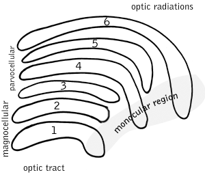
In neuroanatomy, the lateral geniculate nucleus is a structure in the thalamus and a key component of the mammalian visual pathway. It is a small, ovoid, ventral projection of the thalamus where the thalamus connects with the optic nerve. There are two LGNs, one on the left and another on the right side of the thalamus. In humans, both LGNs have six layers of neurons alternating with optic fibers.

<span class="mw-page-title-main">Retinal ganglion cell</span> Type of cell within the eye

A retinal ganglion cell (RGC) is a type of neuron located near the inner surface of the retina of the eye. It receives visual information from photoreceptors via two intermediate neuron types: bipolar cells and retina amacrine cells. Retina amacrine cells, particularly narrow field cells, are important for creating functional subunits within the ganglion cell layer and making it so that ganglion cells can observe a small dot moving a small distance. Retinal ganglion cells collectively transmit image-forming and non-image forming visual information from the retina in the form of action potential to several regions in the thalamus, hypothalamus, and mesencephalon, or midbrain.

The receptive field, or sensory space, is a delimited medium where some physiological stimuli can evoke a sensory neuronal response in specific organisms.

Parvocellular cells, also called P-cells, are neurons located within the parvocellular layers of the lateral geniculate nucleus (LGN) of the thalamus. "Parvus" is Latin for "small", and the name "parvocellular" refers to the small size of the cell compared to the larger magnocellular cells. Phylogenetically, parvocellular neurons are more modern than magnocellular ones.

Magnocellular cells, also called M-cells, are neurons located within the magnocellular layer of the lateral geniculate nucleus of the thalamus. The cells are part of the visual system. They are termed "magnocellular" since they are characterized by their relatively large size compared to parvocellular cells.

The opponent process is a color theory that states that the human visual system interprets information about color by processing signals from photoreceptor cells in an antagonistic manner. The opponent-process theory suggests that there are three opponent channels, each comprising an opposing color pair: red versus green, blue versus yellow, and black versus white (luminance). The theory was first proposed in 1892 by the German physiologist Ewald Hering.

<span class="mw-page-title-main">Motion perception</span> Inferring the speed and direction of objects

Motion perception is the process of inferring the speed and direction of elements in a scene based on visual, vestibular and proprioceptive inputs. Although this process appears straightforward to most observers, it has proven to be a difficult problem from a computational perspective, and difficult to explain in terms of neural processing.

<span class="mw-page-title-main">Koniocellular cell</span>

A koniocellular cell is a neuron with a small cell body that is located in the koniocellular layer of the lateral geniculate nucleus (LGN) in primates, including humans.

Intrinsically photosensitive retinal ganglion cells (ipRGCs), also called photosensitive retinal ganglion cells (pRGC), or melanopsin-containing retinal ganglion cells (mRGCs), are a type of neuron in the retina of the mammalian eye. The presence of ipRGCs was first suspected in 1927 when rodless, coneless mice still responded to a light stimulus through pupil constriction, This implied that rods and cones are not the only light-sensitive neurons in the retina. Yet research on these cells did not advance until the 1980s. Recent research has shown that these retinal ganglion cells, unlike other retinal ganglion cells, are intrinsically photosensitive due to the presence of melanopsin, a light-sensitive protein. Therefore, they constitute a third class of photoreceptors, in addition to rod and cone cells.

<span class="mw-page-title-main">Retinotopy</span> Mapping of visual input from the retina to neurons

Retinotopy is the mapping of visual input from the retina to neurons, particularly those neurons within the visual stream. For clarity, 'retinotopy' can be replaced with 'retinal mapping', and 'retinotopic' with 'retinally mapped'.

Blobs are sections of primary visual cortex above and below layer IV where groups of neurons sensitive to color assemble in cylindrical shapes. They were first identified in 1979 by Margaret Wong-Riley when she used a cytochrome oxidase stain, from which they get their name. These areas receive input from koniocellular cells in the dorsal lateral geniculate nucleus dLGN and output to the thin stripes of area V2. Interblobs are areas between blobs that receive the same input, but are sensitive to orientation instead of color. They output to the pale and thick stripes of area V2.

Robert Shapley is an American neurophysiologist, the Natalie Clews Spencer Professor of the Sciences at New York University, a professor in the Center for Neural Science and an associate member of the Courant Institute of Mathematical Sciences.

<span class="mw-page-title-main">Midget cell</span>

A midget cell is one type of retinal ganglion cell (RGC). Midget cells originate in the ganglion cell layer of the retina, and project to the parvocellular layers of the lateral geniculate nucleus (LGN). The axons of midget cells travel through the optic nerve and optic tract, ultimately synapsing with parvocellular cells in the LGN. These cells are known as midget retinal ganglion cells due to the small sizes of their dendritic trees and cell bodies. About 80% of RGCs are midget cells. They receive inputs from relatively few rods and cones. In many cases, they are connected to midget bipolar cells, which are linked to one cone each.

<span class="mw-page-title-main">Phantom contour</span>

A phantom contour is a type of illusory contour. Most illusory contours are seen in still images, such as the Kanizsa triangle and the Ehrenstein illusion. A phantom contour, however, is perceived in the presence of moving or flickering images with contrast reversal. The rapid, continuous alternation between opposing, but correlated, adjacent images creates the perception of a contour that is not physically present in the still images. Quaid et al. have also authored a PhD thesis on the phantom contour illusion and its spatiotemporal limits which maps out limits and proposes mechanisms for its perception centering around magnocellularly driven visual area MT.

Retinal waves are spontaneous bursts of action potentials that propagate in a wave-like fashion across the developing retina. These waves occur before rod and cone maturation and before vision can occur. The signals from retinal waves drive the activity in the dorsal lateral geniculate nucleus (dLGN) and the primary visual cortex. The waves are thought to propagate across neighboring cells in random directions determined by periods of refractoriness that follow the initial depolarization. Retinal waves are thought to have properties that define early connectivity of circuits and synapses between cells in the retina. There is still much debate about the exact role of retinal waves. Some contend that the waves are instructional in the formation of retinogeniculate pathways, while others argue that the activity is necessary but not instructional in the formation of retinogeniculate pathways.

<span class="mw-page-title-main">Peter Schiller (neuroscientist)</span> Neuroscientist

Peter H. Schiller is a professor emeritus of Neuroscience in the Department of Brain and Cognitive Sciences at the Massachusetts Institute of Technology (MIT). He is well known for his work on the behavioral, neurophysiological and pharmacological studies of the primate visual and oculomotor systems.

<span class="mw-page-title-main">Laura Busse</span> German neuroscientist

Laura Busse is a German neuroscientist and professor of Systemic Neuroscience within the Division of Neurobiology at the Ludwig Maximilian University of Munich. Busse's lab studies context-dependent visual processing in mouse models by performing large scale in vivo electrophysiological recordings in the thalamic and cortical circuits of awake and behaving mice.

References

  1. Brodal, Per (2010). The central nervous system : structure and function (4th ed.). New York: Oxford University Press. pp.  226. ISBN   978-0-19-538115-3.
  2. Gilbert, Scott F (2004). Purves, Dale (ed.). Neuroscience (3rd ed.). Sunderland, Mass.: Sinauer. p.  274. ISBN   978-0-87893-725-7.
  3. 1 2 3 4 5 6 7 8 9 10 11 12 Callaway EM (July 2005). "Structure and function of parallel pathways in the primate early visual system". The Journal of Physiology. 566 (Pt 1): 13–9. doi:10.1113/jphysiol.2005.088047. PMC   1464718 . PMID   15905213.
  4. 1 2 3 4 5 6 7 Stein J (2014-01-01). "Dyslexia: the Role of Vision and Visual Attention". Current Developmental Disorders Reports. 1 (4): 267–280. doi:10.1007/s40474-014-0030-6. PMC   4203994 . PMID   25346883.
  5. 1 2 3 4 Atkinson, J. (1992-01-01). "Early visual development: differential functioning of parvocellular and magnocellular pathways". Eye. 6 ( Pt 2) (2): 129–135. doi: 10.1038/eye.1992.28 . PMID   1624034.
  6. 1 2 3 4 5 6 Nassi JJ, Callaway EM (May 2009). "Parallel processing strategies of the primate visual system". Nature Reviews. Neuroscience. 10 (5): 360–72. doi:10.1038/nrn2619. PMC   2771435 . PMID   19352403.
  7. Yabuta, N. H.; Sawatari, A.; Callaway, E. M. (2001-04-13). "Two functional channels from primary visual cortex to dorsal visual cortical areas". Science. 292 (5515): 297–300. Bibcode:2001Sci...292..297Y. CiteSeerX   10.1.1.224.6907 . doi:10.1126/science.1057916. ISSN   0036-8075. PMID   11303106. S2CID   1360928.
  8. 1 2 3 4 Lee BB (March 1996). "Receptive field structure in the primate retina". Vision Research. 36 (5): 631–44. doi:10.1016/0042-6989(95)00167-0. PMID   8762295.
  9. Hadjikhani N, Tootell RB (2000-01-01). "Projection of rods and cones within human visual cortex". Human Brain Mapping. 9 (1): 55–63. doi:10.1002/(sici)1097-0193(2000)9:1<55::aid-hbm6>3.0.co;2-u. PMC   6871842 . PMID   10643730.
  10. Sincich, Lawrence C.; Horton, Jonathan C. (2005-01-01). "The circuitry of V1 and V2: integration of color, form, and motion". Annual Review of Neuroscience. 28: 303–326. doi:10.1146/annurev.neuro.28.061604.135731. ISSN   0147-006X. PMID   16022598.
  11. 1 2 Jayakumar J, Dreher B, Vidyasagar TR (May 2013). "Tracking blue cone signals in the primate brain". Clinical & Experimental Optometry. 96 (3): 259–66. doi:10.1111/j.1444-0938.2012.00819.x. hdl: 11343/39658 . PMID   23186138. S2CID   32259651.
  12. 1 2 Skottun, Bernt C.; Skoyles, John R. (2011-01-01). "On identifying magnocellular and parvocellular responses on the basis of contrast-response functions". Schizophrenia Bulletin. 37 (1): 23–26. doi:10.1093/schbul/sbq114. PMC   3004196 . PMID   20929967.
  13. Skoyles J, Skottun BC (January 2004). "On the prevalence of magnocellular deficits in the visual system of non-dyslexic individuals". Brain and Language. 88 (1): 79–82. doi:10.1016/s0093-934x(03)00162-7. PMID   14698733. S2CID   19152620.
  14. 1 2 Pokorny, Joel (2011-07-07). "Review: steady and pulsed pedestals, the how and why of post-receptoral pathway separation". Journal of Vision. 11 (5): 7. doi: 10.1167/11.5.7 . PMID   21737512.
  15. 1 2 Cheng, Alicia; Eysel, Ulf T.; Vidyasagar, Trichur R. (2004-10-01). "The role of the magnocellular pathway in serial deployment of visual attention". The European Journal of Neuroscience. 20 (8): 2188–2192. doi:10.1111/j.1460-9568.2004.03675.x. PMID   15450098. S2CID   31456201.
  16. 1 2 Hebart, Martin N.; Hesselmann, Guido (2012-06-13). "What visual information is processed in the human dorsal stream?". The Journal of Neuroscience. 32 (24): 8107–8109. doi:10.1523/JNEUROSCI.1462-12.2012. PMC   6703654 . PMID   22699890.
  17. Poggio, G.F.; Poggio, T. (1984). "The analysis of stereopsis". Annual Review of Neuroscience. 7: 379–412. doi:10.1146/annurev.ne.07.030184.002115. PMID   6370081.
  18. Anderson, Andrew J.; Jiao, Julie; Bui, Bang V. (2015-09-01). "Efficiently Measuring Magnocellular and Parvocellular Function in Human Clinical Studies". Translational Vision Science & Technology. 4 (5): 1. doi:10.1167/tvst.4.5.1. PMC   4559216 . PMID   26346944.
  19. 1 2 Vidyasagar, Trichur R. (2004-01-01). "Neural underpinnings of dyslexia as a disorder of visuo-spatial attention". Clinical & Experimental Optometry. 87 (1): 4–10. doi:10.1111/j.1444-0938.2004.tb03138.x. PMID   14720113. S2CID   45931933.
  20. Goodale, M.A.; Westwood, D.A. (2004). "An evolving view of duplex vision: separate but interacting cortical pathways for perception and action". Current Opinion in Neurobiology. 14 (2): 203–211. doi:10.1016/j.conb.2004.03.002. PMID   15082326. S2CID   15917985.
  21. Stein, John (2014-01-01). "Dyslexia: the Role of Vision and Visual Attention". Current Developmental Disorders Reports. 1 (4): 267–280. doi:10.1007/s40474-014-0030-6. ISSN   2196-2987. PMC   4203994 . PMID   25346883.
  22. Bortolon, Catherine; Capdevielle, Delphine; Raffard, Stéphane (2015-06-01). "Face recognition in schizophrenia disorder: A comprehensive review of behavioral, neuroimaging and neurophysiological studies". Neuroscience and Biobehavioral Reviews. 53: 79–107. doi:10.1016/j.neubiorev.2015.03.006. ISSN   1873-7528. PMID   25800172. S2CID   20887067.