Fovea centralis

Last updated
Fovea centralis
Schematic diagram of the human eye en.svg
Schematic diagram of the human eye, with the fovea at the bottom. It shows a horizontal section through the right eye.
Details
Identifiers
Latin fovea centralis
MeSH D005584
TA98 A15.2.04.022
TA2 6785
FMA 58658
Anatomical terminology

The fovea centralis is a small, central pit composed of closely packed cones in the eye. It is located in the center of the macula lutea of the retina. [1] [2]

Contents

The fovea is responsible for sharp central vision (also called foveal vision), which is necessary in humans for activities for which visual detail is of primary importance, such as reading and driving. The fovea is surrounded by the parafovea belt and the perifovea outer region. [2]

The parafovea is the intermediate belt, where the ganglion cell layer is composed of more than five layers of cells, as well as the highest density of cones; the perifovea is the outermost region where the ganglion cell layer contains two to four layers of cells, and is where visual acuity is below the optimum. The perifovea contains an even more diminished density of cones, having 12 per 100 micrometres versus 50 per 100 micrometres in the most central fovea. That, in turn, is surrounded by a larger peripheral area, which delivers highly compressed information of low resolution following the pattern of compression in foveated imaging.[ citation needed ]

Approximately half the nerve fibers in the optic nerve carry information from the fovea, while the remaining half carry information from the rest of the retina. The parafovea extends to a radius of 1.25 mm from the central fovea, and the ':perifovea is found at a 2.75 mm radius from the fovea centralis. [3]

The term fovea comes from Latin fovea 'pit'.

The fovea centralis was named by German histologist Carl Bergmann. [4]

Structure

The fovea is a depression in the inner retinal surface, about 1.5 mm wide, the photoreceptor layer of which is entirely cones and which is specialized for maximum visual acuity. Within the fovea is a region of 0.5mm diameter called the foveal avascular zone (an area without any blood vessels). This allows the light to be sensed without any dispersion or loss. This anatomy is responsible for the depression in the center of the fovea. The foveal pit is surrounded by the foveal rim that contains the neurons displaced from the pit. This is the thickest part of the retina. [5]

The fovea is located in a small avascular zone and receives most of its oxygen from the vessels in the choroid, which is across the retinal pigment epithelium and Bruch's membrane. The high spatial density of cones along with the absence of blood vessels at the fovea accounts for the high visual acuity capability at the fovea. [6]

The center of the fovea is the foveola – about 0.35 mm in diameter – or central pit where only cone photoreceptors are present and there are virtually no rods. [1] The central fovea consists of very compact cones, thinner and more rod-like in appearance than cones elsewhere. These cones are very densely packed (in a hexagonal pattern). Starting at the outskirts of the fovea, however, rods gradually appear, and the absolute density of cone receptors progressively decreases.

In 2018 the anatomy of the foveola was reinvestigated, and it was discovered that outer segments from the central foveolar cones of monkeys are not straight and twice as long as those from the parafovea. [7]

Size

The size of the fovea is relatively small with regard to the rest of the retina. However, it is the only area in the retina where 20/20 vision is attainable, and is the area where fine detail and colour can be distinguished. [8] [9]

Properties

Time-domain OCT of the macular area of a retina at 800 nm, axial resolution 3 um Retina-OCT800.png
Time-domain OCT of the macular area of a retina at 800 nm, axial resolution 3 µm
Spectral-domain OCT macula cross-section scan SD-OCT Macula Cross-Section.png
Spectral-domain OCT macula cross-section scan
Macula histology (OCT) Macula Histology OCT.jpg
Macula histology (OCT)
Diagram showing the relative acuity of the left human eye (horizontal section) in degrees from the fovea AcuityHumanEye.svg
Diagram showing the relative acuity of the left human eye (horizontal section) in degrees from the fovea
Photograph of the retina of the human eye, with overlay diagrams showing the positions and sizes of the macula, fovea, and optic disc Macula.svg
Photograph of the retina of the human eye, with overlay diagrams showing the positions and sizes of the macula, fovea, and optic disc

Function

Illustration of the distribution of cone cells in the fovea of an individual with normal color vision (left), and a color blind (protanopic) retina. Note that the center of the fovea holds very few blue-sensitive cones. ConeMosaics.jpg
Illustration of the distribution of cone cells in the fovea of an individual with normal color vision (left), and a color blind (protanopic) retina. Note that the center of the fovea holds very few blue-sensitive cones.

In the primate fovea (including humans) the ratios of ganglion cells to photoreceptors is about 2.5; almost every ganglion cell receives data from a single cone, and each cone feeds onto between one and 3 ganglion cells. [11] Therefore, the acuity of foveal vision is limited only by the density of the cone mosaic, and the fovea is the area of the eye with the highest sensitivity to fine details. [12] Cones in the central fovea express opsins that are sensitive to green and red light. These cones are the 'midget' pathways that also underpin high acuity functions of the fovea.

The fovea is employed for accurate vision in the direction where it is pointed. It comprises less than 1% of retinal size but takes up over 50% of the visual cortex in the brain. [13] The fovea sees only the central two degrees of the visual field, (approximately twice the width of your thumbnail at arm's length). [14] [15] If an object is large and thus covers a large angle, the eyes must constantly shift their gaze to subsequently bring different portions of the image into the fovea (as in reading). Foveal fixation is also considered as a overt form of attention which allows to focus sensory processing resources on the most relevant sources of information. [16] [17] [18] [19] Also, foveated vision may allow speeding up learning of specific visual tasks by disregarding not relevant context and focusing on the relevant information only with lower dimensionality. [20] [21]

Distribution of rods and cones along a line passing through the fovea and the blind spot of a human eye Human photoreceptor distribution.svg
Distribution of rods and cones along a line passing through the fovea and the blind spot of a human eye

Since the fovea does not have rods, it is not sensitive to dim lighting. Hence, in order to observe dim stars, astronomers use averted vision, looking out of the side of their eyes where the density of rods is greater, and hence dim objects are more easily visible.

The fovea has a high concentration of the yellow carotenoid pigments lutein and zeaxanthin. They are concentrated in the Henle fiber layer (photoreceptor axons that go radially outward from the fovea) and to a lesser extent in the cones. [23] [24] They are believed to play a protective role against the effects of high intensities of blue light which can damage the sensitive cones. The pigments also enhance the acuity of the fovea by reducing the sensitivity of the fovea to short wavelengths and counteracting the effect of chromatic aberration. [25] This is also accompanied by a lower density of blue cones at the center of the fovea. [26] The maximum density of blue cones occurs in a ring about the fovea. Consequently, the maximum acuity for blue light is lower than that of other colours and occurs approximately 1° off center. [26]

Angular size of foveal cones

On average, each square millimeter (mm) of the fovea contains approximately 147,000 cone cells, [27] or 383 cones per millimeter. The average focal length of the eye, i.e. the distance between the lens and fovea, is 17.1 mm. [28] From these values, one can calculate the average angle of view of a single sensor (cone cell), which is approximately 31.46 arc seconds.

The following is a table of pixel densities required at various distances so that there is one pixel per 31.5 arc seconds:

Example objectDistance from eye assumedAbsolute pixel density to match
avg. foveal cone density
(20/10.5 vision)
in PPI (px/cm)
Phone or tablet10 inches (25.4 cm)655.6 (258.1)
Laptop screen2 feet (61 cm)273.2 (107.6)
42" (1.07 m) 16:9 HDTV, 30° view 5.69 feet (1.73 m)96.0 (37.8)

Peak cone density varies highly between individuals, such that peak values below 100,000 cones/mm2 and above 324,000 cones/mm2 are not uncommon. [29] Assuming average focal lengths, this suggests that individuals with both high cone densities and perfect optics may resolve pixels with an angular size of 21.2 arc seconds, requiring PPI values at least 1.5 times those shown above in order for images not to appear pixelated.

It is worth noting that individuals with 20/20 (6/6 m) vision, defined as the ability to discern a 5x5 pixel letter that has an angular size of 5 arc minutes, cannot see pixels smaller than 60 arc seconds. In order to resolve a pixel the size of 31.5 and 21.2 arc seconds, an individual would need 20/10.5 (6/3.1 m) and 20/7.1 (6/2.1 m) vision, respectively. To find the PPI values discernible at 20/20, simply divide the values in the above table by the visual acuity ratio (e.g. 96 PPI / (20/10.5 vision) = 50.4 PPI for 20/20 vision).

Entoptic effects in the fovea

The presence of the pigment in the radially arranged axons of the Henle fiber layer causes it to be dichroic and birefringent [30] to blue light. This effect is visible through the Haidinger's brush when the fovea is pointed to a polarized light source.

The combined effects of the macular pigment and the distribution of short wavelength cones results in the fovea having a lower sensitivity to blue light (blue light scotoma). Though this is not visible under normal circumstances due to "filling in" of information by the brain, under certain patterns of blue light illumination, a dark spot is visible at the point of focus. [31] Also, if mixture of red and blue light is viewed (by viewing white light through a dichroic filter), the point of foveal focus will have a central red spot surrounded by a few red fringes. [31] [32] This is called the Maxwell's spot after James Clerk Maxwell [33] who discovered it.

Bifoveal fixation

In binocular vision, the two eyes converge to enable bifoveal fixation, which is necessary for achieving high stereoacuity.

In contrast, in a condition known as anomalous retinal correspondence, the brain associates the fovea of one eye with an extrafoveal area of the other eye.

Other animals

The fovea is also a pit in the surface of the retinas of many types of fish, reptiles, and birds. Among mammals, it is found only in simian primates. The retinal fovea takes slightly different forms in different types of animals. For example, in primates, cone photoreceptors line the base of the foveal pit, the cells that elsewhere in the retina form more superficial layers having been displaced away from the foveal region during late fetal and early postnatal life. Other foveae may show only a reduced thickness in the inner cell layers, rather than an almost complete absence.

Most birds have a single fovea, but hawks, swallows, and hummingbirds have a double fovea. The second is called the temporal fovea, which enables them to track slow movements. [34] The density of cones in a typical bird's fovea has 400,000 cones per square millimeter, but some birds can reach a density of 1,000,000 cones per square millimeter (e.g., Common Buzzard). [35]

Additional images

See also

Related Research Articles

<span class="mw-page-title-main">Retina</span> Part of the eye

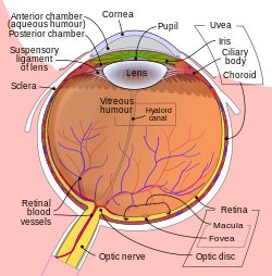
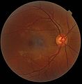
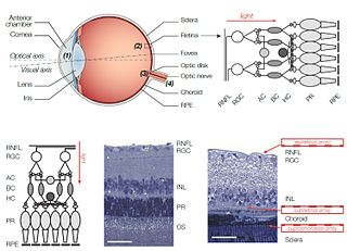
The retina is the innermost, light-sensitive layer of tissue of the eye of most vertebrates and some molluscs. The optics of the eye create a focused two-dimensional image of the visual world on the retina, which then processes that image within the retina and sends nerve impulses along the optic nerve to the visual cortex to create visual perception. The retina serves a function which is in many ways analogous to that of the film or image sensor in a camera.

<span class="mw-page-title-main">Eye</span> Organ that detects light and converts it into electro-chemical impulses in neurons

An eye is a sensory organ that allows an organism to perceive visual information. It detects light and converts it into electro-chemical impulses in neurons (neurones). It is part of an organism's visual system.

<span class="mw-page-title-main">Macula</span> Oval-shaped pigmented area near the center of the retina

The macula (/ˈmakjʊlə/) or macula lutea is an oval-shaped pigmented area in the center of the retina of the human eye and in other animals. The macula in humans has a diameter of around 5.5 mm (0.22 in) and is subdivided into the umbo, foveola, foveal avascular zone, fovea, parafovea, and perifovea areas.

<span class="mw-page-title-main">Peripheral vision</span> Area of ones field of vision outside of the point of fixation

Peripheral vision, or indirect vision, is vision as it occurs outside the point of fixation, i.e. away from the center of gaze or, when viewed at large angles, in the "corner of one's eye". The vast majority of the area in the visual field is included in the notion of peripheral vision. "Far peripheral" vision refers to the area at the edges of the visual field, "mid-peripheral" vision refers to medium eccentricities, and "near-peripheral", sometimes referred to as "para-central" vision, exists adjacent to the center of gaze.

<span class="mw-page-title-main">Photoreceptor cell</span> Type of neuroepithelial cell

A photoreceptor cell is a specialized type of neuroepithelial cell found in the retina that is capable of visual phototransduction. The great biological importance of photoreceptors is that they convert light into signals that can stimulate biological processes. To be more specific, photoreceptor proteins in the cell absorb photons, triggering a change in the cell's membrane potential.

<span class="mw-page-title-main">Retinoschisis</span> Eye disease involving splitting of the retina

Retinoschisis is an eye disease characterized by the abnormal splitting of the retina's neurosensory layers, usually in the outer plexiform layer. Retinoschisis can be divided into degenerative forms which are very common and almost exclusively involve the peripheral retina and hereditary forms which are rare and involve the central retina and sometimes the peripheral retina. The degenerative forms are asymptomatic and involve the peripheral retina only and do not affect the visual acuity. Some rarer forms result in a loss of vision in the corresponding visual field.

<span class="mw-page-title-main">Rod cell</span> Photoreceptor cells that can function in lower light better than cone cells

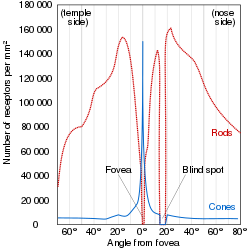
Rod cells are photoreceptor cells in the retina of the eye that can function in lower light better than the other type of visual photoreceptor, cone cells. Rods are usually found concentrated at the outer edges of the retina and are used in peripheral vision. On average, there are approximately 92 million rod cells in the human retina. Rod cells are more sensitive than cone cells and are almost entirely responsible for night vision. However, rods have little role in color vision, which is the main reason why colors are much less apparent in dim light.

<span class="mw-page-title-main">Cone cell</span> Photoreceptor cells responsible for color vision made to function in bright light

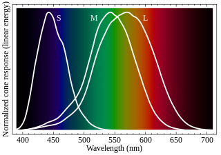
Cone cells or cones are photoreceptor cells in the retinas of vertebrates' eyes. They respond differently to light of different wavelengths, and the combination of their responses is responsible for color vision. Cones function best in relatively bright light, called the photopic region, as opposed to rod cells, which work better in dim light, or the scotopic region. Cone cells are densely packed in the fovea centralis, a 0.3 mm diameter rod-free area with very thin, densely packed cones which quickly reduce in number towards the periphery of the retina. Conversely, they are absent from the optic disc, contributing to the blind spot. There are about six to seven million cones in a human eye, with the highest concentration being towards the macula.

<span class="mw-page-title-main">Visual acuity</span> Clarity of vision

Visual acuity (VA) commonly refers to the clarity of vision, but technically rates an animal's ability to recognize small details with precision. Visual acuity depends on optical and neural factors. Optical factors of the eye influence the sharpness of an image on its retina. Neural factors include the health and functioning of the retina, of the neural pathways to the brain, and of the interpretative faculty of the brain.

<span class="mw-page-title-main">Scanning laser ophthalmoscopy</span>

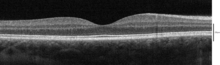
Scanning laser ophthalmoscopy (SLO) is a method of examination of the eye. It uses the technique of confocal laser scanning microscopy for diagnostic imaging of the retina or cornea of the human eye.

<span class="mw-page-title-main">Macular degeneration</span> Medical condition associated with vision loss

Macular degeneration, also known as age-related macular degeneration, is a medical condition which may result in blurred or no vision in the center of the visual field. Early on there are often no symptoms. Over time, however, some people experience a gradual worsening of vision that may affect one or both eyes. While it does not result in complete blindness, loss of central vision can make it hard to recognize faces, drive, read, or perform other activities of daily life. Visual hallucinations may also occur.

<span class="mw-page-title-main">Retinal implant</span>

A retinal implant is a visual prosthesis for restoration of sight to patients blinded by retinal degeneration. The system is meant to partially restore useful vision to those who have lost their photoreceptors due to retinal diseases such as retinitis pigmentosa (RP) or age-related macular degeneration (AMD). Retinal implants are being developed by a number of private companies and research institutions, and three types are in clinical trials: epiretinal, subretinal, and suprachoroidal. The implants introduce visual information into the retina by electrically stimulating the surviving retinal neurons. So far, elicited percepts had rather low resolution, and may be suitable for light perception and recognition of simple objects.

<span class="mw-page-title-main">Retinal pigment epithelium</span>

The pigmented layer of retina or retinal pigment epithelium (RPE) is the pigmented cell layer just outside the neurosensory retina that nourishes retinal visual cells, and is firmly attached to the underlying choroid and overlying retinal visual cells.

<span class="mw-page-title-main">Bird vision</span> Senses for birds

Vision is the most important sense for birds, since good eyesight is essential for safe flight. Birds have a number of adaptations which give visual acuity superior to that of other vertebrate groups; a pigeon has been described as "two eyes with wings". Birds are theropod dinosaurs, and the avian eye resembles that of other reptiles, with ciliary muscles that can change the shape of the lens rapidly and to a greater extent than in the mammals. Birds have the largest eyes relative to their size in the animal kingdom, and movement is consequently limited within the eye's bony socket. In addition to the two eyelids usually found in vertebrates, bird's eyes are protected by a third transparent movable membrane. The eye's internal anatomy is similar to that of other vertebrates, but has a structure, the pecten oculi, unique to birds.

<span class="mw-page-title-main">Mammalian eye</span>

Mammals normally have a pair of eyes. Although mammalian vision is not so excellent as bird vision, it is at least dichromatic for most of mammalian species, with certain families possessing a trichromatic color perception.

<span class="mw-page-title-main">Foveola</span>

The foveola is located within a region called the macula, a yellowish, cone photoreceptor filled portion of the human retina. Approximately 0.35 mm in diameter, the foveola lies in the center of the fovea and contains only cone cells and a cone-shaped zone of Müller cells. In this region the cone receptors are found to be longer, slimmer, and more densely packed than anywhere else in the retina, thus allowing that region to have the potential to have the highest visual acuity in the eye.

Berlin's edema a common condition caused by blunt injury to the eye. It is characterized by decreased vision in the injured eye a few hours after the injury. Under examination the retina appears opaque and white in colour in the periphery but the blood vessels are normally seen along with "cherry red spot" in the foveal region. This whitening is indicative of cell damage, which occurs in the retinal pigment epithelium and outer segment layer of photoreceptors. Damage to the outer segment often results in photoreceptor death through uncertain mechanisms. Usually there is no leakage of fluid and therefore it is not considered a true edema. The choroidal fluorescence in fluorescent angiography is absent. Visual acuity ranges from 20/20 to 20/400.

<span class="mw-page-title-main">Perifovea</span> Region in the retina

Perifovea is a region in the retina that circumscribes the parafovea and fovea and is a part of the macula lutea. The perifovea is a belt that covers a 10° radius around the fovea and is 1.5 mm wide. The perifovea ends when the Henle's fiber layer disappears and the ganglion cells are one-layered.

Occult macular dystrophy (OMD) is a rare inherited degradation of the retina, characterized by progressive loss of function in the most sensitive part of the central retina (macula), the location of the highest concentration of light-sensitive cells (photoreceptors) but presenting no visible abnormality. "Occult" refers to the degradation in the fundus being difficult to discern. The disorder is called "dystrophy" instead of "degradation" to distinguish its genetic origin from other causes, such as age. OMD was first reported by Y. Miyake et al. in 1989.

Drug abuse retinopathy is damage to the retina of the eyes caused by chronic drug abuse. Types of retinopathy caused by drug abuse include maculopathy, Saturday night retinopathy, and talc retinopathy. Common symptoms include temporary and permanent vision loss, blurred vision, and night blindness. Substances commonly associated with this condition include poppers, heroin, cocaine, methamphetamine, tobacco, and alcohol.

References

  1. 1 2 "Simple Anatomy of the Retina". Webvision. University of Utah. Archived from the original on 2011-03-15. Retrieved 2011-09-28.
  2. 1 2 Iwasaki, M; Inomata, H (1986). "Relation between superficial capillaries and foveal structures in the human retina". Investigative Ophthalmology & Visual Science. 27 (12): 1698–705. PMID   3793399.
  3. "eye, human."Encyclopædia Britannica. 2008. Encyclopædia Britannica 2006 Ultimate Reference Suite DVD
  4. Thibos, Larry; Lenner, Katharina; Thibos, Cameron (18 Dec 2023). "Carl Bergmann (1814–1865) and the discovery of the anatomical site in the retina where vision is initiated". Journal of the History of the Neurosciences. 33 (2): 180–203. doi:10.1080/0964704X.2023.2286991. PMID   38109332. S2CID   266361309.
  5. Emmett T. Cunningham; Paul Riordan-Eva (2011). Vaughan & Asbury's general ophthalmology (18th ed.). McGraw-Hill Medical. p. 13. ISBN   978-0-07-163420-5.
  6. Provis, Jan M; Dubis, Adam M; Maddess, Ted; Carroll, Joseph (2013). "Adaptation of the central retina for high acuity vision: Cones, the fovea and the avascular zone". Progress in Retinal and Eye Research. 35: 63–81. doi:10.1016/j.preteyeres.2013.01.005. PMC   3658155 . PMID   23500068.
  7. Tschulakow, Alexander V; Oltrup, Theo; Bende, Thomas; Schmelzle, Sebastian; Schraermeyer, Ulrich (2018). "The anatomy of the foveola reinvestigated". PeerJ. 6: e4482. doi: 10.7717/peerj.4482 . PMC   5853608 . PMID   29576957. CC-BY icon.svg Material was copied from this source, which is available under a Creative Commons Attribution 4.0 International License.
  8. Gregory S. Hageman. "Age-Related Macular Degeneration (AMD)" . Retrieved December 11, 2013.
  9. "Macular Degeneration Frequently Asked Questions". Archived from the original on December 15, 2018. Retrieved December 11, 2013.
  10. Yanoff M, Duker JS. 2014. Ophthalmology. In: Schubert HD, editor. Part 6 Retina and Vitreous, Section 1 Anatomy. 4th ed. China: Elsevier Saunders. p. 420.
  11. Ahmad, Kareem M; Klug, Karl; Herr, Steve; Sterling, Peter; Schein, Stan (2003). "Cell density ratios in a foveal patch in macaque retina" (PDF). Visual Neuroscience. 20 (2): 189–209. CiteSeerX   10.1.1.61.2917 . doi:10.1017/s0952523803202091. PMID   12916740. S2CID   2894449.
  12. Smithsonian/The National Academies, Light:Student Guide and Source Book. Carolina Biological Supply Company, 2002. ISBN   0-89278-892-5.
  13. Krantz, John H. (2012). "Chapter 3: The Stimulus and Anatomy of the Visual System" (PDF). Experiencing Sensation and Perception. Pearson Education. ISBN   978-0-13-097793-9. OCLC   711948862 . Retrieved 6 April 2012.
  14. Fairchild, Mark. (1998), Color Appearance Models. Reading, Mass.: Addison, Wesley, & Longman, p. 7. ISBN   0-201-63464-3
  15. O’Shea, R. P. (1991). Thumb’s rule tested: Visual angle of thumb’s width is about 2 deg. Perception, 20, 415-418. https://doi.org/10.1068/p200415
  16. Yarbus, Alfred L. (1967), "Methods", Eye Movements and Vision, Boston, MA: Springer US, pp. 5–58, doi:10.1007/978-1-4899-5379-7_2, ISBN   978-1-4899-5381-0 , retrieved 2022-01-30
  17. Borji, Ali; Itti, Laurent (2013). "State-of-the-Art in Visual Attention Modeling". IEEE Transactions on Pattern Analysis and Machine Intelligence. 35 (1): 185–207. doi:10.1109/tpami.2012.89. ISSN   0162-8828. PMID   22487985. S2CID   641747.
  18. Tatler, B. W.; Hayhoe, M. M.; Land, M. F.; Ballard, D. H. (2011-05-27). "Eye guidance in natural vision: Reinterpreting salience". Journal of Vision. 11 (5): 5. doi:10.1167/11.5.5. ISSN   1534-7362. PMC   3134223 . PMID   21622729.
  19. Foulsham, Tom; Walker, Esther; Kingstone, Alan (2011). "The where, what and when of gaze allocation in the lab and the natural environment". Vision Research. 51 (17): 1920–1931. doi: 10.1016/j.visres.2011.07.002 . ISSN   0042-6989. PMID   21784095. S2CID   17511680.
  20. Sailer, U. (2005-09-28). "Eye-Hand Coordination during Learning of a Novel Visuomotor Task". Journal of Neuroscience. 25 (39): 8833–8842. doi:10.1523/jneurosci.2658-05.2005. ISSN   0270-6474. PMC   6725583 . PMID   16192373.
  21. Ognibene, Dimitri; Baldassare, Gianluca (2014). "Ecological Active Vision: Four Bioinspired Principles to Integrate Bottom–Up and Adaptive Top–Down Attention Tested With a Simple Camera-Arm Robot". IEEE Transactions on Autonomous Mental Development. 7 (1): 3–25. doi: 10.1109/tamd.2014.2341351 . hdl: 10281/301362 . ISSN   1943-0604. S2CID   1197651.
  22. Foundations of Vision Archived 2013-12-03 at the Wayback Machine , Brian A. Wandell
  23. Krinsky, Norman I; Landrum, John T; Bone, Richard A (2003). "Biologic Mechanisms of the Protective Role of Lutein and Zeaxanthin in the Eye". Annual Review of Nutrition. 23: 171–201. doi:10.1146/annurev.nutr.23.011702.073307. PMID   12626691.
  24. Landrum, John T; Bone, Richard A (2001). "Lutein, Zeaxanthin, and the Macular Pigment". Archives of Biochemistry and Biophysics. 385 (1): 28–40. doi:10.1006/abbi.2000.2171. PMID   11361022.
  25. Beatty, S; Boulton, M; Henson, D; Koh, H-H; Murray, I J (1999). "Macular pigment and age related macular degeneration". British Journal of Ophthalmology. 83 (7): 867–877. doi:10.1136/bjo.83.7.867. PMC   1723114 . PMID   10381676.
  26. 1 2 Curcio, Christine A; Allen, Kimberly A; Sloan, Kenneth R; Lerea, Connie L; Hurley, James B; Klock, Ingrid B; Milam, Ann H (1991). "Distribution and morphology of human cone photoreceptors stained with anti-blue opsin". The Journal of Comparative Neurology. 312 (4): 610–624. doi:10.1002/cne.903120411. PMID   1722224. S2CID   1947541.
  27. Shroff, Anand (2011). An Eye on Numbers: A Ready Reckoner in Ophthalmology. Postscript Media Pvt. p. 97. ISBN   978-81-921123-1-2.
  28. Serpenguzel, Ali; Serpengüzel, Ali; Poon, Andrew W. (2011). Optical Processes in Microparticles and Nanostructures: A Festschrift Dedicated to Richard Kounai Chang on His Retirement from Yale University. World Scientific. ISBN   978-981-4295-77-2.
  29. Curcio, Christine A; Sloan, Kenneth R; Kalina, Robert E; Hendrickson, Anita E (1990). "Human photoreceptor topography". The Journal of Comparative Neurology. 292 (4): 497–523. doi:10.1002/cne.902920402. PMID   2324310. S2CID   24649779.
  30. Vannasdale, D. A; Elsner, A. E; Weber, A; Miura, M; Haggerty, B. P (2009). "Determination of foveal location using scanning laser polarimetry". Journal of Vision. 9 (3): 21.1–17. doi:10.1167/9.3.21. PMC   2970516 . PMID   19757960.
  31. 1 2 Magnussen, Svein; Spillmann, Lothar; Stürzel, Frank; Werner, John S (2001). "Filling-in of the foveal blue scotoma". Vision Research. 41 (23): 2961–2967. doi:10.1016/S0042-6989(01)00178-X. PMC   2715890 . PMID   11704235.
  32. Isobe, Kosaku; Motokawa, Koiti (1955). "Functional Structure of the Retinal Fovea and Maxwell's Spot". Nature. 175 (4450): 306–307. Bibcode:1955Natur.175..306I. doi:10.1038/175306a0. PMID   13235884. S2CID   4181434.
  33. Flom, M. C; Weymouth, F. W (1961). "Centricity of Maxwell's Spot in Strabismus and Amblyopia". Archives of Ophthalmology. 66 (2): 260–268. doi:10.1001/archopht.1961.00960010262018. PMID   13700314.
  34. "Birds Comparative Physiology of Vision" . Retrieved December 29, 2019.
  35. "Avian Eye Tunics" . Retrieved December 29, 2019.