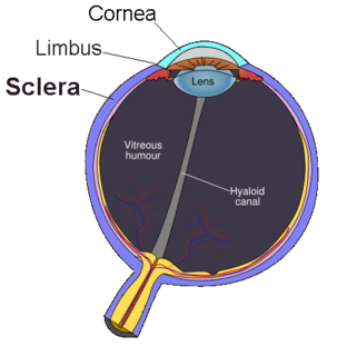
The sclera, also known as the white of the eye or, in older literature, as the tunica albuginea oculi, is the opaque, fibrous, protective, outer layer of the human eye containing mainly collagen and some crucial elastic fiber. In humans, and some other vertebrates, the whole sclera is white, contrasting with the coloured iris, but in most mammals, the visible part of the sclera matches the colour of the iris, so the white part does not normally show while other vertebrates have distinct colors for both of them. In the development of the embryo, the sclera is derived from the neural crest. In children, it is thinner and shows some of the underlying pigment, appearing slightly blue. In the elderly, fatty deposits on the sclera can make it appear slightly yellow. People with dark skin can have naturally darkened sclerae, the result of melanin pigmentation.
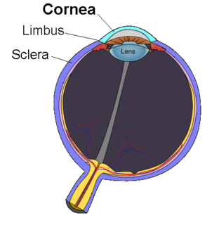
The cornea is the transparent front part of the eye that covers the iris, pupil, and anterior chamber. Along with the anterior chamber and lens, the cornea refracts light, accounting for approximately two-thirds of the eye's total optical power. In humans, the refractive power of the cornea is approximately 43 dioptres. The cornea can be reshaped by surgical procedures such as LASIK.

The aqueous humour is a transparent water-like fluid similar to plasma, but containing low protein concentrations. It is secreted from the ciliary body, a structure supporting the lens of the eyeball. It fills both the anterior and the posterior chambers of the eye, and is not to be confused with the vitreous humour, which is located in the space between the lens and the retina, also known as the posterior cavity or vitreous chamber. Blood cannot normally enter the eyeball.

Kayser–Fleischer rings are dark rings that appear to encircle the iris of the eye. They are due to copper deposition in part of the cornea as a result of particular liver diseases. They are named after German ophthalmologists Bernhard Kayser and Bruno Fleischer who first described them in 1902 and 1903. Initially thought to be due to the accumulation of silver, they were first demonstrated to contain copper in 1934.

The uvea, also called the uveal layer, uveal coat, uveal tract, vascular tunic or vascular layer is the pigmented middle of the three concentric layers that make up an eye.

The human eye is a sense organ, part of the sensory nervous system, that reacts to visible light and allows us to use visual information for various purposes including seeing things, keeping our balance, and maintaining circadian rhythm.
Eye banks recover, prepare and deliver donated eyes for cornea transplants and research. The first successful cornea transplant was performed in 1905 and the first eye bank was founded in 1944. Currently, in the United States, eye banks provide tissue for over 80,000 cornea transplants each year to treat conditions such as keratoconus and corneal scarring. In some cases, the white of the eye (sclera) is used to surgically repair recipient eyes. Unlike other organs and tissues, there is an adequate supply of corneas for transplants in the United States, and excess tissue is exported internationally, where there are shortages in many countries, due to greater demand and a less-developed eye banking infrastructure.

A scleral lens, also known as a scleral contact lens, is a large contact lens that rests on the sclera and creates a tear-filled vault over the cornea. Scleral lenses are designed to treat a variety of eye conditions, many of which do not respond to other forms of treatment.

The long ciliary nerves, two or three in number, are given off from the nasociliary nerve as it crosses the optic nerve. The nasociliary nerve that the long ciliary nerves branch from is itself a branch of the ophthalmic branch (V1) of the trigeminal nerve (CN V).

The short ciliary nerves are nerves of the orbit around the eye. They are branches of the ciliary ganglion. They supply parasympathetic and sympathetic nerve fibers to the ciliary muscle, iris, and cornea. Damage to the short ciliary nerve may result in loss of the pupillary light reflex, or mydriasis.

The anterior ciliary arteries are seven small arteries in each eye-socket that supply the conjunctiva, sclera and the recti muscles. They are derived from the muscular branches of the ophthalmic artery.
The pars plana is part of the ciliary body in the uvea, the middle layer of the three layers that comprise the eye.
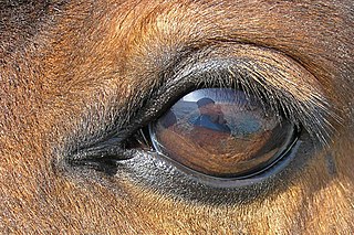
The equine eye is one of the largest of any land mammal. Its visual abilities are directly related to the animal's behavior; for example, it is active during both day and night, and it is a prey animal. Both the strengths and weaknesses of the horse's visual abilities should be taken into consideration when training the animal, as an understanding of the horse's eye can help to discover why the animal behaves the way it does in various situations.
A staphyloma is an abnormal protrusion of the uveal tissue through a weak point in the eyeball. The protrusion is generally black in colour, due to the inner layers of the eye. It occurs due to weakening of outer layer of eye by an inflammatory or degenerative condition. It may be of five types, depending on the location on the eyeball.

Mammals normally have a pair of eyes. Although mammalian vision is not so excellent as bird vision, it is at least dichromatic for most of mammalian species, with certain families possessing a trichromatic color perception.
Sclerocornea is a congenital anomaly of the eye in which the cornea blends with sclera, having no clear-cut boundary. The extent of the resulting opacity varies from peripheral to total. The severe form is thought to be inherited in an autosomal recessive manner, but there may be another, milder form that is expressed in a dominant fashion. In some cases the patients also have abnormalities beyond the eye (systemic), such as limb deformities and craniofacial and genitourinary defects.
The globe of the eye, or bulbus oculi, is the eyeball apart from its appendages. A hollow structure, the bulbus oculi is composed of a wall enclosing a cavity filled with fluid with three coats: the sclera, choroid, and the retina. Normally, the bulbus oculi is bulb-like structure. However, the bulbus oculi is not completely spherical. Its anterior surface, transparent and more curved, is known as the cornea of the bulbus oculi.

The accessory visual structures are the protecting and supporting structures (adnexa) of the eye, including the eyebrow, eyelids, and lacrimal apparatus. The eyebrows, eyelids, eyelashes, lacrimal gland and drainage apparatus all play a crucial role with regards to globe protection, lubrication, and minimizing the risk of ocular infection. The adnexal structures also help to keep the cornea moist and clean.
In biology, a tunica is a layer, coat, sheath, or similar covering. The word came to English from the New Latin of science and medicine. Its literal sense is about the same as that of the word tunic, with which it is cognate. In biology one of its senses used to be the taxonomic name of a genus of plants, but the nomenclature has been revised and those plants are now included in the genus Petrorhagia.

The eagle eye is among the sharpest in the animal kingdom, with an eyesight estimated at 4 to 8 times stronger than that of the average human. Although an eagle may only weigh 10 pounds (4.5 kg), its eyes are roughly the same size as those of a human. Eagle weight varies: a small eagle could weigh 700 grams (1.5 lb), while a larger one could weigh 6.5 kilograms (14 lb); an eagle of about 10 kilograms (22 lb) weight could have eyes as big as that of a human being who weighs 200 pounds (91 kg). Although the size of the eagle eye is about the same as that of a human being, the back side shape of the eagle eye is flatter. Their eyes are stated to be larger in size than their brain, by weight. Color vision with resolution and clarity are the most prominent features of eagles' eyes, hence sharp-sighted people are sometimes referred to as "eagle-eyed". Eagles can identify five distinctly colored squirrels and locate their prey even if hidden.

