Phallolysin

Last updated
This image depicts the various types of mushrooms within the Amanita genus. Phallolysin is most commonly found in the Amanita phalloides species. Amanita phalloides group.jpg
This image depicts the various types of mushrooms within the Amanita genus. Phallolysin is most commonly found in the Amanita phalloides species.

Phallolysin is a protein found the Amanita phalloides species of the Amanita genus of mushrooms, the species commonly known as the death cap mushroom. The protein is toxic and causes cytolysis in many cells found in animals and is noted for its hemolytic properties. [1] It was one of the first toxins discovered in Amanita phalloides when the various toxins in the species where first being researched. [2] The protein itself is observed to come in 3 variations, with observed differences in isoelectric point. [3] Cytolysis can be best described as being the destruction of cells, likely due to exposure from an external source such as pathogens and toxins. Hemolysis then follows a similar destructive pathway, but instead focuses specifically on the destruction of red blood cells. Phallolysin is known to be thermolabile, meaning that it is destroyed at high temperatures, and acid labile, meaning that it is easily broken down in acidic environments.

Contents

History

The toxic properties of death cap mushrooms have been known for most of recorded history, with historical accounts implicating it in the deaths of emperors. [4] Attempts to isolate the toxic compounds began in the late 19th century, with the cytolytic elements of A. phalloides being isolated in 1891. [5] [2] It has been thought that the Roman Emperor Claudius, in 54 AD, and the Holy Roman Emperor Charles VI, in 1740, were some of the earliest victims of death cap poisoning. Due to this, the death cap mushroom has gained the nickname the ‘killer of kings.’ The beginning of this research into the hemolytic properties of the Amanita phalloides, or the Death Cap Mushroom, began with Eduard Rudolf Kobert in 1891, who originally denoted it ‘phallin,’ and was continued by John Jacob Abel and William Webber Ford in 1908. [6] These mushrooms have been attributed to greater than 90% of all cases of mushroom poisoning, in which no active treatment of intoxication cases currently exists. This toxin targets mainly the liver, but may also impact the kidneys and central nervous system as well. [7] As a result of the hemolytic and cytolytic properties, this toxin has been considered for anti-tumor treatments in the early 1970s, where the osmotic lysis of cell membranes was hoped to treat the uncontrollable cell division that tumor cells are notorious for. However, in addition to the non-specificity of the toxin, these trials resulted in the development of an increased potassium concentration in the bloodstream due to the extreme intravascular hemolysis and cytolysis of multiple cell types. Due to the discovery of these lethal side effects, this antitumor treatment route was halted to make room for more sophisticated treatment strategies. [8]

Physical properties

Image depicting the basis of cytolysis, in which an external factor causes an influx of water causing the volume of the cell to exceed the allowed capacity, further causing it to rupture, or lyse, and expel the internal cellular components. Citolisi.png
Image depicting the basis of cytolysis, in which an external factor causes an influx of water causing the volume of the cell to exceed the allowed capacity, further causing it to rupture, or lyse, and expel the internal cellular components.

Phallolysin has three variations, which differ in observed isoelectric point. The variations have differences in the amino acids that make up the protein structure, with identical amounts of some amino acids while varying in others. They have near identical molecular weights of 34 kDa. [1] This protein has been found to be relatively stable in alkaline solutions. The structure of this toxin is a combination of two to three cytolytic proteins. Two of the three proteins have been found to be composed of amino acids with high solubility in water, and each containing one tryptophan residue. [9] This protein is composed of roughly 25% neutral sugars such as galactose, glucose, and mannose, but lack amino sugars. Although they are inactivated by temperatures above 65 °C and acidic environments, they are able to remain stable when coming in contact with proteases or glycosidic enzymes. Such proteases range from pepsin, trypsin, alpha-chymotrypsin, subtilisin, pronase E, bromelin, proteinase K, alpha-amylase, and pancreatin. [10] ,. [11] The cytolytic properties of Phallolysin are able to be attributed by the capability for the toxin to produce protrusions on the plasma membrane, and further rupture these protrusions, resulting in the formation of transmembrane ion channels in the cell membrane lipid bilayers. These openings then allow for water to diffuse into the cell at a rate that the cell is unable to withstand, further destroying the cell via cytolysis, or osmotic lysis. [12] Each of the three types of Phallolysin are denoted as being Phallolysin A, B, and C. Phallolysin A maintains an isoelectric point of 8.1, Phallolysin B maintains an isoelectric point range of 7.5 - 7.6, and Phallolysin C maintains an isoelectric point of 7.0. [1] This protein functions best within a weakly acidic environment, as a result of being denatured by more acidic environments. Temperatures of 65 °C sustained for roughly 30 minutes have the ability to destroy the toxin’s hemolytic capabilities. [13] , [14]

Effects on animal cells

Phallolysin has been observed to have hemolytic properties toward a variety of animal cells, with it primarily being observed in mammals. The toxic effects are reduced at higher temperatures. [1] , [10] These properties are believed to be instigated by ion permeable membrane channels that form as a result of the hemolytic capabilities of phallolysin. In addition to hemolysis, phallolysin in high concentrations are also thought to cause damage to bovine phospholipids with a negative net charge, phosphatidylcholine, and sphingomyelin containing liposomes. However, phospholipid-membranes are only susceptible to phallolysin without receptor proteins being present. [15] These effects are similar to those of staphylococcal 𝛼-toxin. [1] , [6] Cytolysis can go into effect at concentrations beginning at 10-8 M, with a lag time of roughly only 2 to 3 minutes. This is accompanied by the rapid movement of Na+ ions into the cell, and the rapid movement of K+ ions out of the cell. [16] This rapid rate of cytolysis occurs primarily in human erythrocytes, or human red blood cells, due to the presence of glycoproteins or glycolipids that act as specific receptors. This interaction further backs up the claim in which phallolysin does not target the cell’s plasma membrane, but rather the glycoprotein receptors. [17] This protein has also been known to increase levels of cellular phospholipase, which is a lipolytic enzyme that functions as a phospholipid hydrolyzer to break ester bonds in phospholipids. This has been discovered in the specific cellular phospholipase A2 in 3T3 Swiss mouse fibroblasts, which are key components in the structural formation of connective tissues. This study suggests that phallolysin additionally acts by hydrolyzing membrane phospholipids in fibroblasts. [18] Such results also suggest that these cell surfaces in which phallolysin acts upon are also Ca2+ enzyme dependent, however the protein itself is not Ca2+ dependent. [18] , [14] Phallolysin has additionally been discovered to interact mostly with D-galactose and the 𝛽-derivatives, with no glycosylation preferences between O-glycosylation and N-glycosylation. [14] When performing studies on the treatment of rat mast cells with multiple fungal cytolysins, phallolysin was found to interact greatly with lecithin, a fatty substance found in the mice’s tissue. It was also found to cause degranulation, or the release of histamine, of the mast cells, depending on the dosage. [19] Various mammals were additionally tested to determine the sensitivity of red blood cells to this toxin. From this, it was determined that mice are more sensitive than rabbits, and rabbits and guinea pigs are roughly equal in sensitivity. Rabbits and guinea pigs are more sensitive than rats, rats are more sensitive than humans, humans are more sensitive than dogs and pigs, and dogs and pigs are more sensitive than sheep and cattle. This is further displayed in the order: mouse > rabbit = guinea pig > rat > man > dog ≃ pig > sheep-cattle. [13]

See also

Related Research Articles

<i>Amanita phalloides</i> Poisonous mushroom (death cap)

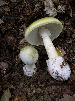
Amanita phalloides, commonly known as the death cap, is a deadly poisonous basidiomycete fungus, one of many in the genus Amanita. Widely distributed across Europe, but introduced to other parts of the world since the late twentieth century, A. phalloides forms ectomycorrhizas with various broadleaved trees. In some cases, the death cap has been introduced to new regions with the cultivation of non-native species of oak, chestnut, and pine. The large fruiting bodies (mushrooms) appear in summer and autumn; the caps are generally greenish in colour with a white stipe and gills. The cap colour is variable, including white forms, and is thus not a reliable identifier.

<span class="mw-page-title-main">Destroying angel</span> Deadly poisonous fungus

The name destroying angel applies to several similar, closely related species of deadly all-white mushrooms in the genus Amanita. They are Amanita virosa in Europe and A. bisporigera and A. ocreata in eastern and western North America, respectively. Another European species of Amanita referred to as the destroying angel, Amanita verna - also referred to as the 'Fool's mushroom' - was first described in France in 1780.

α-Amanitin Chemical compound

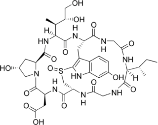
α-Amanitin (alpha-Amanitin) is a cyclic peptide of eight amino acids. It is possibly the most deadly of all the amatoxins, toxins found in several species of the mushroom genus Amanita, one being the death cap as well as the destroying angel, a complex of similar species, principally A. virosa and A. bisporigera. It is also found in the mushrooms Galerina marginata and Conocybe filaris. The oral LD50 of amanitin is 100 μg/kg for rats.

<span class="mw-page-title-main">Oligopeptide</span>

An oligopeptide, often just called peptide, consists of two to twenty amino acids and can include dipeptides, tripeptides, tetrapeptides, and pentapeptides. Some of the major classes of naturally occurring oligopeptides include aeruginosins, cyanopeptolins, microcystins, microviridins, microginins, anabaenopeptins, and cyclamides. Microcystins are best studied, because of their potential toxicity impact in drinking water. A review of some oligopeptides found that the largest class are the cyanopeptolins (40.1%), followed by microcystins (13.4%).

<span class="mw-page-title-main">Phalloidin</span> Chemical compound

Phalloidin belongs to a class of toxins called phallotoxins, which are found in the death cap mushroom (Amanita phalloides). It is a rigid bicyclic heptapeptide that is lethal after a few days when injected into the bloodstream. The major symptom of phalloidin poisoning is acute hunger due to the destruction of liver cells. It functions by binding and stabilizing filamentous actin (F-actin) and effectively prevents the depolymerization of actin fibers. Due to its tight and selective binding to F-actin, derivatives of phalloidin containing fluorescent tags are used widely in microscopy to visualize F-actin in biomedical research.

<span class="mw-page-title-main">Mushroom poisoning</span> Harmful effects from ingestion of toxic substances present in a mushroom

Mushroom poisoning is poisoning resulting from the ingestion of mushrooms that contain toxic substances. Symptoms can vary from slight gastrointestinal discomfort to death in about 10 days. Mushroom toxins are secondary metabolites produced by the fungus.

<i>Amanita virosa</i> Species of fungus

Amanita virosa, commonly known in Europe as the destroying angel or the European destroying angel amanita, is a deadly poisonous basidiomycete fungus, one of many in the genus Amanita. Occurring in Europe, A. virosa associates with various deciduous and coniferous trees. The large fruiting bodies appear in summer and autumn; the caps, stipes and gills are all white in colour.

Amatoxin is the collective name of a subgroup of at least nine related toxic compounds found in three genera of poisonous mushrooms and one species of the genus Pholiotina. Amatoxins are very potent, as little as half a mushroom cap can cause severe liver injury if swallowed.

Cytolysin refers to the substance secreted by microorganisms, plants or animals that is specifically toxic to individual cells, in many cases causing their dissolution through lysis. Cytolysins that have a specific action for certain cells are named accordingly. For instance, the cytolysins responsible for the destruction of red blood cells, thereby liberating hemoglobins, are named hemolysins, and so on. Cytolysins may be involved in immunity as well as in venoms.

<i>Amanita ocreata</i> Species of poisonous fungus in the genus Amanita endemic to western North America

Amanita ocreata, commonly known as the death angel, destroying angel, angel of death or more precisely western North American destroying angel, is a deadly poisonous basidiomycete fungus, one of many in the genus Amanita. Occurring in the Pacific Northwest and California floristic provinces of North America, A. ocreata associates with oak trees. The large fruiting bodies generally appear in spring; the cap may be white or ochre and often develops a brownish centre, while the stipe, ring, gill and volva are all white.

<span class="mw-page-title-main">Hemolysin</span> Molecule destroying the membrane of red blood cells

Hemolysins or haemolysins are lipids and proteins that cause lysis of red blood cells by disrupting the cell membrane. Although the lytic activity of some microbe-derived hemolysins on red blood cells may be of great importance for nutrient acquisition, many hemolysins produced by pathogens do not cause significant destruction of red blood cells during infection. However, hemolysins are often capable of lysing red blood cells in vitro.

β-Amanitin Cyclic peptide part of a group of toxins present in Amanita mushrooms

β-Amanitin (beta-Amanitin) is a cyclic peptide comprising eight amino acids. It is part of a group of toxins called amatoxins, which can be found in several mushrooms belonging to the genus Amanita. Some examples are the death cap and members of the destroying angel complex, which includes A. virosa and A. bisporigera. Due to the presence of α-Amanitin, β-Amanitin, γ-Amanitin and epsilon-Amanitin these mushrooms are highly lethal to human beings.

The phallotoxins consist of at least seven compounds, all of which are bicyclic heptapeptides, isolated from the death cap mushroom (Amanita phalloides). They differ from the closely related amatoxins by being one residue smaller, both in the final product and the precursor protein.

<span class="mw-page-title-main">Antamanide</span> Chemical compound

Antamanide is a cyclic decapeptide isolated from a fungus, the death cap: Amanita phalloides. It is being studied as a potential anti-toxin against the effects of phalloidin and for its potential for treating edema. It contains 1 valine residue, 4 proline residues, 1 alanine residue, and 4 phenylalanine residues with a structure of c(Val-Pro-Pro-Ala-Phe-Phe-Pro-Pro-Phe-Phe). It was isolated by determining the source of the anti-phalloidin activity from a lipophillic extraction from the organism. It has been shown that antamanide can react to form alkali metal ion complexes. These include complexes with sodium and calcium ions. When these complexes are formed, the cyclopeptide structure undergoes a conformational change.

'Staphylococcus aureus delta toxin is a toxin produced by Staphylococcus aureus. It has a wide spectrum of cytolytic activity.

<span class="mw-page-title-main">Amanullinic acid</span> Chemical compound

Amanullinic acid is a cyclic nonribosomal peptide. It is an amatoxin, all of which are found in several members of the mushroom genus Amanita. Amanullinic acid is relatively non-toxic( oral LD50 >20 mg/kg in mice).

<i>Amanita bisporigera</i> Poisonous species of fungus in the family Amanitaceae endemic to North America

Amanita bisporigera is a deadly poisonous species of fungus in the family Amanitaceae. It is commonly known as the eastern destroying angel amanita, the eastern North American destroying angel or just as the destroying angel, although the fungus shares this latter name with three other lethal white Amanita species, A. ocreata, A. verna and A. virosa. The fruit bodies are found on the ground in mixed coniferous and deciduous forests of eastern North America south to Mexico, but are rare in western North America; the fungus has also been found in pine plantations in Colombia. The mushroom has a smooth white cap that can reach up to 10 cm (4 in) across, and a stipe, up to 14 cm (5.5 in) long by 1.8 cm (0.7 in) thick, that has a delicate white skirt-like ring near the top. The bulbous stipe base is covered with a membranous sac-like volva. The white gills are free from attachment to the stalk and crowded closely together. As the species name suggests, A. bisporigera typically bears two spores on the basidia, although this characteristic is not as immutable as was once thought.

<i>Amanita exitialis</i> Species of fungus

Amanita exitialis, also known as the Guangzhou destroying angel, is a mushroom of the large genus Amanita. It is distributed in eastern Asia, and probably also in India where it has been misidentified as A. verna. Deadly poisonous, it is a member of section Phalloideae and related to the death cap A. phalloides. The fruit bodies (mushrooms) are white, small to medium-sized with caps up to 7 cm (2.8 in) in diameter, a somewhat friable ring and a firm volva. Unlike most agaric mushrooms which typically have four-spored basidia, the basidia of A. exitialis are almost entirely two-spored. Eight people were fatally poisoned in China after consuming the mushroom in 2000, and another 20 have been fatally poisoned since that incident. Molecular analysis shows that the species has a close phylogenetic relationship with three other toxic white Amanitas: A. subjunquillea var. alba, A. virosa and A. bisporigera.

Virotoxins are monocyclic peptides formed by at least five different compounds: alaviroidin, viroisin, deoxoviroisin, viroidin, and deoxoviroidin. The structure and biological activity of virotoxins are similar to that of phallotoxins, thus suggesting that virotoxins are biosynthetically derived from phallotoxins or share common precursor pathways. As with phallotoxins, virotoxins are not considered to have significant toxic effects after oral exposure. At the molecular level, like phallotoxins, they interact with actin, stabilizing the bonds between actin monomers and preventing microfilaments depolymerization. However, the ultraviolet-spectra of interaction between actin and virotoxins is different from that of actin-phallotoxins, suggesting a different molecular interaction. Virotoxins have a more flexible structure when compared with phallotoxins and the presence of two additional hydroxyl groups may provide different reactivity. The intraperitoneal LD50 of virotoxins in mice ranges from 1.0 to 5.1 mg/kg and their main toxicological feature is hemorrhagic hepatic necrosis caused by an interaction of the virotoxins with outer surface of the hepatocyte through unknown mechanisms. At this point, the role of virotoxins in human toxicity remains unclear, although due to its poor oral absorption, little clinical importance is given to this class of toxins.

Phoratoxins are a group of peptide toxins that belong to the family of thionins, a subdivision of small plant toxins. Phoratoxins are proteins present in the leaves and branches of the Phoradendron, commonly known as the American variant of the mistletoe, a plant commonly used as decoration during the festive season. The berries of the mistletoe do not contain phoratoxins, making them less toxic compared to other parts of the plant. The toxicity of the mistletoe is dependent on the host tree, since mistletoe is known to be a semi-parasite. The host tree provides fixed inorganic nitrogen compounds necessary for the mistletoe to synthesize phoratoxins.

References

  1. 1 2 3 4 5 Faulstich H, Bühring HJ, Seitz J (September 1983). "Physical properties and function of phallolysin". Biochemistry. 22 (19): 4574–4580. doi:10.1021/bi00288a035. PMID   6626515.
  2. 1 2 Wieland T (1996). "Toxins and Psychoactive Compounds from Mushrooms". In Howard DH, Miller JD (eds.). Human and Animal Relationships. Berlin, Heidelberg: Springer Berlin Heidelberg. pp. 229–248. doi:10.1007/978-3-662-10373-9_12. ISBN   978-3-662-10375-3.
  3. Seeger R (1975-09-01). "Demonstration and isolation of phallolysin, a haemolytic toxin from Amanita phalloides". Naunyn-Schmiedeberg's Archives of Pharmacology. 287 (3): 277–287. doi:10.1007/BF00501473. PMID   1171383. S2CID   20334083.
  4. Marmion VJ, Wiedemann TE (May 2002). "The death of Claudius". Journal of the Royal Society of Medicine. 95 (5): 260–261. doi:10.1177/014107680209500515. PMC   1279685 . PMID   11983773.
  5. Kobert R (1891). "Über Pilzvergiftung". St. Petersburger Med Wochenschr. 16: 463–471.
  6. 1 2 Wieland T (1986). "Phallolysin". In Wieland T (ed.). Peptides of Poisonous Amanita Mushrooms. Springer Series in Molecular Biology. Berlin, Heidelberg: Springer. pp. 207–210. doi:10.1007/978-3-642-71295-1_10. ISBN   978-3-642-71295-1.
  7. Ye Y, Liu Z (August 2018). "Management of Amanita phalloides poisoning: A literature review and update". Journal of Critical Care. 46: 17–22. doi:10.1016/j.jcrc.2018.03.028. PMID   29627659. S2CID   4717865.
  8. Lemaire M, Halperin ML (August 2009). "Rapid tumor cell swelling and bursting: beware of collateral damage". Molecular Therapy. 17 (8): 1310–1311, author reply 1311–1312. doi:10.1038/mt.2009.161. PMC   2835251 . PMID   19644494.
  9. Faulstich H, Bühring HJ, Seitz J (September 1983). "Physical properties and function of phallolysin". Biochemistry. 22 (19): 4574–4580. doi:10.1021/bi00288a035. PMID   6626515.
  10. 1 2 Faulstich H, Weckauf-Bloching M (December 1974). "Isolation and toxicity of two cytolytic glycoproteins from Amanita phalloides mushrooms". Hoppe-Seyler's Zeitschrift Fur Physiologische Chemie. 355 (12): 1489–1494. doi:10.1515/bchm2.1974.355.2.1489. PMID   4461647.
  11. Seeger R (1975). "Some physico-chemical properties of phallolysin obtained from Amanita phalloides". Naunyn-Schmiedeberg's Archives of Pharmacology. 288 (2–3): 155–162. doi:10.1007/BF00500523. PMID   1172197. S2CID   11518543.
  12. Wilmsen HU, Faulstich H, Eibl H, Boheim G (1985-09-01). "Phallolysin. A mushroom toxin, forms proton and voltage gated membrane channels". European Biophysics Journal. 12 (4): 199–209. doi:10.1007/BF00253846. PMID   2412811. S2CID   11991766.
  13. 1 2 Seeger R, Burkhardt M, Haupt M, Feulner L (May 1976). "The haemolytic effect of phallolysin". Naunyn-Schmiedeberg's Archives of Pharmacology. 293 (2): 163–170. doi:10.1007/BF00499222. PMID   8736. S2CID   25332224.
  14. 1 2 3 Antonyuk VO (2005). "Study on carbohydrate specificity of hemolytic lectin from death-cap mushroom (Amanita phalloides (Vaill. Fr.) Secr)". Biopolymers and Cell. 21 (4): 319–325. doi: 10.7124/bc.0006F8 . ISSN   0233-7657.
  15. Bühring HJ, Vaisius AC, Faulstich H (August 1983). "Membrane damage of liposomes by the mushroom toxin phallolysin". Biochimica et Biophysica Acta (BBA) - Biomembranes. 733 (1): 117–123. doi:10.1016/0005-2736(83)90097-4. PMID   6882750.
  16. Seitz J, Adler G, Stofft E, Faulstich H (August 1981). "The mechanism of cytolysis of erythrocytes by the mushroom toxin phallolysin. Morphological and biochemical evidence for sodium influx and swelling". European Journal of Cell Biology. 25 (1): 46–53. PMID   7285957.
  17. Shier WT, DuBourdieu DJ, Kawaguchi H (1983-01-01). "Selection for resistance to phallolysin, a cytolytic toxin from the death-cap mushroom (Amanita phalloides)". Toxicon. 21 (3): 445–448. doi:10.1016/0041-0101(83)90103-4. PMID   6684808.
  18. 1 2 Shier WT, Trotter JT (August 1980). "Stimulation of cell surface phospholipase A2 and prostaglandin synthesis in 3T2 mouse fibroblasts by phallolysin, a toxin from Amanita phalloides". Biochimica et Biophysica Acta (BBA) - Lipids and Lipid Metabolism. 619 (2): 235–246. doi:10.1016/0005-2760(80)90072-7. PMID   7190848.
  19. Seeger R, Bunsen E (1980-12-01). "Degranulation of rat mast cells in vitro by the fungal cytolysins phallolysin, rubescenslysin and fascicularelysin". Naunyn-Schmiedeberg's Archives of Pharmacology. 315 (2): 163–166. doi:10.1007/BF00499259. PMID   7207644. S2CID   2378095.