Scanning electrochemical microscopy

Last updated

Scanning electrochemical microscopy (SECM) is a technique within the broader class of scanning probe microscopy (SPM) that is used to measure the local electrochemical behavior of liquid/solid, liquid/gas and liquid/liquid interfaces. [1] [2] [3] [4] [5] Initial characterization of the technique was credited to University of Texas electrochemist, Allen J. Bard, in 1989. [6] Since then, the theoretical underpinnings have matured to allow widespread use of the technique in chemistry, biology and materials science. Spatially resolved electrochemical signals can be acquired by measuring the current at an ultramicroelectrode (UME) tip as a function of precise tip position over a substrate region of interest. Interpretation of the SECM signal is based on the concept of diffusion-limited current. [7] Two-dimensional raster scan information can be compiled to generate images of surface reactivity and chemical kinetics.

Contents

The technique is complementary to other surface characterization methods such as surface plasmon resonance (SPR), [8] electrochemical scanning tunneling microscopy (ESTM), [9] and atomic force microscopy (AFM) [10] in the interrogation of various interfacial phenomena. In addition to yielding topographic information, SECM is often used to probe the surface reactivity of solid-state materials, electrocatalyst materials, enzymes and other biophysical systems. [11] SECM and variations of the technique have also found use in microfabrication, surface patterning, and microstructuring. [12]

History

The emergence of ultramicroelectrodes (UMEs) around 1980 was pivotal to the development of sensitive electroanalytical techniques like SECM. UMEs employed as probes enabled the study of quick or localized electrochemical reactions. The first SECM-like experiment was performed in 1986 by Engstrom to yield direct observation of reaction profiles and short-lived intermediates. [13] Simultaneous experiments by Allen J. Bard using an Electrochemical Scanning Tunneling Microscope (ESTM) demonstrated current at large tip-to-sample distances that was inconsistent with electron tunneling. This phenomenon was attributed to Faradaic current, compelling a more thorough analysis of electrochemical microscopy. [14] The theoretical basis was presented in 1989 by Bard, where he also coined the term Scanning Electrochemical Microscopy. In addition to the simple collection modes used at the time, Bard illustrated the widespread utility of SECM through the implementation of various feedback modes. [6] As the theoretical foundation developed, annual SECM-related publications steadily rose from 10 to around 80 in 1999, when the first commercial SECM became available. [15] SECM continues to increase in popularity due to theoretical and technological advances that expand experimental modes while broadening substrate scope and enhancing sensitivity. [16]

Principles of operation

Electric potential is manipulated through the UME tip in a bulk solution containing a redox-active couple (e.g. Fe2+/Fe3+). When a sufficiently negative potential is applied, (Fe3+) is reduced to (Fe2+) at the UME tip, generating a diffusion-limited current. [13] The steady-state current is governed by the flux of oxidized species in solution to the UME disc and is given by:

where iT,∞ is the diffusion-limited current, n is the number of electrons transferred at the electrode tip (O + ne → R), F is Faraday's constant, C is the concentration of the oxidized species in solution, D is the diffusion coefficient and a is the radius of the UME disc. In order to probe a surface of interest, the tip is moved closer to the surface and changes in current are measured.

There are two predominant modes of operation, which are feedback mode and collection-generation mode.

Feedback mode

Feedback mode and approach curve Fig3 SECM.jpg
Feedback mode and approach curve

In a bulk solution, the oxidized species is reduced at the tip, producing a steady-state current that is limited by hemispherical diffusion. As the tip approaches a conductive substrate in the solution, the reduced species formed at the tip is oxidized at the conductive surface, yielding an increase in the tip current and creating a regenerative "positive" feedback loop. [6] The opposite effect is observed when probing insulating surfaces, as the oxidized species cannot be regenerated and diffusion to the electrode is inhibited as a result of physical obstruction as the tip approaches the substrate, creating a "negative" feedback loop and decreasing the tip current. An additional parameter to consider when probing insulating surfaces is the electrode sheath diameter, rg, since it contributes to the physical obstruction of diffusion.

The change in tip current as a function of distance d can be plotted as an "approach curve" as shown.

Due to the rate dependent nature of SECM measurements, it is also employed to study electron-transfer kinetics. [17]

Collection-generation modes

Another mode of operation that is employed is tip generation/substrate collection (TG/SC). In TG/SC mode, the tip is held at a potential sufficient for an electrode reaction to occur and "generate" a product while the substrate is held at a potential sufficient for the electrode product to react with or be "collected" by the substrate. [6] The reciprocal to this method is substrate generation/tip collection (SG/TC), where the substrate acts to generate a species that is measured at the tip. Both TG/SC and SG/TC variations are also categorized as "direct" modes. [7]

Two currents are generated: the tip current, iT, and the substrate current, iS. Since the substrate is generally much larger than the tip, the efficiency of collection, iS/iT, is 1 if no reactions occur during the transfer of tip-generated species to the substrate. As the distance between tip and substrate, d, decreases, the collection efficiency, iS/iT, approaches 1.

Alternating Current (ac)-SECM

In ac-SECM a sinusoidal bias is applied to the dc bias of the SECM probe allowing the impedance of a sample to be measured, as is the case in electrochemical impedance spectroscopy. [18] Unlike dc-SECM techniques ac-SECM does not require the use of a redox mediator. This is particularly advantageous for measurements where the redox mediator could affect the chemistry of the system under study. [19] Examples include corrosion studies where a redox mediator may act to inhibit or enhance the rate of corrosion, and biological studies where a redox mediator may be toxic to the living cell under study.

In ac-SECM the feedback response measured is dependent on both the sample type and the experimental conditions. [20] When a sample is insulating the measured impedance will always increase with decreasing probe to sample distance. This is not the case for a conductive sample however. For a conductive sample measured in a high conductivity electrolyte, or measured with a low ac frequency, decreasing the probe to sample distance will lead to an increase in impedance. If, however, a conductive sample is measured in a low conductivity electrolyte, or with a high ac frequency, decreasing the probe to sample distance will result in a lower measured impedance.

SECM Electrode scanning in constant-height mode Fig5 SECM Modded.jpg
SECM Electrode scanning in constant-height mode

SECM imaging

Changes in current as a function of distance between electrode tip and substrate surface allow imaging of insulating and conducting surfaces for topology and reactivity information by moving the tip across surfaces and measuring tip current.

The most common scanning mode is constant-height mode, [7] where the tip height is unchanging and is scanned across the surface in the x-y plane. Alternatively, constant distance measurements are possible, which change the z position to maintain the probe to sample distance as the probe is scanned across the surface in the x-y plane. The constant distance measurement can be based on an electrical signal as is the case in the constant-current mode, [7] where the device attempts to maintain a constant current by changing the substrate to tip distance, d, and recording the change in d. A mechanical signal can also be used to control the probe to sample distance. Examples of this are the intermittent contact (ic)-SECM [21] and shear force [22] techniques which use changes in probe vibration to maintain the probe to sample distance.

Spatial resolution is dependent on the tip radius, the substrate to tip distance, the precision of the electronics, and other considerations.

Instrumentation

Schematic of the fundamental devices involved in most SECM experiments. Experimental control is exercised through manipulation of the piezo controller and potential programmer, while data is collected through the bipotentiostat. SECM Circuitry in pdf.pdf
Schematic of the fundamental devices involved in most SECM experiments. Experimental control is exercised through manipulation of the piezo controller and potential programmer, while data is collected through the bipotentiostat.

Early SECMs were constructed solely by individual lab groups from a set of common components including potentiostat (or bipotentiostat) and potential programmer, current amplifier, piezoelectric positioner and controller, computer, and UME. [4] Many SECM experiments are highly specific in nature, and in-house assembly of SECMs remains common. The development of new techniques toward the reliable nanofabrication of electrodes has been a primary focus in the literature due to several distinct advantages including high mass-transfer rates and low levels of reactant adsorption in kinetic experiments. [23] [24] Additionally, enhanced spatial resolution afforded by reduced tip size expands the scope of SECM studies to smaller and faster phenomena. The following methods encompass an abbreviated summary of fabrication techniques in a rapidly developing field.

Preparation of electrodes

(A) Schematics of an ultramicroelectrode. The exposed metal is the active part of the electrode. (B) Optical micrograph of a tip. The platinum wire (orange) is sealed inside a glass sheath. Fig2 SECM Modded.jpg
(A) Schematics of an ultramicroelectrode. The exposed metal is the active part of the electrode. (B) Optical micrograph of a tip. The platinum wire (orange) is sealed inside a glass sheath.

SECM probes use platinum as the active core material, however carbon, gold, mercury, and silver have all been used. [25] Typical preparation of a microscale electrode is performed by heat sealing a microwire or carbon fiber in a glass capillary under vacuum. This tip can be connected to a larger copper electrode through the use of silver epoxy then polished to yield a sharpened tip. Nanofabrication of electrodes can be performed by etching a metal wire with sodium cyanide and sodium hydroxide. Etched metal wires can then be coated with wax, varnish, molten paraffin or glass, poly(a-methylstyrene), polyimide, [26] electropolymerized phenol, and electrophoretic paint. [27] Nanotips produced by these methods are conical, however disc-shaped tips can be obtained by micropipette pulling of glass sealed electrodes. Nanoscale electrodes allow for high resolution experiments of biological features of sub micron scale or single molecule analysis. "Penetration" experiments, where the tip is inserted into a microstructure (such as a thin polymer film with fixed redox centers) to probe kinetic and concentration parameters, also require the use of nanoscale electrodes. [28] However, microelectrodes remain ideal for quantitative kinetic and feedback mode experiments due to their increased surface area.

Modification of electrodes has developed beyond the size parameter. SECM-AFM probes can act as both a force sensor and electrode through the utilization of a flattened, etched metal wire coated by electrophoretic paint. In this system, the flattened wire acts as a flexible cantilever to measure the force against a sample (AFM) as the wire electrode measures the current (SECM). [2] Similarly, SECM functionality can be imparted into standard AFM probes by sputtering the surface with a conductive metal or by milling an insulated tip with a focused ion beam (FIB). Electron-beam lithography has also been demonstrated to reproducibly generate SECM-AFM probes using silicon wafers. [29] AFM probe manufacturers, such as Scuba Probe Technologies fabricate SECM-AFM probes with reliable electrical contacts for operation in liquids. [30]

Images of the chemical environment that is decoupled from localized topographies are also desirable to study larger or uneven surfaces. "Soft stylus probes" were recently developed by filling a microfabricated track on a polyethylene terephthalate sheet with a conductive carbon ink. Lamination with a polymer film produced v-shaped stylus that was cut to expose the carbon tip. The flexibility inherent in the probe design allows for constant contact with the substrate that bends the probe. When dragged across a sample, probe bending accommodates for topographical differences in the substrate and provides a quasi-constant tip-to-substrate distance, d. [31]

Micro-ITIES probes represent another type of specialty probe that utilizes the Interface between Two Immiscible Electrolyte Solutions (ITIES). These tips feature a tapered pipette containing a solution containing a metal counter electrode, and are used to measure electron and ion transfer events when immersed in a second, immiscible liquid phase containing a counter-reference electrode. [1]

Often the probing of liquid/liquid and air/liquid interfaces via SECM require the use of a submarine electrode. [32] In this configuration, the electrode is fashioned into a hook shape where the electrode can be inverted and submerged within the liquid layer. The UME tip points upwards and can be positioned directly beneath the liquid/liquid or air/liquid interface. The portion of the electrode passing through the interface region is electrically insulated to prevent indirect interfacial perturbations.

Increases in the complexity of electrodes along with decreases in size have prompted the need for high resolution characterization techniques. Scanning electron microscopy (SEM), cyclic voltammetry (CV), and SECM approach curve measurements are frequently applied to identify the dimension and geometry of fabricated probes.

Potentiostat

The potentiostat biases and measures the voltage using the standard three electrode system of voltammetry experiments. The UME acts as the working electrode to apply a controlled potential to the substrate. The auxiliary electrode (or counter electrode) acts to balance the current generated at the working electrode, often through a redox reaction with the solvent or supporting electrolyte. Voltage measured with regard to the well defined reduction potential of the reference electrode, although this electrode itself does not pass any current.

Positioners and translators

SECM utilizes many of the same positioning components that are available to other materials characterization techniques. Precise positioning between the tip and sample is an important factor that is complementary to tip size. The position of the probe relative to a given point on the material surface in the x, y, and z directions is typically controlled by a motor for rough positioning coupled with a piezoelectric motor for finer control. More specifically, systems may feature an inchworm motor that directs coarse positioning with additional z control governed by a PZT piezo pusher. Stepper motors with XYZ piezo block positioner or closed-loop controller systems have also been used. [15]

Applications

SECM has been employed to probe the topography and surface reactivity of solid-state materials, track the dissolution kinetics of ionic crystals in aqueous environments, screen electrocatalytic prospects, elucidate enzymatic activities, and investigate dynamic transport across synthetic/natural membranes and other biophysical systems. Early experiments focused on these solid/liquid interfaces and the characterization of typical solution-based electrochemical systems at higher spatial resolution and sensitivities than bulk electrochemical experiments typically afford. More recently the SECM technique has been adapted to explore the chemical transfer dynamics at liquid/liquid and liquid/gas interfaces.

Solid/Liquid Interface

Microstructuring

SECM and variations of the technique have also found use in microfabrication, surface patterning, and microstructuring. [12] A multitude of surface reactions within this context have been explored including metal deposition, etching and patterning of surfaces by enzymes. Scanning probe lithography (SPL) of surfaces can be performed using the SECM configuration. Due to size limitations in the microfabrication procedures for the UMEs, spatial resolution is decreased, affording larger feature sizes compared to other SPL techniques. An early example demonstrated patterning of dodecylthiolate self-assembled monolayers (SAMs) by moving the UME in a two-dimensional array in close proximity to the surface while applying an oxidative or reductive potential, thus locally desorbing the chemical species. [12] Micron-sized features were effectively patterned into the SAM. An inherent benefit of SECM over other SPL techniques for surface patterning can be attributed to its ability to simultaneously acquire surface-related electrochemical information while performing lithography. Other studies have demonstrated the utility of SECM for the deposition of local gold islands as templates for attachment of biomolecules and fluorescent dyes. [33] Such studies are suggestive of the technique’s potential for the fabrication of nanoscale assemblies, making it particularly suited to explore previously studied systems tethered to small gold clusters.

Varieties of SECM employing the micropipet tip geometry have been used to generate spatially resolved microcrystals of a solid solution. [34] Here, glass microcapillaries with sub-micron sized orifices replace the standard UME allowing femtoliter-sized droplets to be suspended from the capillary over a conductive surface acting as the working electrode. Upon contact with the positively biased surface, the droplets of salt solutions achieve supersaturation and crystallize with well-defined, microscale geometries. Such technology could lend itself well to solid-state electrochemical sensors on microdevices.

Ionic dissolution

Ionic dissolution of crystal by local reduction of M Fig10 SECM.jpg
Ionic dissolution of crystal by local reduction of M

The dissolution of ionic crystals in aqueous environments is fundamentally important to the characterization of a host of naturally occurring and synthetic systems. [35] The high spatial resolution and three-dimensional mobility provided by the UME allows one to probe the dissolution kinetics on specific faces of single ionic crystals, whereas previous characterization techniques relied on a bulk or ensemble average measurement. Due to the high mass transfer rates associated with UMEs in the SECM configuration, it is possible to quantify systems defined by very fast reaction kinetics. In addition, UMEs allow monitoring over a wide dynamic range, making possible the study of ionic solids with large differences in solubility.

Early examples demonstrating the utility of SECM to extract quantitative rate data from such systems was carried out on CuSO4 crystals in an aqueous solution saturated with Cu2+ and SO2−
4
ions. [36] By positioning an UME in the SECM configuration approximately one-electrode radius away from the (100) face of a CuSO4 crystal, it was possible to perturb the dissolution equilibrium by locally reducing Cu2+ at the UME surface. As the crystal face locally dissolved into copper and sulfate ions, a visible pit was formed and the chronoamperometric signal could be monitored as a function of distance between the UME and the crystal. Assuming first or second order kinetic behavior, the dissolution rate constant could then be extracted from the data. Similar studies have been performed on additional crystal systems without a supporting electrolyte. [37]

Electrocatalysis investigation

Approaching the search for novel catalytic materials to replace precious metals used in fuel cells demands extensive knowledge of the oxygen reduction reaction (ORR) occurring at the metal surface. Often even more pressing are the physical limitations imposed by the need to survey and assess the electrocatalytic viability of large numbers of potential catalytic candidates. Some groups studying electrocatalysis have demonstrated the use of SECM as a rapid screening technique that provides local quantitative electrochemical information about catalytic mixtures and materials. [38] [39]

A variety of approaches have been suggested for high throughput assessment of novel metallic electrocatalysts. One functional, non-SECM approach, enabled the electrocatalytic activities of a large number of catalysts to be assessed optically by employing a technique that detected proton production on deposited arrays of proton-sensitive fluorescent dyes. [40] Though of certain utility, the technique suffers from the failure to extract quantitative electrochemical information from any catalytic system of interest, thus requiring the quantitative electrochemical information to be obtained off-line from the array experiment. Bard et al. have demonstrated assessment of electrocatalytic activities at high volume using the SECM configuration. [38] With this approach, direct quantitative electrochemical information from multicomponent systems can be acquired on a rapid screening platform. Such high throughput screening significantly assists the search for abundant, efficient and cost-effective electrocatalytic materials as substitutes for platinum and other precious metals.

Biological analysis

Substrate imaging (constant current mode). (A) SECM image of a portion (10 um x 10 um) of a human breast cell using a 120 nm radius tip. (B) Optical micrograph of the same cell showing the SECM image area delimited by a white square. SECM Fig6 SECM.jpg
Substrate imaging (constant current mode). (A) SECM image of a portion (10 µm × 10 µm) of a human breast cell using a 120 nm radius tip. (B) Optical micrograph of the same cell showing the SECM image area delimited by a white square. SECM

The ability to probe non-conductive surfaces makes SECM a feasible method for analyzing membranes, redox active enzymes, and other biophysical systems.

Changes in intracellular redox activity may be related to conditions such as oxidative stress and cancer. Redox processes of individual living cells can be probed by SECM, which serves as a non-invasive method for monitoring intracellular charge transfer. In such measurements, the cell of interest is immobilized on a surface submerged in a solution with the oxidized form of the redox mediator and feedback mode is employed. A potential is applied to the tip, which reduces the oxidized species, generating a steady-state current, iT. When the tip product enters the cell, it is re-oxidized by processes within the cell and sent back out. Depending on the rate at which tip product is regenerated by the cell, the tip current will change. A study by Liu et al. [41] employed this method and showed that the redox states within three human breast cell lines (nonmotile, motile, and metastatic) were consistently different. SECM can not only examine immobilized cells, but also be used to study the kinetics of immobilized redox-active enzymes. [42]

Transport of ions such as K+ and Na+ across membranes or other biological interfaces is vital to many cell processes; SECM has been employed in studying transport of redox active species across cell membranes. In feedback mode, the transfer of molecules across a membrane can be induced by collecting the transferred species at the tip and forming a concentration gradient. [4] The changes in current can be measured as a function of molecule transport rate.

Liquid/liquid interface

Electrocatalysis

The interface between two immiscible electrolyte solutions (ITIES) can be studied using SECM with a micro-ITIES probe. The probe lies in one layer, and is moved closer to the junction while applying a potential. Oxidation or reduction depletes the substrate concentration, resulting in diffusion from either layer. At close tip-interface distances, rates of diffusion between the organic/aqueous layer for a substrate or ionic species are observed. [43] Electron transfer rates have also been studied extensively at the ITIES. In such experiments, redox couples are dissolved in separate phases and the current at the ITIES is recorded. [1] This is also the fundamental principle in studying transport across membranes.

Liquid/gas interface

The transfer of chemical species across air/liquid interfaces is integral to almost every physical, physiological, biological and environmental system on some level. Thus far, a major thrust in the field has been the quantification of molecular transfer dynamics across monolayer films in order to gain insight into chemical transport properties of cellular membrane systems and chemical diffusion at environmental interfaces. [44]

Though much work has been done in the area of evaporation through monolayers at air/water interfaces, it was the introduction of SECM that provided researchers an alternative method for exploring the permeability of monolayers to small solute molecules across such interfaces. By precisely positioning a submarine electrode beneath an organic monolayer that separates an air/water interface, researchers were able to perturb the oxygen diffusion equilibrium by local reduction of oxygen in the aqueous layer, thereby eliciting diffusion across the monolayer. [45] Diffusion dynamics of the system can be elucidated by measuring the current response at the UME with high spatial and temporal resolution. SECM is quite amenable to such kinetics studies since the current response can be monitored with high sensitivity due to the rapid mass transfer rates associated with UMEs in the SECM configuration. The three dimensional mobility of the UME also affords spatial probing of membranes to identify points of high flux or permeability. A very similar approach has been employed for diffusion studies at liquid/liquid and solid/liquid interfaces.

Related Research Articles

<span class="mw-page-title-main">Surface science</span> Study of physical and chemical phenomena that occur at the interface of two phases

Surface science is the study of physical and chemical phenomena that occur at the interface of two phases, including solid–liquid interfaces, solid–gas interfaces, solid–vacuum interfaces, and liquid–gas interfaces. It includes the fields of surface chemistry and surface physics. Some related practical applications are classed as surface engineering. The science encompasses concepts such as heterogeneous catalysis, semiconductor device fabrication, fuel cells, self-assembled monolayers, and adhesives. Surface science is closely related to interface and colloid science. Interfacial chemistry and physics are common subjects for both. The methods are different. In addition, interface and colloid science studies macroscopic phenomena that occur in heterogeneous systems due to peculiarities of interfaces.

Scanning probe microscopy (SPM) is a branch of microscopy that forms images of surfaces using a physical probe that scans the specimen. SPM was founded in 1981, with the invention of the scanning tunneling microscope, an instrument for imaging surfaces at the atomic level. The first successful scanning tunneling microscope experiment was done by Gerd Binnig and Heinrich Rohrer. The key to their success was using a feedback loop to regulate gap distance between the sample and the probe.

<span class="mw-page-title-main">Cyclic voltammetry</span> Method of analyzing electrochemical reactions

In electrochemistry, cyclic voltammetry (CV) is a type of potentiodynamic measurement. In a cyclic voltammetry experiment, the working electrode potential is ramped linearly versus time. Unlike in linear sweep voltammetry, after the set potential is reached in a CV experiment, the working electrode's potential is ramped in the opposite direction to return to the initial potential. These cycles of ramps in potential may be repeated as many times as needed. The current at the working electrode is plotted versus the applied voltage to give the cyclic voltammogram trace. Cyclic voltammetry is generally used to study the electrochemical properties of an analyte in solution or of a molecule that is adsorbed onto the electrode.

<span class="mw-page-title-main">Self-assembled monolayer</span>

Self-assembled monolayers (SAM) of organic molecules are molecular assemblies formed spontaneously on surfaces by adsorption and are organized into more or less large ordered domains. In some cases molecules that form the monolayer do not interact strongly with the substrate. This is the case for instance of the two-dimensional supramolecular networks of e.g. perylenetetracarboxylic dianhydride (PTCDA) on gold or of e.g. porphyrins on highly oriented pyrolitic graphite (HOPG). In other cases the molecules possess a head group that has a strong affinity to the substrate and anchors the molecule to it. Such a SAM consisting of a head group, tail and functional end group is depicted in Figure 1. Common head groups include thiols, silanes, phosphonates, etc.

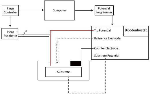
The electrochemical scanning tunneling microscope (EC-STM) is a scanning tunneling microscope that measures the structures of surfaces and electrochemical reactions in solid-liquid interfaces at atomic or molecular scales.

<span class="mw-page-title-main">Voltammetry</span>

Voltammetry is a category of electroanalytical methods used in analytical chemistry and various industrial processes. In voltammetry, information about an analyte is obtained by measuring the current as the potential is varied. The analytical data for a voltammetric experiment comes in the form of a voltammogram which plots the current produced by the analyte versus the potential of the working electrode.

Nanoelectrochemistry is a branch of electrochemistry that investigates the electrical and electrochemical properties of materials at the nanometer size regime. Nanoelectrochemistry plays significant role in the fabrication of various sensors, and devices for detecting molecules at very low concentrations.

<span class="mw-page-title-main">Polarography</span> Type of voltammetry

Polarography is a type of voltammetry where the working electrode is a dropping mercury electrode (DME) or a static mercury drop electrode (SMDE), which are useful for their wide cathodic ranges and renewable surfaces. It was invented in 1922 by Czech chemist Jaroslav Heyrovský, for which he won the Nobel prize in 1959. The main advantages of mercury as electrode material are as follows: 1) a large voltage window: ca. from +0.2 V to -1.8 V vs reversible hydrogen electrode (RHE). Hg electrode is particularly well-suited for studying electroreduction reactions. 2) very reproducible electrode surface, since mercury is liquid. 3) very easy cleaning of the electrode surface by making a new drop of mercury from a large Hg pool connected by a glass capillary.

In electrochemistry, overpotential is the potential difference (voltage) between a half-reaction's thermodynamically-determined reduction potential and the potential at which the redox event is experimentally observed. The term is directly related to a cell's voltage efficiency. In an electrolytic cell the existence of overpotential implies that the cell requires more energy than thermodynamically expected to drive a reaction. In a galvanic cell the existence of overpotential means less energy is recovered than thermodynamics predicts. In each case the extra/missing energy is lost as heat. The quantity of overpotential is specific to each cell design and varies across cells and operational conditions, even for the same reaction. Overpotential is experimentally determined by measuring the potential at which a given current density is achieved.

<span class="mw-page-title-main">Scanning ion-conductance microscopy</span>

Scanning ion-conductance microscopy (SICM) is a scanning probe microscopy technique that uses an electrode as the probe tip. SICM allows for the determination of the surface topography of micrometer and even nanometer-range structures in aqueous media conducting electrolytes. The samples can be hard or soft, are generally non-conducting, and the non-destructive nature of the measurement allows for the observation of living tissues and cells, and biological samples in general.

<span class="mw-page-title-main">Electrochemiluminescence</span> Emission of light from electrochemical reactions

Electrochemiluminescence or electrogenerated chemiluminescence (ECL) is a kind of luminescence produced during electrochemical reactions in solutions. In electrogenerated chemiluminescence, electrochemically generated intermediates undergo a highly exergonic reaction to produce an electronically excited state that then emits light upon relaxation to a lower-level state. This wavelength of the emitted photon of light corresponds to the energy gap between these two states. ECL excitation can be caused by energetic electron transfer (redox) reactions of electrogenerated species. Such luminescence excitation is a form of chemiluminescence where one/all reactants are produced electrochemically on the electrodes.

In analytical chemistry, a rotating disk electrode (RDE) is a working electrode used in three-electrode systems for hydrodynamic voltammetry. The electrode rotates during experiments, inducing a flux of analyte to the electrode. These working electrodes are used in electrochemical studies when investigating reaction mechanisms related to redox chemistry, among other chemical phenomena. The more complex rotating ring-disk electrode can be used as a rotating disk electrode if the ring is left inactive during the experiment.

An ultramicroelectrode (UME) is a working electrode used in a voltammetry. The small size of UME give them large diffusion layers and small overall currents. These features allow UME to achieve useful steady-state conditions and very high scan rates (V/s) with limited distortion. UME were developed independently by Wightman and Fleischmann around 1980. Small current at UME enables electrochemical measurements in low conductive media, where voltage drop associated with high solution resistance makes these experiments difficult for conventional electrodes. Furthermore, small voltage drop at UME leads to a very small voltage distortion at the electrode-solution interface which allows using two-electrode setup in voltammetric experiment instead of conventional three-electrode setup.

A model lipid bilayer is any bilayer assembled in vitro, as opposed to the bilayer of natural cell membranes or covering various sub-cellular structures like the nucleus. They are used to study the fundamental properties of biological membranes in a simplified and well-controlled environment, and increasingly in bottom-up synthetic biology for the construction of artificial cells. A model bilayer can be made with either synthetic or natural lipids. The simplest model systems contain only a single pure synthetic lipid. More physiologically relevant model bilayers can be made with mixtures of several synthetic or natural lipids.

A liquid metal electrode is an electrode that uses a liquid metal, such as mercury, Galinstan, and NaK. They can be used in electrocapillarity, voltammetry, and impedance measurements.

<span class="mw-page-title-main">Fast-scan cyclic voltammetry</span>

Fast-scan cyclic voltammetry (FSCV) is cyclic voltammetry with a very high scan rate (up to 1×10⁶ V·s−1). Application of high scan rate allows rapid acquisition of a voltammogram within several milliseconds and ensures high temporal resolution of this electroanalytical technique. An acquisition rate of 10 Hz is routinely employed.

In electrochemistry, protein film voltammetry is a technique for examining the behavior of proteins immobilized on an electrode. The technique is applicable to proteins and enzymes that engage in electron transfer reactions and it is part of the methods available to study enzyme kinetics.

A probe tip is an instrument used in scanning probe microscopes (SPMs) to scan the surface of a sample and make nano-scale images of surfaces and structures. The probe tip is mounted on the end of a cantilever and can be as sharp as a single atom. In microscopy, probe tip geometry and the composition of both the tip and the surface being probed directly affect resolution and imaging quality. Tip size and shape are extremely important in monitoring and detecting interactions between surfaces. SPMs can precisely measure electrostatic forces, magnetic forces, chemical bonding, Van der Waals forces, and capillary forces. SPMs can also reveal the morphology and topography of a surface.

Electrochemical AFM (EC-AFM) is a particular type of Scanning probe microscopy (SPM), which combines the classical Atomic force microscopy (AFM) together with electrochemical measurements. EC-AFM allows to perform in-situ AFM measurements in an electrochemical cell, in order to investigate the actual changes in the electrode surface morphology during electrochemical reactions. The solid-liquid interface is thus investigated. This technique was developed for the first time in 1996 by Kouzeki et al., who studied amorphous and polycrystalline thin films of Naphthalocyanine on Indium tin oxide in a solution of 0.1 M Potassium chloride (KCl). Unlike the Electrochemical scanning tunneling microscope, previously developed by Itaya and Tomita in 1988, the tip is non-conductive and it is easily steered in a liquid environment.

Single-Entity Electrochemistry (SEE) refers to the electroanalysis of an individual unit of interest. A unique feature of SEE is that it unifies multiple different branches of electrochemistry. Single-Entity Electrochemistry pushes the bounds of the field as it can measure entities on a scale of 100 microns to angstroms. Single-Entity Electrochemistry is important because it gives the ability to view how a single molecule, or cell, or "thing" affects the bulk response, and thus the chemistry that might have gone unknown otherwise. The ability to monitor the movement of one electron or ion from one unit to another is valuable, as many vital reactions and mechanisms undergo this process. Electrochemistry is well suited for this measurement due to its incredible sensitivity. Single-Entity Electrochemistry can be used to investigate nanoparticles, wires, vesicles, nanobubbles, nanotubes, cells, and viruses, and other small molecules and ions. Single-entity electrochemistry has been successfully used to determine the size distribution of particles as well as the number of particles present inside a vesicle or other similar structures

References

  1. 1 2 3 Unwin, Patrick; Barker, Gonsalves; Macpherson, Slevin (1999). "Scanning electrochemical microscopy: beyond the solid/liquid interface". 385: 223–240.{{cite journal}}: Cite journal requires |journal= (help)
  2. 1 2 Zhang, Jie; Barker, Unwin (2001). "Measurement of the forward and back rate constants for electron transfer at the interface between two immiscible electrolyte solutions using scanning electrochemical microscopy (SECM): Theory and experiment". Electrochemistry Communications. 3 (7): 372–378. doi:10.1016/s1388-2481(01)00173-4.
  3. Mirkin, Michael; Sun (2006). "Kinetics of electron transfer reactions at nanoelectrodes". Analytical Chemistry. 78 (18): 6526–6534. doi:10.1021/ac060924q. PMID   16970330.
  4. 1 2 3 Mirkin, Michael; Peng Sun; Francois O. Laforge (2007). "Scanning electrochemical microscopy in the 21st century". Physical Chemistry Chemical Physics. 9 (7): 802–823. Bibcode:2007PCCP....9..802S. doi:10.1039/b612259k. PMID   17287874.
  5. Wittstock, Gunther (2003). "Imaging Localized Reactivities of Surfaces by Scanning Electrochemical Microscopy". Topics in Applied Physics. 85: 335–366. doi:10.1007/3-540-44817-9_11. ISBN   978-3-540-42583-0.
  6. 1 2 3 4 Bard, Allen J.; Fan, Fu Ren F.; Kwak, Juhyoun.; Lev, Ovadia. (1989). "Scanning electrochemical microscopy. Introduction and principles". Analytical Chemistry. 61 (2): 132–138. doi:10.1021/ac00177a011. ISSN   0003-2700.
  7. 1 2 3 4 Bard, Allen (2001). Scanning Electrochemical Microscopy. New York: Marcel Dekker. ISBN   0-8247-0471-1.
  8. Szunerits, Sabine; Knorr, Nikolaus; Calemczuk, Roberto; Livache, Thierry (2004). "New Approach to Writing and Simultaneous Reading of Micropatterns: Combining Surface Plasmon Resonance Imaging with Scanning Electrochemical Microscopy (SECM)". Langmuir. 20 (21): 9236–9241. doi:10.1021/la0492557. ISSN   0743-7463. PMID   15461512.
  9. Wittstock, Gunther; Thomas H. Treutler (2003). "Combination of an electrochemical tunneling microscope (ECSTM) and a scanning electrochemical microscope (SECM): application for tip-induced modification of self-assembled monolayers". Electrochimica Acta. 48 (20–22): 2923–2932. doi:10.1016/s0013-4686(03)00357-8.
  10. Mizaikoff, B; Bertagnolli, Lugstein; Kueng, Kranz (2004). "Mapping of enzyme activity by detection of enzymatic products during AFM imaging with integrated SECM–AFM probes". Ultramicroscopy. 100 (3–4): 127–134. doi:10.1016/j.ultramic.2003.10.004. PMID   15231302.
  11. Wittstock, Gunther; Burchardt, Malte; Pust, SaschaE.; Shen, Yan; Zhao, Chuan (2007). "Scanning Electrochemical Microscopy for Direct Imaging of Reaction Rates". Angewandte Chemie International Edition. 46 (10): 1584–1617. doi:10.1002/anie.200602750. ISSN   1433-7851. PMID   17285666.
  12. 1 2 3 Gorman, Christopher; Stephan Kramer; Ryan R. Fuierer (2003). "Scanning Probe Lithography Using Self-Assembled Monolayers". Chemical Reviews. 103 (11): 4367–4418. doi:10.1021/cr020704m. PMID   14611266.
  13. 1 2 Engstrom, R.C.; M. Weber; D. J. Wunder; R. Burgess; S. Winguist (April 1986). "Measurements within the diffusion layer using a microelectrode probe". Anal. Chem. 58 (4): 844–848. doi:10.1021/ac00295a044.
  14. Bard, Allen; Hsue-Yang Liu; Fu-Ren F. Fan; Charles W. Lin (1986). "Scanning Electrochemical and Tunneling Ultramicroelectrode Microscope for High-Resolution Examination of Electrode Surfaces in Solution". J. Am. Chem. Soc. 108 (13): 3838–3839. doi:10.1021/ja00273a054.
  15. 1 2 Mirkin, Michael; Peng Sun; Francois Laforge (30 November 2006). "Scanning electrochemical microscopy in the 21st century". Physical Chemistry Chemical Physics. 9 (7): 802–23. Bibcode:2007PCCP....9..802S. doi:10.1039/b612259k. PMID   17287874 . Retrieved 5 October 2011.
  16. Mirkin, Michael V.; Nogala, Wojciech; Velmurugan, Jeyavel; Wang, Yixian (2011). "Scanning electrochemical microscopy in the 21st century. Update 1: five years after". Physical Chemistry Chemical Physics. 13 (48): 21196–21312. Bibcode:2011PCCP...1321196M. doi:10.1039/c1cp22376c. PMID   22031463.
  17. Bard, Allen J.; David O. Wipf (1991). "Effect of Heterogeneous Electron-Transfer Rate at the Substrate on the Tip Feedback Current". J. Electrochem. Soc. 138 (2): 469–474. doi:10.1149/1.2085612.
  18. "Introduction to ac-SECM" (PDF). Bio-Logic Science Instruments. Retrieved 2019-05-29.
  19. Horrocks, Benjamin R.; Schmidtke, David.; Heller, Adam.; Bard, Allen J. (1993-12-15). "Scanning electrochemical microscopy. 24. Enzyme ultramicroelectrodes for the measurement of hydrogen peroxide at surfaces". Analytical Chemistry. 65 (24): 3605–3614. doi:10.1021/ac00072a013. ISSN   0003-2700. PMID   8311247.
  20. Diakowski, Piotr M.; Baranski, Andrzej S. (2006). "Positive and negative AC impedance feedback observed above conductive substrates under SECM conditions". Electrochimica Acta. 52 (3): 854–862. doi:10.1016/j.electacta.2006.06.020. ISSN   0013-4686.
  21. McKelvey, Kim; Edwards, Martin A.; Unwin, Patrick R. (August 2010). "Intermittent Contact−Scanning Electrochemical Microscopy (IC−SECM): A New Approach for Tip Positioning and Simultaneous Imaging of Interfacial Topography and Activity". Analytical Chemistry. 82 (15): 6334–6337. doi:10.1021/ac101099e. ISSN   0003-2700. PMID   20583818.
  22. Ballesteros Katemann, Bernardo; Schulte, Albert; Schuhmann, Wolfgang (2003-05-09). "Constant-Distance Mode Scanning Electrochemical Microscopy (SECM)—Part I: Adaptation of a Non-Optical Shear-Force-Based Positioning Mode for SECM Tips". Chemistry - A European Journal. 9 (9): 2025–2033. doi:10.1002/chem.200204267. PMID   12740850.
  23. Schuhmann, Wolfgang; Bernardo Ballesteros Katemann; Albert Schulte (2004). "Constant-Distance Mode Scanning Electrochemical Microscopy. Part II: High-Resolution SECM Imaging Employing Pt Nanoelectrodes as Miniaturized Scanning Probes". Electroanalysis. 16 (1–2): 60–65. doi:10.1002/elan.200302918.
  24. Unwin, Patrick; Martin A Edwards; Sophie Martin; Anna L Whitworth; Julie V Macpherson (2006). "Scanning electrochemical microscopy: principles and applications to biophysical systems". Physiological Measurement. 27 (12): R63–R108. Bibcode:2006PhyM...27R..63E. doi:10.1088/0967-3334/27/12/R01. PMID   17135697. S2CID   18506777 . Retrieved 5 October 2011.
  25. Polcari, David; Dauphin-Ducharme, Philippe; Mauzeroll, Janine (2016-11-23). "Scanning Electrochemical Microscopy: A Comprehensive Review of Experimental Parameters from 1989 to 2015". Chemical Reviews. 116 (22): 13234–13278. doi:10.1021/acs.chemrev.6b00067. ISSN   0009-2665. PMID   27736057.
  26. P. Sun, Z. Zhang, J. Guo and Y. Shao, Anal. Chem., 2001, 73, 5346.
  27. C. J. Slevin, N. J. Gray, J. V. Macpherson, M. A. Webb and P. R. Unwin, Electrochem. Commun., 1999, 1, 282.
  28. Amemiya S, Bard AJ, Fan FR, Mirkin MV, Unwin PR. Annu Rev Anal Chem (Palo Alto Calif). 2008;1:95-131.
  29. Dobson P S, Weaver J M R, Holder M N, Unwin P R and Macpherson J V 2005 Characterization of batch microfabricated scanning electrochemical–atomic force microscopy probes Anal. Chem. 77 424–34
  30. "Electrochemical Cantilevers | Scuba Probe Technologies". 7 September 2017.
  31. Fernando Cortés-Salazar, Markus Träuble, Fei Li, Jean-Marc Busnel, Anne-Laure Gassner, Mohamad Hojeij, Gunther Wittstock, Hubert H Girault. "Soft Stylus Probes for Scanning Electrochemical Microscopy" Analytical Chemistry Vol. 18, Issue 16. Date: 08/15/2009 Start Page: 6889.
  32. Unwin, Patrick; Jie Zhang; Christopher J. Slevin; Colin Morton; Peter Scott; David J. Walton (2001). "New Approach for Measuring Lateral Diffusion in Langmuir Monolayers by Scanning Electrochemical Microscopy (SECM): Theory and Application". The Journal of Physical Chemistry B. 105 (45): 11120–11130. doi:10.1021/jp004592j.
  33. Mandler, Daniel; Tomokazue Matsue; Iva Turyan (2000). "Patterning and Characterization of Surfaces with Organic and Biological Molecules by the Scanning Electrochemical Microscope". Analytical Chemistry. 72 (15): 3431–3435. doi:10.1021/ac000046a. PMID   10952523.
  34. Tian, Zhong-Qun; Dezhi Yang; Lianhuan Han; Yang Yang; Liu-Bin Zhao; Cheng Zong; Yi-Fan Huang; Dongping Zhan (2011). "Solid-State Redox Solutions: Microfabrication and Electrochemistry". Angewandte Chemie. 50 (37): 8679–8682. doi:10.1002/anie.201103386. PMID   21793140.
  35. Unwin, Patrick; Julie Macpherson (1994). "Oscillatory Dissolution of an Ionic Single Crystal Surface Observed with the Scanning Electrochemical Microscope". The Journal of Physical Chemistry. 98 (45): 11764–11770. doi:10.1021/j100096a022.
  36. Unwin, Patrick; Julie Macpherson (1993). "A Novel Approach to the Study of Dissolution Kinetics Using the Scanning Electrochemical Microscope: Theory and Application to Copper Sulfate Pentahydrate Dissolution in Aqueous Sulfuric Acid Solutions". The Journal of Physical Chemistry. 98 (6): 1704–1713. doi:10.1021/j100057a026.
  37. Unwin, Patrick; Julie Macpherson (1996). "Scanning Electrochemical Microscope-Induced Dissolution: Theory and Experiment for Silver Chloride Dissolution Kinetics in Aqueous Solution without Supporting Electrolyte". The Journal of Physical Chemistry. 100 (50): 19475–19483. doi:10.1021/jp9614862.
  38. 1 2 Bard, Allen; Walsh, Fernandez (2005). "Thermodynamic Guidelines for the Design of Bimetallic Catalysts for Oxygen Electroreduction and Rapid Screening by Scanning Electrochemical Microscopy. M-Co (M: Pd, Ag, Au)". Journal of the American Chemical Society. 127 (1): 357–365. doi:10.1021/ja0449729. hdl: 11336/102572 . PMID   15631486.
  39. Bard, Allen; Aguilar, Zoski (2003). "Scanning Electrochemical Microscopy. 46. Shielding Effects on Reversible and Quasireversible Reactions". Analytical Chemistry. 75 (13): 2959–2966. doi:10.1021/ac034011x. PMID   12964739.
  40. Mallouk, Thomas; Erik Reddington; Anthony Sapienza; Bogdan Gurau; Rameshkrishnan Viswanathan; S. Sarangapani; Eugene S. Smotkin (1998). "Combinatorial Electrochemistry: A Highly Parallel, Optical Screening Method for Discovery of Better Electrocatalysts". Science. 280 (5370): 1735–1737. Bibcode:1998Sci...280.1735R. doi:10.1126/science.280.5370.1735. PMID   9624047.
  41. Liu, Biao; Susan A. Rotenberg; Michael V. Mirkin (August 2000). "Scanning electrochemical microscopy of living cells: Different redox activities of nonmetastatic and metastatic human breast cells". PNAS. 97 (18): 9855–9860. Bibcode:2000PNAS...97.9855L. doi: 10.1073/pnas.97.18.9855 . PMC   27604 . PMID   10963658.
  42. Pierce, David T.; Patrick R. Unwin; Allen J. Bard (1992). "Scanning Electrochemical Microscopy: 17. Studies of Enzyme-Mediator Kinetics for Membrane and Surface Immobilized Glucose Oxidase". Anal. Chem. 64 (17): 1795–1804. doi:10.1021/ac00041a011.
  43. Mirkin, Michael; Yuanhua Shao (30 October 1998). "Probing Ion Transfer at the Liquid/Liquid Interface by Scanning Electrochemical Microscopy (SECM)". Journal of Physical Chemistry B. 102 (49): 9915–9921. doi:10.1021/jp9828282.
  44. Thibodeaux, L.J. (1996). Environmental Chemodynamics: Movement of Chemicals in Air, Water and Soil . New York. ISBN   9780471612957.{{cite book}}: CS1 maint: location missing publisher (link)
  45. Unwin, Patrick; Christopher J. Slevin; Steve Ryley; David J. Walton (1998). "A New Approach for Measuring the Effect of a Monolayer on Molecular Transfer across an Air/Water Interface Using Scanning Electrochemical Microscopy". Langmuir. 14 (19): 5331–5334. doi:10.1021/la980320k.